. 2024 Dec 23;192(1):104-117.
doi: 10.1093/bjd/ljae295.

Chondroitin sulfate proteoglycan 4 increases invasion of recessive dystrophic epidermolysis bullosa-associated cutaneous squamous cell carcinoma by modifying transforming growth factor-β signalling

Affiliations

Chondroitin sulfate proteoglycan 4 increases invasion of recessive dystrophic epidermolysis bullosa-associated cutaneous squamous cell carcinoma by modifying transforming growth factor-β signalling

Allison R K Macaulay et al. Br J Dermatol. .

Abstract

Background: Recessive dystrophic epidermolysis bullosa (RDEB) is a rare genetic skin-blistering disorder that often progresses to metastatic cutaneous squamous cell carcinoma (cSCC) at chronic wound sites. Chondroitin sulfate proteoglycan 4 (CSPG4) is a cell-surface proteoglycan that is an oncoantigen in multiple malignancies, where it modulates oncogenic signalling, drives epithelial-to-mesenchymal transition (EMT) and enables cell motility.

Objectives: To evaluate CSPG4 expression and function in RDEB cSCC.

Methods: RDEB cSCC cell lines were used to assess CSPG4-dependent changes in invasive potential, transforming growth factor (TGF)-β1-stimulated signal activation and clinically relevant cytopathology metrics in an in vitro full-thickness tumour model. CSPG4 expression in RDEB cSCC and non-RDEB cSCC tumours was analysed via immunohistochemistry and single-cell RNA sequencing (scRNA-Seq), respectively.

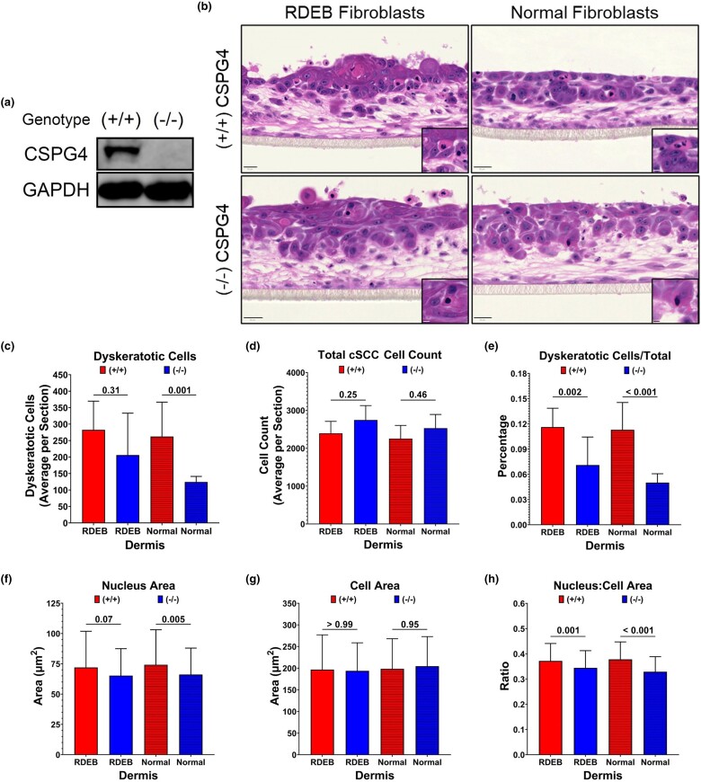
Results: Inhibiting CSPG4 expression reduced invasive potential in multiple RDEB cSCC cell lines and altered membrane-proximal TGF-β signal activation via changes in SMAD3 phosphorylation. CSPG4 expression was uniformly localized to basal layer keratinocytes in fibrotic RDEB skin and tumour cells at the tumour-stroma interface at the invasive front in RDEB cSCC tumours in vivo. Analysis of published scRNA-Seq data revealed that CSPG4 expression was correlated with an enhanced EMT transcriptomic signature in cells at the tumour-stroma interface of non-RDEB cSCC tumours. Cytopathological metrics, for example nucleus : cell area ratio, were influenced by CSPG4 expression in in vitro tumour models.

Conclusions: We determined that CSPG4 expression in RDEB cSCC cell lines enhanced the invasive potential of tumours. Mechanistically, CSPG4 was found to enhance membrane-proximal TGF-β-stimulated signalling via SMAD3, which is a key mediator of EMT in RDEB cSCC. The implication of these studies is that CSPG4 may represent a therapeutic target that can be leveraged for the clinical management of patients with RDEB cSCC.

Plain language summary

Recessive dystrophic epidermolysis bullosa (or ‘RDEB’) is a rare skin disease that affects 3 in 1 million children worldwide. People with RDEB lack an essential protein that allows the skin to resist tearing. This results in widespread blistering on the body and chronic wounds. People with RDEB can develop a lethal form of ‘metastatic’ skin cancer (where cancer cells spread to other parts of the body). However, before a cancer becomes metastatic, a cell must ‘invade’ its original tissue for it to access other parts of the body. This USA-based study investigated a protein called ‘CSPG4’. This protein is present on the surfaces of cancer cells but not in most normal tissues. Our aim was to look at whether CSPG4 enhances the invasion of skin cancer associated with RDEB. This mechanism could be targeted by anti-cancer treatments. We used skin cancer cells from people with RDEB to demonstrate that CSPG4 increases the invasive ability of cancer cells. It does this by changing how the cells respond to a signal (called ‘TGF-β’) that is present in high levels in RDEB skin. We used cancer tissue from people with RDEB to show that CSPG4 is produced by cells at the edges of tumours. These cells are responsible for invading the surrounding normal tissue. Our findings suggest that CSPG4 could be a therapeutic target. Targeting this protein may limit the ability of RDEB-associated skin cancer to invade and metastasize.


Conflict of interest statement

Conflicts of interest The authors declare no conflicts of interest.

Figures

Figure 1
Figure 1
Primary keratinocytes, recessive dystrophic epidermolysis (RDEB) cutaneous squamous cell carcinoma (cSCC) cell lines and RDEB tissues express chondroitin sulfate proteoglycan 4 (CSPG4) at variable levels. (a) Western blot showing cultured primary keratinocytes isolated from punch biopsies from healthy donors (N), from RDEB donors (R) or from RDEB cSCC cell lines derived from whole tumours (Table S1). The CSPG4 banding pattern shows a non-chondroitin sulfate-modified core protein at 250 kDa (the dominant isoform present in all samples) and a chondroitin sulfate-modified isoform that migrates as a diffuse band near 400 kDa. (b) Quantification of both 250- and 400-kDa CSPG4 bands in Western blot shown in (a) normalized to glyceraldehyde 3 phosphate dehydrogenase (GAPDH). (c) Flow cytometry showing the percentage of total population expressing CSPG4 on the cell surface in RDEB cSCC cell lines 2, 4 and 53 (n = 3 biologic replicates). Bars indicate mean, with error bars representing the SD. (d) Fresh frozen paraffin-embedded sections of healthy skin, RDEB skin with a histopathological finding of extensive dermal fibrosis and RDEB cSCC with a histopathological finding of extensive dermal fibrosis stained with haematoxylin and eosin (H&E; top panels). Immunohistochemistry (IHC) against CSPG4 (bottom panels) was evaluated in a semiquantitative manner: healthy skin = +1 (weak and focal); fibrotic RDEB skin = +2 (membranous); RDEB cSCC tumour = +2 (membranous and cytoplasmic). Epidermis (E) and dermis (D) are indicated. Pockets of CSPG4-expressing cells in healthy skin are highlighted with black arrows (bottom left panel). CSPG4 expression on the invasive front of RDEB cSCC are indicated by black arrows (bottom right panel). Inset IHC images show membranous staining of CSPG4 (bottom panels). Scale bars range from 200 to 250 µm. (e) Immunofluorescence (IF) and haematoxylin and eosin staining of fresh frozen samples from healthy skin and clear RDEB skin without a clinical history of wounding. Scale bars = 50 µm in IF images. Haematoxylin and eosin images are at ×40 magnification.
Figure 2
Figure 2
In vitro full-thickness models of recessive dystrophic epidermolysis (RDEB) cutaneous squamous cell carcinoma (cSCC) reveal chondroitin sulfate proteoglycan 4 (CSPG4)-dependent changes in tumour cytopathology. (a) Western blot showing total loss of CSPG4 protein expression in SCC4-derived knockout cells (–/–). (b) Representative haematoxylin and eosin-stained images of a full-thickness model assembled from parental SCC4 (+/+) or knockout (–/–) SCC4 cell lines and RDEB or healthy primary fibroblasts (scale bars = 20 µm). Insets show dyskeratotic cells (scale bars = 5 µm; n≥ 3 models per cSCC/fibroblast combination). (c) Average number of dyskeratotic cells per image (n≥ 9 images per SCC/fibroblast combination). (d) Average total cSCC cell count, including dyskeratotic cells, per image (n≥ 9 images per cSCC/fibroblast combination). (e) Average percentage of dyskeratotic cells per total cSCC cells per image (n≥ 9 images per cSCC/fibroblast combination). (f) Average nucleus area (µm2) and (g) average cell area (µm2) of randomly selected cSCC cells, excluding dyskeratotic cells, for each cSCC/fibroblast combination (n ≥ 180 cells). (h) Ratio of nucleus : cell areas for each randomly selected cSCC cell. Bars represent mean (SD) values. P-values were calculated using one-way Anova with Šídák correction for multiple comparisons. GAPDH, glyceraldehyde 3 phosphate dehydrogenase.
Figure 3
Figure 3
CSPG4 expression in cutaneous squamous cell carcinoma (cSCC) tumour cells is enriched in the tumour-specific keratinocyte (TSK) population and is correlated with an epithelial-to-mesenchymal transition (EMT) transcriptomic phenotype. Analyses of healthy skin (n = 10 participants; n = 9779 total cells) and cSCC tumour (n = 7 patients; n = 4210 total cells) datasets originally published in Ji et al. were replicated as per their methods. (a) Uniform Manifold Approximation and Projection (UMAP) plots showing keratinocyte populations in healthy (top panel) and tumour (centre panel) tissue. Bottom panel shows distribution of CSPG4-expressing cells (n = 229 cells) in the tumour dataset. (b) Heatmap showing the average expression level of hallmark gene markers and CSPG4 (left text) for each keratinocyte cluster (top text). (c) Cell counts in each tumour keratinocyte population (left panel) split by negative (grey) or positive (red) CSPG4 expression status. Proportion of CSPG4+ cells in each population (right panel). (d) CSPG4 expression level [log(normalized counts)] for every cell (left panel) or only CSPG4-expressing cells (right panel) in each tumour keratinocyte population. (e) EMT score for bulk tumour keratinocyte populations (left panel) or split by CSPG4 expression status (right panel). (f) Correlation plot between EMT score and CSPG4 expression level [log(normalized counts)] for all CSPG4+ cells. P-values determined by Student’s t-test with Bonferroni correction.
Figure 4
Figure 4
Chondroitin sulfate proteoglycan 4 (CSPG4) enhances invasive potential in recessive dystrophic epidermolysis bullosa (RDEB) cutaneous squamous cell carcinoma (cSCC) cell lines. (a) Western blot showing knockdown of CSPG4 following transfection with CSPG4-targeted or negative control small interfering RNA (siRNA). (b) Invasion assay using SCC2 and SCC4 cell lines transfected with negative control (+ CSPG4) or CSPG4-targeted (– CSPG4) siRNAs. Plot shows the invasive capacity in response to transforming growth factor (TGF)-β1 (10 ng mL–1). Bars represent the mean (SD) number of invaded cells from five random fields/well from ≥ 6 replicates. P-values were determined by a Student’s t-test with Holm–Šídák correction. (c) Proliferation data from SCC2 (left panel) and SCC4 (right panel) after transfection with negative control (+ CSPG4) or CSPG4-target (– CSPG4) siRNAs, serum-starved to stall proliferation, and then stimulated with or without TGF-β1 in complete growth medium for 48 h. Absorbance at 492 nm is directly proportional to cell count. Bars represent mean (SD) absorbance values (n = 6 replicates). P-values determined with a Student’s t-test. Ctrl, control; GAPDH, glyceraldehyde 3 phosphate dehydrogenase; Neg, negative.
Figure 5
Figure 5
SMAD3 phosphorylation is enhanced in chondroitin sulfate proteoglycan 4 (CSPG4)-expressing SCC2 and SCC4 cell lines. Representative Western blots against phosphorylated SMAD3 (pSMAD3), total SMAD3 and glyceraldehyde 3 phosphate dehydrogenase (GAPDH) loading control in (a) SCC2 cells and (b) SCC4 cells 0.5, 2 and 6 h after transforming growth factor (TGF)-β1 stimulation. Cells were transfected with either negative control (Neg Ctrl) or CSPG4-targeted (CSPG4) small interfering RNAs (siRNAs); three replicates for each siRNA and TGF-β1 stimulation (+/–) condition. Quantification of pSMAD3 : SMAD3 band intensity ratios in (c) SCC2 cells and (d) SCC4 cells. pSMAD3 and SMAD3 band intensities were first normalized to GAPDH loading control, and then a ratio of pSMAD3 : SMAD3 was calculated for each sample. Samples transfected with Neg Ctrl siRNA (+ CSPG4) are indicated by red bars and CSPG4-targeted siRNA (– CSPG4) are indicated by blue bars, with each siRNA set containing samples without (‘SFM’) or with (‘TGF-β1’) TGF-β1 stimulation. Bars represent mean (SEM) band intensity ratio from three replicates. The magnitude of SMAD3 activation in TGF-β1-stimulated samples (‘TGF-β1’) relative to baseline (‘SFM’) was calculated at each timepoint for Neg Ctrl siRNA (+ CSPG4) and CSPG4-targeted siRNA (– CSPG4) conditions in (e) SCC2 cells and (f) SCC4 cells. These calculations were performed by fitting a linear regression model to log-transformed pSMAD3 : SMAD3 band intensity ratios. A magnitude value of 1 indicates no change in SMAD3 activation after TGF-β1 stimulation relative to baseline. Using the linear regression model, estimated marginal means were calculated for each siRNA and stimulation condition and post hoc statistical comparisons were performed to obtain P-values. Bars represent back-transformed means (SEM).
Figure 6
Figure 6
Chronic inflammation and fibrosis increase chondroitin sulfate proteoglycan 4 (CSPG4) expression in recessive dystrophic epidermolysis bullosa (RDEB) skin and promotes RDEB cutaneous squamous cell carcinoma (cSCC) progression. RDEB skin without a history of chronic injury has similar CSPG4 expression patterns to healthy skin, with expression limited to clusters of basal-layer epidermal stem cells (left panel). However, upon cycles of chronic injury at the dermal–epidermal junction (orange bolts) and inflammation because of innate defects in adhesion and wound-healing programmes, RDEB skin becomes increasingly fibrotic (centre panel). This dermal phenotype is marked by a stiff extracellular matrix (collagens and α-smooth muscle actin) assembled primarily by myofibroblasts and excessive secretion of inflammatory cytokines [e.g. transforming growth factor (TGF)-β1, interleukin (IL)-6, tumour necrosis factor (TNF)-α] by infiltrating immune cells. At the onset of fibrosis, CSPG4 expression in basal-layer keratinocytes transitions from heterogeneous to homogeneous. This change may be because CSPG4 expression is upregulated in response to factors and conditions present in fibrotic tissue,, or due to expansion/proliferation of CSPG4-expressing cells in response to inflammatory factors (see Figure 4c). When RDEB skin with underlying fibrosis develops RDEB cSCC, a strong uniform expression of CSPG4 by stroma-interfacing cells is retained (right panel) – potentially due to epigenetic modifications, or stimulation by inflammatory factors. The increased bioavailability of TGF-β1 in RDEB stroma before and after RDEB cSCC development functions to promote tumour cell invasion and/or metastasis through, in part, CSPG4-dependent mechanisms described in this work.

References

    1. Has C, Bauer JW, Bodemer C et al. Consensus reclassification of inherited epidermolysis bullosa and other disorders with skin fragility. Br J Dermatol 2020; 183:614–27. - PubMed
    1. Kim M, Li M, Intong-Wheeler L et al. Epidemiology and outcome of squamous cell carcinoma in epidermolysis bullosa in Australia and New Zealand. Acta Derm Venereol 2018; 98:70–6. - PubMed
    1. Fine J-D, Johnson LB, Weiner M et al. Epidermolysis bullosa and the risk of life-threatening cancers: the National EB Registry experience, 1986–2006. J Am Acad Dermatol 2009; 60:203–11. - PubMed
    1. Mellerio JE, Robertson SJ, Bernardis C et al. Management of cutaneous squamous cell carcinoma in patients with epidermolysis bullosa: best clinical practice guidelines. Br J Dermatol 2016; 174:56–67. - PubMed
    1. Tartaglia G, Cao Q, Padron ZM, South AP. Impaired wound healing, fibrosis, and cancer: the paradigm of recessive dystrophic epidermolysis bullosa. Int J Mol Sci 2021; 22:5104. - PMC - PubMed

MeSH terms