. 2024 Dec;19(1):2380145.
doi: 10.1080/15592294.2024.2380145. Epub 2024 Jul 17.

Circ-0006332 stimulates cardiomyocyte pyroptosis via the miR-143/TLR2 axis to promote doxorubicin-induced cardiac damage

Affiliations

Circ-0006332 stimulates cardiomyocyte pyroptosis via the miR-143/TLR2 axis to promote doxorubicin-induced cardiac damage

Ping Zhang et al. Epigenetics. 2024 Dec.

Abstract

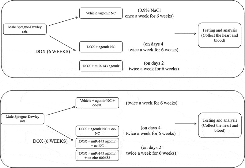
Doxorubicin (DOX)-mediated cardiotoxicity can impair the clinical efficacy of chemotherapy, leading to heart failure (HF). Given the importance of circRNAs and miRNAs in HF, this paper intended to delineate the mechanism of the circular RNA 0006332 (circ -0,006,332)/microRNA (miR)-143/Toll-like receptor 2 (TLR2) axis in doxorubicin (DOX)-induced HF. The binding of miR-143 to circ -0,006,332 and TLR2 was assessed with the dual-luciferase assay, and the binding between miR-143 and circ -0,006,332 was determined with FISH, RIP, and RNA pull-down assays. miR-143 and/or circ -0,006,332 were overexpressed in rats and cardiomyocytes, followed by DOX treatment. In cardiomyocytes, miR-143 and TLR2 expression, cell viability, LDH release, ATP contents, and levels of IL-1β, IL-18, TNF-α, and pyroptosis-related molecules were examined. In rats, cardiac function, serum levels of cardiac enzymes, apoptosis, myocardial fibrosis, and levels of IL-1β, IL-18, TNF-α, TLR2, and pyroptosis-related molecules were detected. miR-143 diminished TLR2 expression by binding to TLR2, and circ -0,006,332 bound to miR-143 to downregulate miR-143 expression. miR-143 expression was reduced and TLR2 expression was augmented in DOX-induced cardiomyocytes. miR-143 inhibited DOX-induced cytotoxicity by suppressing pyroptosis in H9C2 cardiomyocytes. In DOX-induced rats, miR-143 reduced cardiac dysfunction, myocardial apoptosis, myocardial fibrosis, TLR2 levels, and pyroptosis. Furthermore, overexpression of circ -0,006,332 blocked these effects of miR-143 on DOX-induced cardiomyocytes and rats. Circ -0,006,332 stimulates cardiomyocyte pyroptosis by downregulating miR-143 and upregulating TLR2, thus promoting DOX-induced cardiac injury.

Keywords: TLR2; circular RNA-0006332; doxorubicin; heart failure; microRNA-143; pyroptosis.


Conflict of interest statement

No potential conflict of interest was reported by the author(s).

Figures

Figure 1.
Figure 1.
miR-143 reduces injury and pyroptosis in DOX-treated cardiomyocytes. a: dual-luciferase assay to analyse the targeting relationship between miR-143 and TLR2; b: RT-qPCR to detect the levels of miR-143 and TLR2; c: CCK-8 assay to detect cell viability; d: the LDH release kit to detect LDH release; e: ATP production; f: ELISA to detect IL-18 levels in cell supernatant; g: ELISA to detect IL-1β levels in cell supernatant; h: ELISA to detect TNF-α levels in cell supernatant; i-j: Western blotting to detect protein levels of TLR2 and pyroptosis-related molecules. In panels b-h and j, *p < 0.05 compared with the vehicle + mimic NC group, #P < 0.05 compared with the DOX + mimic NC group. Measurement data were expressed as mean ± standard deviation. The independent sample t-test was used for comparisons between two groups, and one-way ANOVA with Tukey’s post hoc test was used for comparisons among multiple groups. The experiments were repeated three times.
Figure 2.
Figure 2.
miR-143 improves myocardial injury in DOX-induced (2.5 mg/kg) rats (n = 5). a-d: quantitative analysis of echocardiographic detection of cardiac function (LVEF, LVFS, LVEDD, and LVESD); e-f: TUNEL staining to detect apoptosis in myocardial tissues; g-i: ELISA to detect levels of CK-MB, cTnT, and LDH in myocardial tissues; j-k: Masson staining to observe myocardial tissue fibrosis, scale bar = 50 μm; *p < 0.05 compared with the vehicle + agomir NC group, #P < 0.05 compared with the DOX + agomir NC group. Values in the figure were measurement data and expressed as mean ± standard deviation. One-way ANOVA with Tukey’s post hoc test (a-d, f-i, k) was used for comparisons among multiple groups.
Figure 3.
Figure 3.
miR-143 prevents DOX-induced cardiomyocyte pyroptosis in vivo. a-g: Western blotting to detect levels of TLR2 protein and pyroptosis-related proteins (ASC, cleave-caspase-1, GSDMD-N, NLRP3, and HMGB1) in cardiac tissues; h-j: ELISA to detect IL-1β, IL-18, and TNF-α in rat serum; k: RT-qPCR to detect miR-143 expression in myocardial tissues; *p < 0.05 compared with the vehicle + agomir NC group, #P < 0.05 compared with the DOX + agomir NC group. Values in the figure were measurement data and expressed as mean ± standard deviation. One-way ANOVA and Tukey’s post hoc test were used for comparison among multiple groups (b-k).
Figure 4.
Figure 4.
Circ -0,006,332 overexpression accelerates DOX-induced cardiomyocyte pyroptosis through miR-143 downregulation. a: RT-qPCR to detect miR-143 and circ -0,006,332 expression in rat heart tissues; b: the FISH assay to detect intracellular co-localization of circ -0,006,332 and miR-143 in cardiomyocytes; c: dual-luciferase assay to detect the binding relationship between miR-143 and circ -0,006,332 in H9C2 cells; d: circ 0006332 and miR-143 expression measured by RT-qPCR. e: RNA pull-down assay to detect the binding of miR-143 to circ -0,006,332; f: CCK-8 assay to detect cardiomyocyte viability; g: LDH release in cardiomyocyte supernatants; h: intracellular ATP contents; i-k: ELISA to detect IL-1β, IL-18, and TNF-α levels in cell supernatants; l-m: protein levels of TLR2 and pyroptosis-related molecules (ASC, cleave-caspase-1, GSDMD-N, NLRP3, and HMGB1) in cardiomyocytes detected by western blotting. In panel A, *p < 0.05 compared with the vehicle group; in panel C, *p < 0.05 compared with the WT + mimic NC group; in panel D, *p < 0.05 compared with the IgG group; in panels F-M, *p < 0.05 compared with the vehicle + mimic NC + oe-NC group, #P < 0.05 compared with the DOX + mimic NC + oe-NC group, and $ p < 0.05 compared with the DOX+ miR-143 mimic + oe-NC group. Values in the figures were measurement data and expressed as mean ± standard deviation. Two-group comparisons were analysed with the independent sample t-test (A, D, E), and one-way ANOVA with Tukey’s post hoc test (C-F-K, M) was used for comparisons among multiple groups. The cell experiments were repeated three times.
Figure 5.
Figure 5.
Overexpression of circ -0,006,332 reverses the improvement effect of miR-143 on DIC in rats. a-dd quantitative analysis of echocardiographic detection of cardiac function (LVEF, LVFS, LVEDD, and LVESD); e-f: TUNEL staining to detect apoptosis in myocardial tissues; g-i: ELISA to detect levels of CK-MB, cTnT, and LDH in myocardial tissues; j-k: Masson staining to observe myocardial tissue fibrosis, scale bar = 50 μm; *p < 0.05 compared with the vehicle + agomir NC + oe-NC group, #P < 0.05 compared with the DOX + agomir NC + oe-NC group, $P < 0.05 compared with the DOX + miR-143 agomir + oe-NC. Values in the figure were measurement data and expressed as mean ± standard deviation. One-way ANOVA with Tukey’s post hoc test (a-d, f-i, k) was used for comparisons among multiple groups.
Figure 6.
Figure 6.
Overexpression of circ -0,006,332 nullifies the inhibition of miR-143 on DOX-induced cardiomyocyte pyroptosis in rats. a-b: Western blotting to detect levels of TLR2 protein and pyroptosis-related proteins (ASC, cleave-caspase-1, GSDMD-N, NLRP3, and HMGB1) in cardiac tissues; c-e: ELISA to detect IL-1β, IL-18, and TNF-α in rat serum; *p < 0.05 compared with the vehicle + agomir NC + oe-NC group, #P < 0.05 compared with the DOX + agomir NC + oe-NC group, $P < 0.05 compared with the DOX + miR-143 agomir + oe-NC group. Values in the figure were measurement data and expressed as mean ± standard deviation. One-way ANOVA and Tukey’s post hoc test were used for comparison among multiple groups (b-e).
Figure 7.
Figure 7.
The schematic diagram in the rat experimental protocol.

Similar articles

Cited by

References

    1. McGowan JV, Chung R, Maulik A, et al. Anthracycline chemotherapy and Cardiotoxicity. Cardiovasc Drugs Ther. 2017;31(1):63–18. doi: 10.1007/s10557-016-6711-0 - DOI - PMC - PubMed
    1. Kim H, Kang HJ, Park KD, et al. Risk factor analysis for secondary malignancy in Dexrazoxane-treated pediatric cancer patients. Cancer Res Treat. 2019;51(1):357–367. doi: 10.4143/crt.2017.457 - DOI - PMC - PubMed
    1. Schirone L, D’Ambrosio L, Forte M, et al. Mitochondria and doxorubicin-induced cardiomyopathy: a complex interplay. Cells. 2022;11(13):2000. doi: 10.3390/cells11132000 - DOI - PMC - PubMed
    1. Kemp CD, Conte JV.. The pathophysiology of heart failure. Cardiovasc Pathol. 2012;21(5):365–371. doi: 10.1016/j.carpath.2011.11.007 - DOI - PubMed
    1. Rawat PS, Jaiswal A, Khurana A, et al. Doxorubicin-induced cardiotoxicity: an update on the molecular mechanism and novel therapeutic strategies for effective management. Biomed Pharmacother. 2021;139:139111708. doi: 10.1016/j.biopha.2021.111708 - DOI - PubMed

LinkOut - more resources