. 2024 Aug 7;138(15):941-962.
doi: 10.1042/CS20240178.

Distinct functional and molecular profiles between physiological and pathological atrial enlargement offer potential new therapeutic opportunities for atrial fibrillation

Affiliations

Distinct functional and molecular profiles between physiological and pathological atrial enlargement offer potential new therapeutic opportunities for atrial fibrillation

Yi Ching Chen et al. Clin Sci (Lond). .

Abstract

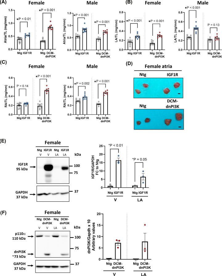
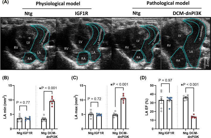
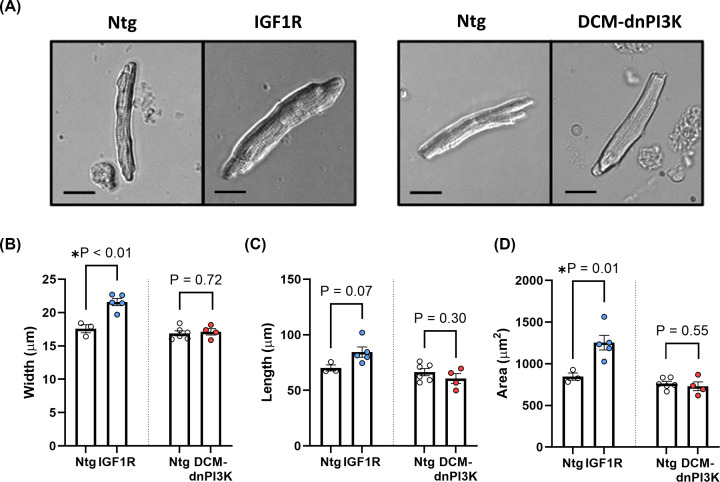
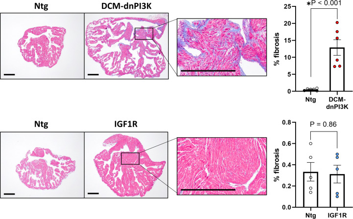
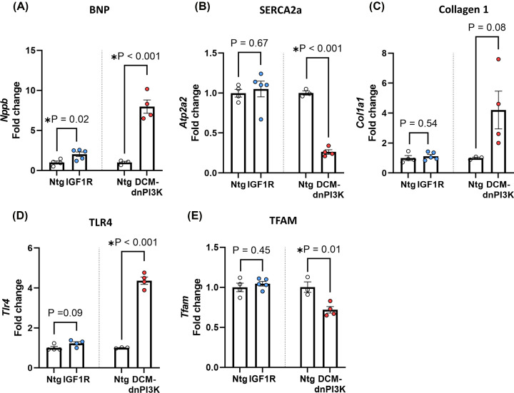
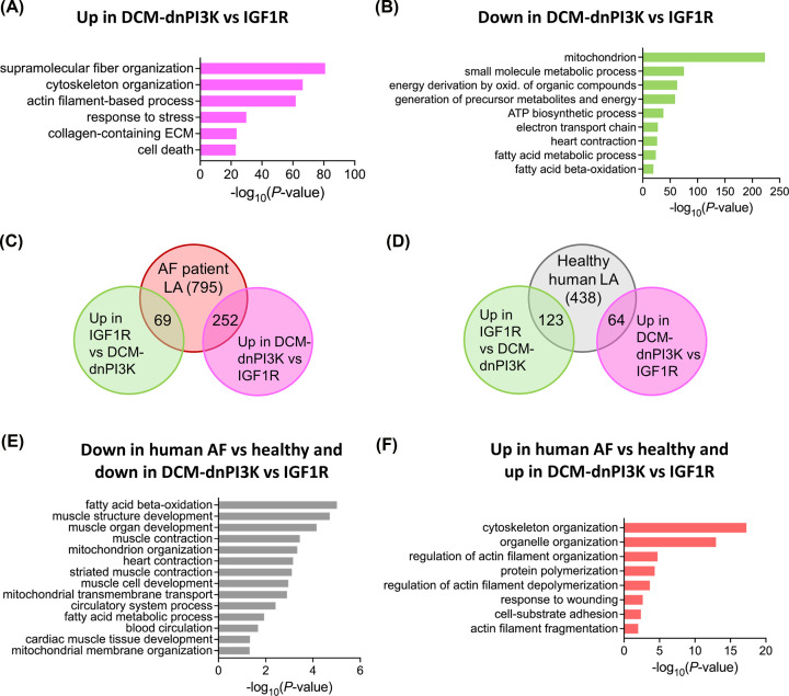
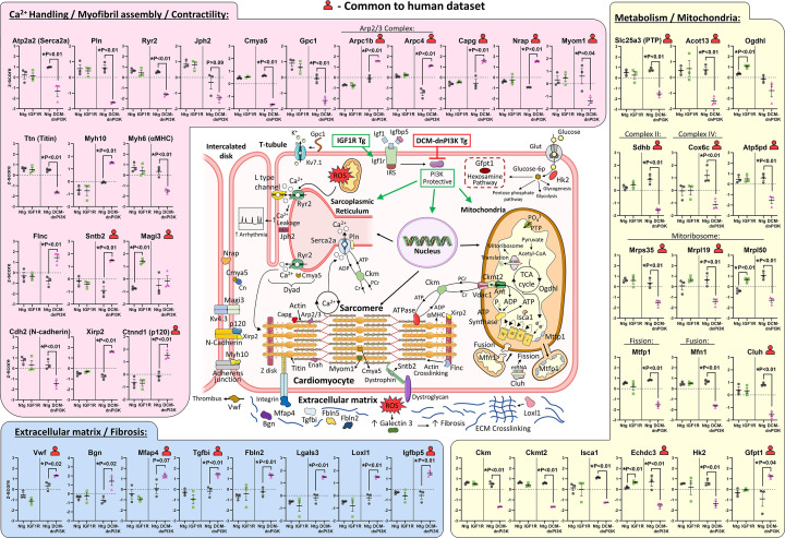
Atrial fibrillation (AF) remains challenging to prevent and treat. A key feature of AF is atrial enlargement. However, not all atrial enlargement progresses to AF. Atrial enlargement in response to physiological stimuli such as exercise is typically benign and reversible. Understanding the differences in atrial function and molecular profile underpinning pathological and physiological atrial remodelling will be critical for identifying new strategies for AF. The discovery of molecular mechanisms responsible for pathological and physiological ventricular hypertrophy has uncovered new drug targets for heart failure. Studies in the atria have been limited in comparison. Here, we characterised mouse atria from (1) a pathological model (cardiomyocyte-specific transgenic (Tg) that develops dilated cardiomyopathy [DCM] and AF due to reduced protective signalling [PI3K]; DCM-dnPI3K), and (2) a physiological model (cardiomyocyte-specific Tg with an enlarged heart due to increased insulin-like growth factor 1 receptor; IGF1R). Both models presented with an increase in atrial mass, but displayed distinct functional, cellular, histological and molecular phenotypes. Atrial enlargement in the DCM-dnPI3K Tg, but not IGF1R Tg, was associated with atrial dysfunction, fibrosis and a heart failure gene expression pattern. Atrial proteomics identified protein networks related to cardiac contractility, sarcomere assembly, metabolism, mitochondria, and extracellular matrix which were differentially regulated in the models; many co-identified in atrial proteomics data sets from human AF. In summary, physiological and pathological atrial enlargement are associated with distinct features, and the proteomic dataset provides a resource to study potential new regulators of atrial biology and function, drug targets and biomarkers for AF.

Keywords: biochemical techniques and resources; cardiac arrhythmia; drug discovery and design; myocardium; proteomics.


Conflict of interest statement

The authors declare that there are no competing interests associated with the manuscript.

Figures

Figure 1
Figure 1. Atria characterisation in mouse models of physiological and pathological cardiac hypertrophy
(A) Total atria weight, (B) left atrial (LA) weight and (C) right atrial (RA) weight normalised to tibia length (TL) in the physiological model (IGF1R Tg) and pathological model (DCM-dnPI3K Tg) at ∼20 weeks of age. Ntg = non-transgenic controls. Female mice: n = 7–10. Male mice: n = 6–9. Data are presented as mean ± SEM. All data passed the normality test. Groups compared using unpaired t-test. (D) Representative atria of female mice. Scale bar = 0.1 cm. (E) Representative Western blot and quantitation of IGF1R protein expression (normalised to GAPDH) in ventricles (V) and left LA of female IGF1R and Ntg mice. Unpaired t-tests (n = 3/group). (F) Representative Western blot and quantitation of dnPI3K protein expression in ventricles (V) and LA of female DCM-dnPI3K and Ntg mice (n = 3/group). N.B. Statistics are not presented on panel F because the dnPI3K band is an ‘all or none’ response given Ntg mice don’t endogenously express the mutant dnPI3K. The relative difference is in reference to the background signal/noise on the blot.
Figure 2
Figure 2. Echocardiographic assessment of left atrial dimensions and function in female mice with physiological or pathological cardiac hypertrophy
(A) Representative echocardiograms from female mice in the physiological model (IGF1R Tg) and pathological model (DCM-dnPI3K Tg) versus Ntg at ∼20 weeks of age. (B–D) LA systolic area (LA min), LA diastolic area (LA max), LA ejection fraction (LA EF), n = 6/group. Data are presented as mean ± SEM. All data passed the normality test. Unpaired t-test.
Figure 3
Figure 3. Atrial myocyte dimensions in female mice with physiological or pathological cardiac hypertrophy
(A) Atrial myocytes isolated from hearts of mouse models of IGF1R Tg and DCM-dnPI3K Tg versus Ntg at ∼20 weeks of age. Atrial myocyte width (B), length (C) and area (D). IGF1R model: 40–170 myocytes were measured from 3 to 5 animals/group. DCM-dnPI3K model: 28–86 myocytes were measured from 4 to 6 animals/group. Data are mean ± SEM. Unpaired t-tests. Scale bar: 20 μm.
Figure 4
Figure 4. Histological assessment of left atrial fibrosis in female mice with physiological or pathological cardiac hypertrophy
Histological examination of LA sections stained with Masson’s Trichrome and quantitation of LA fibrosis in 20 week-old female mouse models of physiological (IGF1R Tg) and pathological cardiac hypertrophy (DCM-dnPI3K Tg) vs. Ntg. Data are mean ± SEM. Unpaired t-tests. n = 5–6/group. Scale bar: 0.5 mm.
Figure 5
Figure 5. Left atrial gene expression in female mice with physiological or pathological cardiac hypertrophy
qPCR assessment of gene expression in female mouse models of physiological (IGF1R Tg) and pathological cardiac hypertrophy (DCM-dnPI3K Tg) vs. Ntg at ∼20 weeks of age. (A) B-type natriuretic peptide (BNP, Nppb), (B) sarcoplasmic/endoplasmic reticulum calcium-ATPase 2a (SERCA2a, Atp2a2) Serca2a, (C) collagen 1 (Col1a1), (D) toll-like receptor 4 (Tlr4), and (E) transcription factor A, mitochondrial (TFAM). Unpaired t-test (A, C–E). For data that failed the normality test (TLR4 gene expression Ntg vs. IGF1R, (D)), a Mann–Whitney test was performed.
Figure 6
Figure 6. Proteomic characterisation of left atria from mouse models of physiological and pathological cardiac hypertrophy
Quantitative proteomic profiling was performed on proteins extracted from female LA tissue of mouse models of physiological (IGF1R Tg) and pathological cardiac hypertrophy (DCM-dnPI3K Tg) versus Ntg at 8 weeks of age (A) (i) Venn diagram depicting total number of proteins quantified in all three biological replicates for each group. (ii) Venn diagram depicting number of proteins uniquely identified within each group (all replicates) and differentially expressed abundant proteins in IGF1R vs Ntg (±0.5 log2 fold change, P<0.05). (iii) Volcano plot with dysregulated protein abundance (±0.5 log2 fold change) in IGF1R versus Ntg, P<0.05 by unpaired t-test. (B) (i) Venn diagram depicting total number of proteins quantified in all three biological replicates for each group. (ii) Venn diagram depicting number of proteins uniquely identified within each group and differentially abundant proteins in DCM-dnPI3K versus Ntg. (iii) Volcano plot with dysregulated protein abundance (±0.5 log2 fold change) in DCM-dnPI3K versus Ntg, P<0.05 by unpaired t-test. (C,D) Clustered protein expression heatmaps for the physiological and pathological models (P<0.05 by unpaired t-test; z-score normalised). Significant biological processes, molecular functions and cellular components (Gene Ontology Enrichment Analysis using gProfiler, term size 5-5000) associated with each cluster shown (blue, down-regulation; red, up-regulation).
Figure 7
Figure 7. Differential mouse proteome analysis and comparative analysis with human atrial proteome
Gene ontology enrichment of significant proteins up- (A) or down-regulated (B) in DCM-dnPI3K vs IGF1R (P<0.05, FC +/- 0.5). Biological processes, molecular functions and cellular components (Gene Ontology Enrichment Analysis using gProfiler, term size 5-5000). (C, D) Comparative analysis between mouse models and human data set (Doll et al.) [33]. Hierarchical clustering of LA from AF and healthy group revealed differentially expressed proteins in AF patients (795 proteins up-regulated in AF) compared with healthy human LA (438 proteins in healthy) based on ANOVA, FDR < 0.05. Based on similarity in co-identified proteins, the DCM-dnPI3K model comprises a greater proportion of proteins to human AF LA (252) than healthy human LA (69) (C), and the IGF1R model comprises a greater proportion of proteins to healthy LA (123) than AF LA (64) (D). N.B. UP in IGF1R vs DCM-dnPI3K is equivalent to DOWN in DCM-dnPI3K versus IGF1R (Supplementary Table S3.15). UP, P<0.05 (+0.5). (E, F) Gene ontology enrichment related to comparative differential proteome analysis of AF from human tissue with mouse models (Supplementary Table S3.15). (E) DOWN, P<0.05 (-0.5); (Supplementary Table S15, column P). (F) UP, P<0.05 (+0.5); (Supplementary Table S15, column Q).
Figure 8
Figure 8. Overview of proteins differentially regulated in atria from the pathological and physiological model, and co-identification in human AF
Regulation of proteins in LA from the physiological (IGF1R) and pathological model (DCM-dnPI3K). Proteins have been grouped into three main categories which are critical for atrial function: (1) Ca2+ handling/Myofibril assembly/Contractility (highlighted with pink shading), (2) Metabolism/Mitochondria (yellow shading), (3) Extracellular matrix/Fibrosis (blue shading). The majority of proteins were dysregulated in the pathological model but not the physiological model. Data points represent individual mice, presented as mean ±SEM. N = 3/group (normalised intensity, z-score). Unpaired t-test. Upper red human torso highlights those proteins also shown to be dysregulated in atrial tissue from patients with human AF. Protein names are defined in Supplementary Table S3.

Similar articles

Cited by

References

    1. Lippi G., Sanchis-Gomar F. and Cervellin G. (2021) Global epidemiology of atrial fibrillation: An increasing epidemic and public health challenge. Int. J. Stroke: Off. J. Int. Stroke Soc. 16, 217–221 10.1177/1747493019897870 - DOI - PubMed
    1. Elliott A.D., Middeldorp M.E., Van Gelder I.C., Albert C.M. and Sanders P. (2023) Epidemiology and modifiable risk factors for atrial fibrillation. Nat. Rev. Cardiol.20, 404-417 36600003 10.1038/s41569-022-00820-8 - DOI - PubMed
    1. Verma A., Jiang C.Y., Betts T.R., Chen J., Deisenhofer I., Mantovan R.et al. . (2015) Approaches to catheter ablation for persistent atrial fibrillation. N. Engl. J. Med. 372, 1812–1822 10.1056/NEJMoa1408288 - DOI - PubMed
    1. Fuster V., Ryden L.E., Cannom D.S., Crijns H.J., Curtis A.B., Ellenbogen K.A.et al. . (2011) 2011 ACCF/AHA/HRS focused updates incorporated into the ACC/AHA/ESC 2006 Guidelines for the management of patients with atrial fibrillation: a report of the American College of Cardiology Foundation/American Heart Association Task Force on Practice Guidelines developed in partnership with the European Society of Cardiology and in collaboration with the European Heart Rhythm Association and the Heart Rhythm Society. J. Am. Coll. Cardiol. 57, e101–e198 10.1016/j.jacc.2010.09.013 - DOI - PubMed
    1. Rienstra M., Damman K., Mulder B.A., Van Gelder I.C., McMurray J.J. and Van Veldhuisen D.J. (2013) Beta-blockers and outcome in heart failure and atrial fibrillation: a meta-analysis. JACC Heart Failure 1, 21–28 10.1016/j.jchf.2012.09.002 - DOI - PubMed

MeSH terms

Substances