. 2024 Sep 1;213(5):678-689.
doi: 10.4049/jimmunol.2300675.

Protective Non-neutralizing anti-N-terminal Domain mAb Maintains Fc-mediated Function against SARS-COV-2 Variants up to BA.2.86-JN.1 with Superfluous In Vivo Protection against JN.1 Due to Attenuated Virulence

Affiliations

Protective Non-neutralizing anti-N-terminal Domain mAb Maintains Fc-mediated Function against SARS-COV-2 Variants up to BA.2.86-JN.1 with Superfluous In Vivo Protection against JN.1 Due to Attenuated Virulence

Arman Izadi et al. J Immunol. .

Abstract

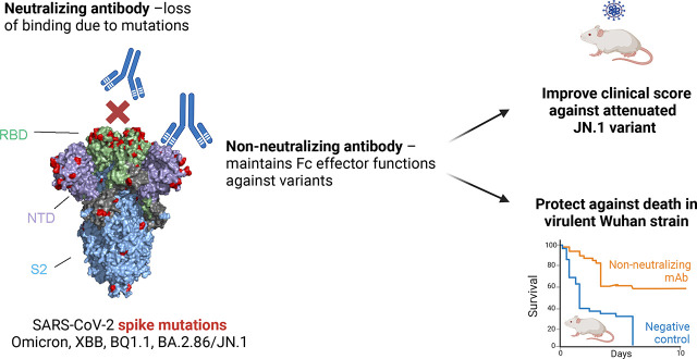
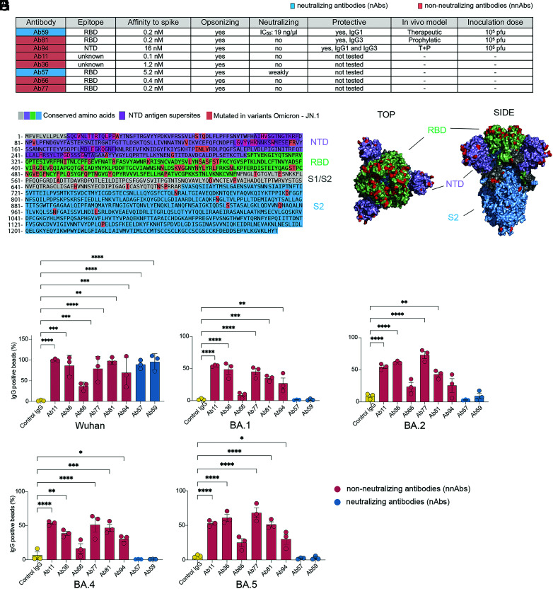
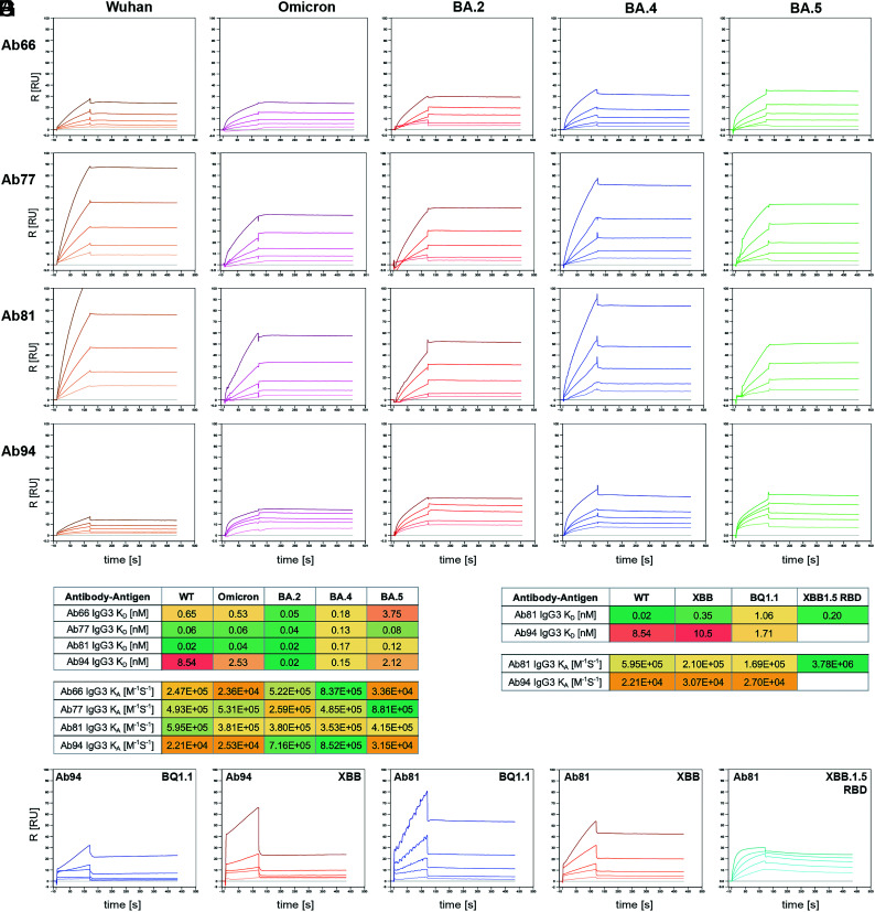
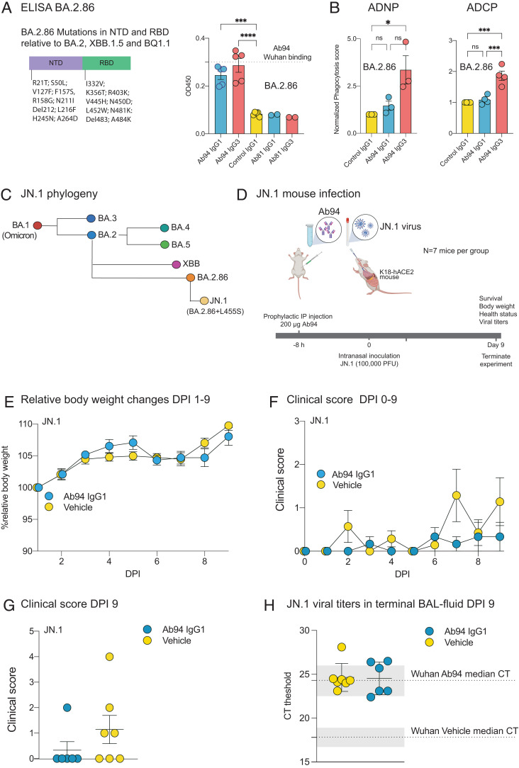
Substantial evidence supports that Fc-mediated effector functions of anti-spike Abs contribute to anti-SARS-Cov-2 protection. We have previously shown that two non-neutralizing but opsonic mAbs targeting the receptor-binding domain and N-terminal domain (NTD), Ab81 and Ab94, respectively, are protective against lethal Wuhan SARS-CoV-2 infection in K18-hACE2 mice. In this article, we investigated whether these protective non-neutralizing Abs maintain Fc-mediated function and Ag binding against mutated SARS-CoV-2 variants. Ab81 and Ab94 retained their nanomolar affinity and Fc-mediated function toward Omicron and its subvariants, such as BA.2, BA.4, BA.5, XBB, XBB1.5, and BQ1.1. However, when encountering the more heavily mutated BA.2.86, Ab81 lost its function, whereas the 10 new mutations in the NTD did not affect Ab94. In vivo experiments with Ab94 in K18-hACE2 mice inoculated with a stringent dose of 100,000 PFU of the JN.1 variant revealed unexpected results. Surprisingly, this variant exhibited low disease manifestation in this animal model with no weight loss or death in the control group. Still, assessment of mice using a clinical scoring system showed better protection for Ab94-treated mice, indicating that Fc-mediated functions are still beneficial. Our work shows that a protective anti-receptor-binding domain non-neutralizing mAb lost reactivity when BA.2.86 emerged, whereas the anti-NTD mAb was still functional. Finally, this work adds new insight into the evolution of the SARS-CoV-2 virus by reporting that JN.1 is substantially less virulent in vivo than previous strains.


Conflict of interest statement

A.I. and P.N. have filed a patent pending for the Abs described in this article. The other authors have no financial conflicts of interest.

Figures

None
Graphical abstract
FIGURE 1.
FIGURE 1.
Protective non-neutralizing Abs Ab81 and Ab94 retain binding against Omicron and subvariants up until BQ.1.1, XBB, and XBB.1.5. (A) Table showing the characteristics of each Ab clone in terms of neutralizing ability, affinity to spike protein, opsonizing ability, and whether tested and protective in an animal model. (B) Spike amino acid sequence divided into different domains (NTD, RBD, S1/2, and S2 domain). Mutations emerging from Omicron to JN.1, according to the GISAID database, are highlighted in red. (C) Spike trimer protein model with the RBD colored in green, NTD in purple, and the S2 domain in light blue (Protein Data Bank code 6vxx) (53). Amino acids of the spike protein of the Wuhan strain with mutated residues, also seen in (B), are highlighted in red on the spike model. (D) Wuhan spike protein was conjugated to microsphere beads and used as a model for SARS-CoV-2 virions. The spike-coated beads were opsonized with anti-spike mAbs. Detection of IgG-positive beads was done by adding an anti-IgG secondary, which was labeled with Alexa Fluor 488. The binding of anti-spike mAbs to spike-coated beads was done by flow cytometry. The y-axis shows the percentage of IgG+ beads. The red Abs are non-neutralizing Abs (nnAbs), and the blue are neutralizing Abs (nAbs). (D–H) Reactivity of anti-spike Abs against Wuhan (D), (E) against Omicron, (F) against BA.2, (G) against BA.4, and (H) against BA.5. Statistical analysis was performed with one-way ANOVA with multiple comparisons against IgG control and corrected with Dunne’s correction test. **p < 0.01, *p < 0.05. ns, p > 0.05.
FIGURE 2.
FIGURE 2.
Binding kinetics with surface plasmon resonance reveal intact nnAb binding to spike variants. Surface plasmon resonance (SPR)–based binding kinetics assays were performed with nnAbs against the spike trimer (WT, Omicron, BA.2, BA.4, and BA.5) as ligand. IgG concentration was kept constant on the sensor chip. (A) IgG3 Ab66. (B) IgG3 Ab77. (C) IgG3 Ab81. (D) IgG3 Ab94. The spike concentration used ranged from 20 to 1.25 nM. (E) Table with KD values and kA for the nnAb Abs against the spike variants. (F) SPR kinetic assay with clones 81 and 94 against XBB and BQ.1.1 spike trimer and XBB.1.5 RBD. (G) Table with KD values and kA for the nnAb Abs against the spike variants. The experiments were performed at least two times, and a representative graph is shown in the figure.
FIGURE 3.
FIGURE 3.
IgG3 subclasses of clones 81 and 94 show potent Fc-mediated phagocytosis against the BQ1.1, XBB, and XBB1.5 variants. Phagocytosis of spike-coated beads by the THP-1 cells was analyzed by flow cytometry. In (A–C, E and F), experiments were done at MOP 12.5 (100,000 THP-1 cells, 1.25 million beads) with 2.5 μg/ml of Abs to opsonize the spike beads. In short, THP-1 cells were gated for bead signal (Supplemental Fig. 3) to generate a percentage of bead-positive cells (dubbed association) seen in (A). The bead-positive cells were further analyzed for bead signal (median fluorescence signal [MFI]), a metric for the quantity of beads. By using association and MFI, a phagocytosis score was produced (percentage of association multiplied by MFI/100). (A) The percentage of bead-positive cells on the y-axis with the treatments on the x-axis. (B) The bead signal (APC) for the bead-positive population on the Y-axis. (C) Phagocytosis score is shown on the y-axis. (D) The phagocytosis score is shown for IgG subclass controls at MOP 25; no difference was observed. (E) The percentage of bead-positive cells is shown at MOP 12.5 (100,000 cells, 2.5 million beads). (F) The phagocytosis score is shown at MOP 25. (G and I) The graphs show association (bead+ cells) as a function of MOP (ranging from 25-6.25), and MOP50 was generated by a nonlinear regression analysis. (G) The differences between IgG3 single mAbs versus mixture are shown. (H) Comparison of the differences between IgG1 single mAbs versus IgG1 cocktails. (I) The differences between IgG1 and IgG3 cocktails are shown. (J). MOP50 for each treatment is shown in addition to the Hill-slope value. (K) Association and phagocytosis score by THP-1 cells incubated with XBB and XBB.1.5 spike-coated beads at MOP 25 is shown. In (A–C and E–J), the data are from five independent experiments. In (D), the data are from three independent experiments, although for (K), the data are from four experiments (XBB) and five (XBB.1.5), respectively. In all panels, the mean is shown, and the error bars are SEM. Statistical analysis was performed with one-way ANOVA with multiple comparisons corrected with Dunne correction test for (A–C, E, and F) compared against IgG control, whereas for (K), comparisons were made for all treatments and corrected with Tukey correction test. **p < 0.01, *p < 0.05. ns, p > 0.05.
FIGURE 4.
FIGURE 4.
JN.1 strain exhibits significantly attenuated virulence in K18-hACE2 mice. (A) Mutations in BA.2.86 NTD and RBD domain compared BA.2, XBB 1.5, and BQ1.1 variants using GISAID database sequence alignment. Ab81 and Ab94 binding to BA.2.86 spike trimer by ELISA with Wuhan signal used as a control highlighted in the graph. (B) Ab-dependent cellular phagocytosis (ADCP) by THP-1 cells (MOP 30, 2.5 µg per mL) and Ab-dependent neutrophil phagocytosis (ADNP) of BA.2.86 spike trimer coated beads (MOP, 30; 2.5 μg/mL mAb and 1% Ab-depleted serum). (C) Evolutionary tree on SARS-CoV-2 variants development focusing on BA.2.86-JN.1. (D) Human ACE2-K18 mice (n = 7 per group) were prophylactically injected i.p. with Ab treatment or vehicle. Eight hours post-treatment, the mice were infected intranasally with SARS-CoV-2 (JN.1). Body weights were measured over 9 d. A clinical scoring system was employed in which body posture, piloerection, abnormal respiration (shortness of breath, wheeze, cough), and weakness/fatigue were assessed on a scale where 0 indicates unaffected, 1 indicates mild, 2 indicates moderate, and 3 indicates severe symptoms. At day 9, terminal BAL fluid was extracted and analyzed by qPCR for viral titers. (E) The mean relative body weight for each group from days 0 to 9 is shown, whereas (F) shows the mean clinical score. (G) Clinical score at day 9 for all mice in each group. (H) Terminal titers of viral load were analyzed from BAL fluid from mice. qPCR was done on BAL fluid, and the cycle threshold was recorded and graphed. The mean value is shown, and previous titers from Ab94 and vehicle against the Wuhan strain are highlighted as gray-shaded areas in the graph as a reference. The mean value is shown for (A, B, and E–H), with error bars being SEM. One mouse in the Ab94 group was omitted from analysis because it was interpreted as an outlier due to unexpected rapid weight loss at day 5 and had to be euthanized at day 7, whereas none of the other 13 mice experienced any weight loss over 9 d. Statistical analysis was performed using one-way ANOVA with multiple comparisons corrections corrected by Dunnett’s post hoc test. The illustrations in (C and D) were made using BioRender. DPI, days postinfection.

Similar articles

References

    1. Hu, B., Guo H., Zhou P., Shi Z.-L.. 2021. Characteristics of SARS-CoV-2 and COVID-19. Nat. Rev. Microbiol. 19: 141–154. - PMC - PubMed
    1. Chen, P., Nirula A., Heller B., Gottlieb R. L., Boscia J., Morris J., Huhn G., Cardona J., Mocherla B., Stosor V., et al. ; BLAZE-1 Investigators . 2020. SARS-CoV-2 neutralizing antibody LY-CoV555 in outpatients with COVID-19. N. Engl. J. Med. 384: 229–237. - PMC - PubMed
    1. Tortorici, M. A., Beltramello M., Lempp F. A., Pinto D., Dang H. V., Rosen L. E., McCallum M., Bowen J., Minola A., Jaconi S., et al. . 2020. Ultrapotent human antibodies protect against SARS-CoV-2 challenge via multiple mechanisms. Science 370: 950–957. - PMC - PubMed
    1. VanBlargan, L. A., Errico J. M., Halfmann P. J., Zost S. J., Crowe J. E., Purcell L. A., Kawaoka Y., Corti D., Fremont D. H., Diamond M. S.. 2022. An infectious SARS-CoV-2 B.1.1.529 Omicron virus escapes neutralization by therapeutic monoclonal antibodies. Nat. Med. 28: 490–495. - PMC - PubMed
    1. Cao, Y., Wang J., Jian F., Xiao T., Song W., Yisimayi A., Huang W., Li Q., Wang P., An R., et al. . 2022. Omicron escapes the majority of existing SARS-CoV-2 neutralizing antibodies. Nature 602: 657–663. - PMC - PubMed

MeSH terms

Supplementary concepts