. 2024 Jul 16;5(7):101653.
doi: 10.1016/j.xcrm.2024.101653.

Neddylation inhibition prevents acetaminophen-induced liver damage by enhancing the anabolic cardiolipin pathway

Affiliations

Neddylation inhibition prevents acetaminophen-induced liver damage by enhancing the anabolic cardiolipin pathway

Clàudia Gil-Pitarch et al. Cell Rep Med. .

Abstract

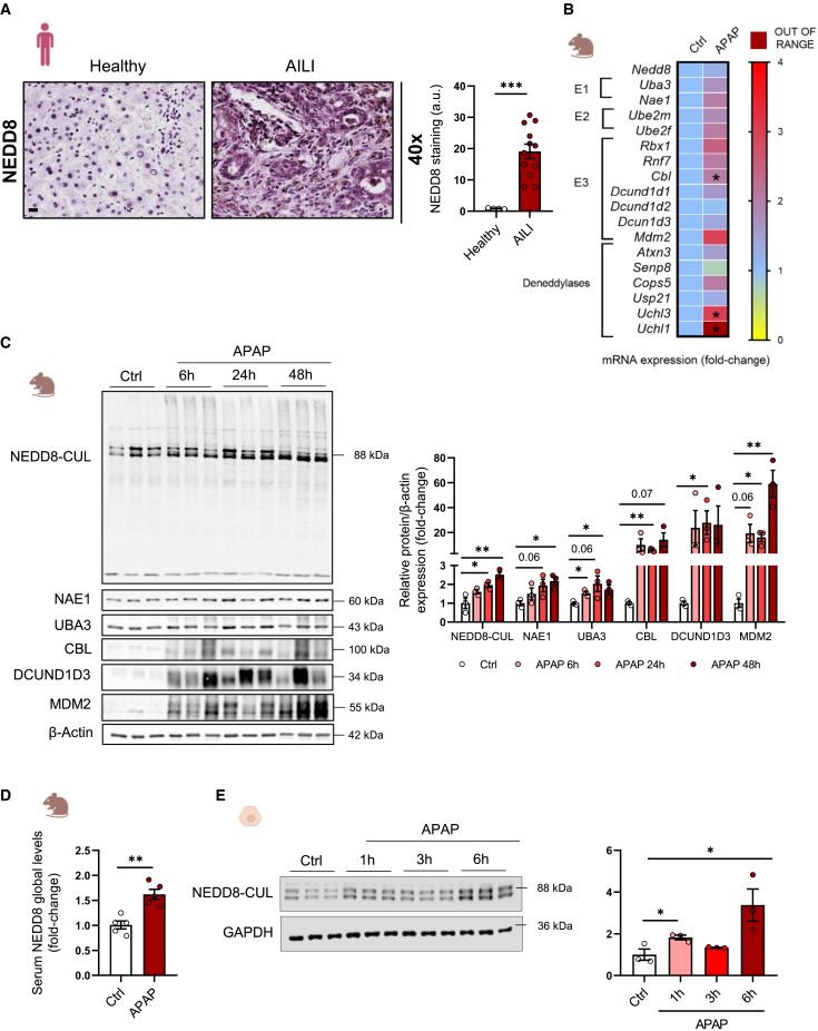
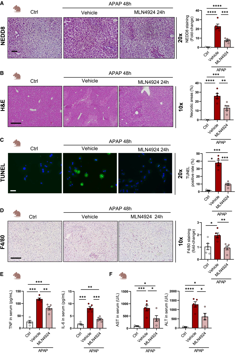
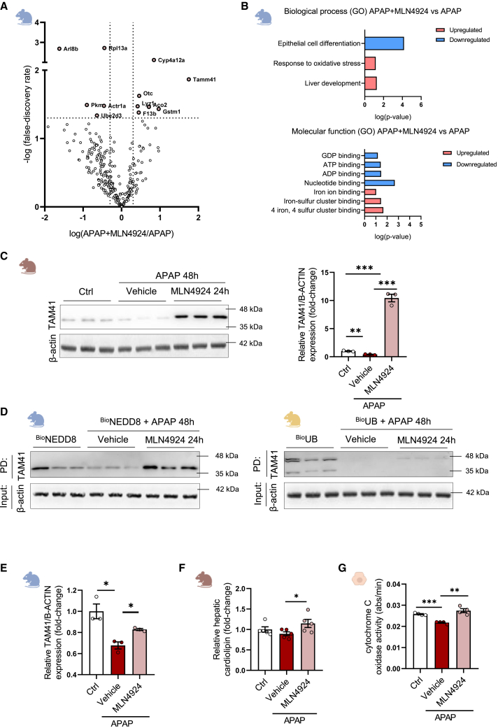
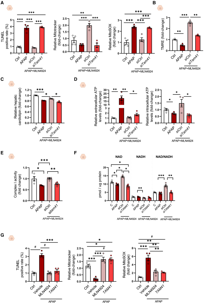
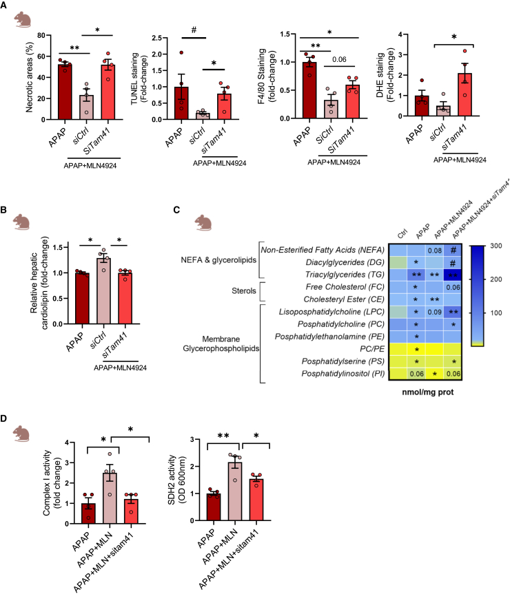
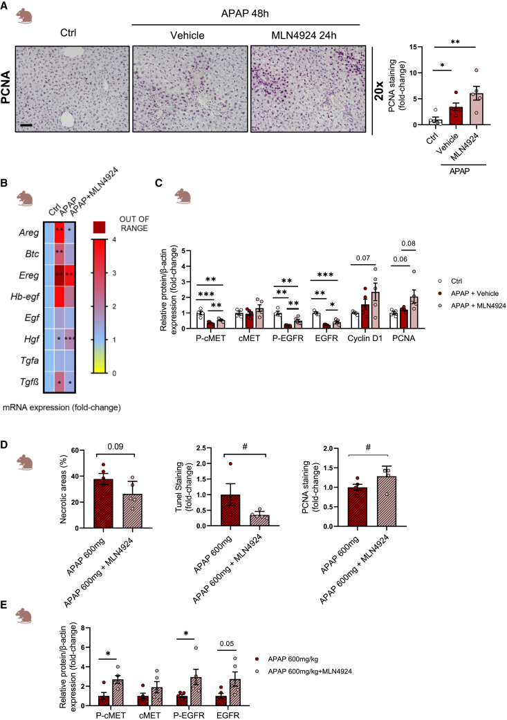
Drug-induced liver injury (DILI) is a significant cause of acute liver failure (ALF) and liver transplantation in the Western world. Acetaminophen (APAP) overdose is a main contributor of DILI, leading to hepatocyte cell death through necrosis. Here, we identified that neddylation, an essential post-translational modification involved in the mitochondria function, was upregulated in liver biopsies from patients with APAP-induced liver injury (AILI) and in mice treated with an APAP overdose. MLN4924, an inhibitor of the neuronal precursor cell-expressed developmentally downregulated protein 8 (NEDD8)-activating enzyme (NAE-1), ameliorated necrosis and boosted liver regeneration in AILI. To understand how neddylation interferes in AILI, whole-body biotinylated NEDD8 (bioNEDD8) and ubiquitin (bioUB) transgenic mice were investigated under APAP overdose with and without MLN4924. The cytidine diphosphate diacylglycerol (CDP-DAG) synthase TAM41, responsible for producing cardiolipin essential for mitochondrial activity, was found modulated under AILI and restored its levels by inhibiting neddylation. Understanding this ubiquitin-like crosstalk in AILI is essential for developing promising targeted inhibitors for DILI treatment.

Keywords: APAP; DILI; NEDD8; mitochondria; necrosis.


Conflict of interest statement

Declaration of interests The authors declare no competing interests.

Figures

None
Graphical abstract
Figure 1
Figure 1
Global NEDD8 is characterized in patients with AILI and in preclinical mice models with an APAP overdose (A) Liver immunohistochemical staining and respective quantification of NEDD8 in a cohort of patients with AILI (N = 12) compared to a healthy group (n = 4). Scale bar corresponds to 50 μm. (B) mRNA expression levels of NEDD8 pathway (Nedd8, Uba3, Nae-1, Ube2m, Ube2f, Rbx1, Rnf7, Cbl, Dcund1d1, Dcund1d2, Dcun1d3, Mdm2, Atxn3, Senp8, Cops5, Usp21, Uchl3, and Uchl1) in mice treated with 360 mg/kg of APAP (n = 4) for 48 h and compared with a control group (n = 4). (C) Protein expression levels of NEDD8 and the enzymes involved in the NEDDylation pathway NAE-1, UBA3, CBL, DCUN1D3, and MDM2 in liver from mice treated with a single dose of 360 mg/kg of APAP (n = 3) for 6, 24, and 48 h and compared with a control group (n = 3). β-Actin was used as a loading control. (D) NEDD8 serum levels was determined in APAP overdose mice models (n = 5) for 48 h and compared to a control group (n = 5). (E) Protein expression levels of NEDD8 in primary hepatocytes treated with 10 mM of APAP overdose for 1, 3, and 6 h. GAPDH was used as a loading control. Triplicates were used for experimental condition. Data are shown as mean ± SEM. ∗p < 0.05, ∗∗p < 0.01, and ∗∗∗p < 0.001 are shown (Student’s test).
Figure 2
Figure 2
Pharmacological neddylation inhibition reduces liver damage Comparison of control mice (n = 5) versus mice treated with 360 mg/kg of APAP overdose (n = 5) versus with 360 mg/kg of APAP overdose and 24 h later with 60 mg/kg MLN4924 (n = 5). (A) Liver immunohistochemical staining and respective quantification of NEDD8 (n = 5). Scale bar corresponds to 100 μm. (B) Liver necrosis areas over the total area in percentage was assessed by H&E staining and a total of 5 pictures per animal were evaluated in a total of 5 mice per group (n = 5). Scale bar corresponds to 200 μm. (C) Cell death was determined by TUNEL assay liver tissue (n = 5). Scale bar corresponds to 100 μm. (D) Inflammation was assessed by F4/80 staining (n = 5). Scale bar corresponds to 200 μm. (E) Tumor necrosis factor (TNF) and interleukin-6 (IL-6) levels were determined by ELISA assay in mice serum after 48 h of APAP overdose (n = 5 per group). (F) Transaminase ALT and AST levels were measured in mice serum after 48 h of APAP overdose (n = 5 per group). Data are shown as mean ± SEM. ∗p < 0.05, ∗∗p < 0.01, ∗∗∗p < 0.001 and ∗∗∗∗p < 0.0001 are shown (Student’s test).
Figure 3
Figure 3
Proteomic characterization of neddylated proteins in transgenic BioNEDD8 mice treated with 360 mg/kg of APAP overdose and 24 h later administered MLN4924 treatment (A) Volcano plot representation of specific proteins regulated in BioNEDD8 mice treated with APAP overdose and 24 h later with 60 mg/kg MLN4924 (n = 4) in comparison with APAP overdose mice group (n = 3) after 24 h of APAP overdose. (B) Gene Ontology (GO) biological processes and GO molecular functions upregulated and downregulated in BioNEDD8 mice treated with APAP overdose and 24 h later with MLN4924 (n = 4) in comparison with APAP overdose mice group (n = 3). (C) Protein expression levels of TAM41 in total liver homogenate of WT mice (n = 3), treated with APAP overdose and 24 h later with MLN4924 (n = 3) compared with a control group (n = 3). β-Actin was used as a loading control. (D) Protein expression levels of TAM41 after pull-down enrichment of biotinylated proteins in BioNEDD8 mice treated with APAP overdose (n = 3) and 24 h later with MLN4924 (n = 3) compared with a control group (n = 3). β-Actin was used as a loading control from the input protein extract. Protein expression levels of TAM41 after pull-down enrichment of biotinylated proteins in BioUB mice treated with APAP overdose (n = 3) and 24 h later with MLN4924 (n = 3) compared with a control group (n = 3). β-Actin was used as a loading control from the input protein extract. (E) Quantification of the western blot obtained from the immunoprecipitation of NEDD8 and western blot against TAM41 in in WT mice treated with APAP overdose (n = 3) and 24 h later with MLN4924 (n = 3) compared with a control group (n = 3). (F) Hepatic cardiolipin levels in WT mice treated with APAP overdose (n = 5) and 24 h later with MLN4924 (n = 5) compared with a control group (n = 5). (G) Cytochrome c oxidase activity was determined in WT mice hepatocytes treated with APAP overdose (n = 4) and 24 h later with MLN4924 (n = 4) compared with a control group (n = 4). Data are shown as mean ± SEM. ∗p < 0.05, ∗∗p < 0.01 and ∗∗∗p < 0.001 are shown (Student’s test).
Figure 4
Figure 4
Pharmacological neddylation inhibition by MLN4924 reduces cell death and increases mitochondrial function in primary hepatocytes and mice model treated with APAP overdose In WT hepatocytes under 10 mM APAP overdose for 6 h and treated with 3 μM MLN4924 for 3 h (n = 4). (A) Cell death was evaluated by TUNEL assay, mitochondrial activity was determined by MitoTracker, and the mitochondrial ROS was determined by MitoSOX. Scale bar corresponds to 50 μm. (B) mRNA expression levels of Mfn1, Opa1, Fis1, Mff, Pink1, Sqstm1, Parkin, and Polg. (C) GSH, GSSG, and GSH/GSSG levels (nmol/mg of protein) were measured by liquid chromatography-mass spectrometry (HPLC-MS). (D) mRNA expression levels of Ho-1 and Nos2. Data are shown as mean ± SEM. ∗p < 0.05, ∗∗p < 0.01, ∗∗∗p < 0.001, and #p < 0.00005 are shown (Student’s test).
Figure 5
Figure 5
Tam41 silencing in primary hepatocytes abolishes the beneficial effect of MLN4924 treatment and recovering TAM41 levels by overexpression resembled MLN4924 treatment (A) Cell death was evaluated by TUNEL assay, mitochondrial activity was determined by MitoTracker, and the mitochondrial ROS was determined by MitoSOX. (B–F) Mitochondrial potential membrane, (C) hepatic cardiolipin, (D) extracellular and intracellular ATP levels, (E) complex I mitochondrial activity, and (F) NADH and NAD+ levels (pmol/mg of protein) in WT hepatocytes under 10 mM APAP overdose for 6 h and treated with 3 μM MLN4924 + siTam41 (n = 4). (G) Cell death was evaluated by TUNEL assay, mitochondrial activity was determined by MitoTracker, and the mitochondrial ROS was determined by MitoSOX in WT hepatocytes under 10 mM APAP overdose for 6 h and treated with 3 μM MLN4924 + overexpression of Tam41 (n = 4). Data are shown as mean ± SEM. ∗p < 0.05, ∗∗p < 0.01, ∗∗∗p < 0.001, and #p < 0.00005 are shown (Student’s test).
Figure 6
Figure 6
Tam41 silencing in mice suppresses the positive effect of MLN4924 treatment In the preclinical mice model, mice were treated with a toxic dose of APAP (n = 4), and 24 h later these mice received MLN4924 in a single dose (n = 4). Twelve hours before MLN4924 treatment, mice were silenced Tam41 (n = 4). Mice were sacrificed 48 h after APAP overdose. (A) Liver necrosis was assessed by H&E staining. Cell death was evaluated by TUNEL. Inflammation was assessed by F4/80 staining. Oxidative stress was evaluated by dihydroethidium (DHE). (B) Hepatic cardiolipin quantification. (C) Liver quantification of lipids that belong to non-esterified fatty acids, glycerolipids, sterols, and membrane glycerophospholipids groups (nmol/mg protein). The statistical significance in the APAP column corresponds to Ctrl vs. APAP comparison, in the APAP+MLN4924 column, APAP vs. APAP+MLN4924, and in the APAP+MLN4924+siTam41 column, APAP+MLN4924 vs. APAP+MLN4924+siTam41, respectively. (D) Complex I and complex II (succinate dehydrogenase) mitochondrial activity assay. Data are shown as mean ± SEM. ∗p < 0.05, ∗∗p < 0.01, ∗∗∗p < 0.001, and #p < 0.00005 are shown (Student’s test).
Figure 7
Figure 7
Pharmacological neddylation inhibition improves mitochondrial function and regenerative response in preclinical mice model treated with an APAP overdose and 24 h later with MLN4924 treatment In the preclinical mice model under APAP-induced toxicity of 360 mg/kg (n = 5), 24 h after APAP administration, mice received MLN4924 in a single dose (n = 5). Mice were sacrificed 48 h after APAP overdose. (A) PCNA expression by immunohistochemistry (n = 5). Scale bar corresponds to 100 μm. (B) mRNA expression levels of Areg, Btc, Ereg, Hb-egf, Egf, Hgf, Tgfa, and Tgfβ (n = 4). (C) Protein expression levels of P-cMET, cMET, p-EGFR, EGFR, cyclin D1, and PCNA. β-Actin was used as a loading control (n = 5). In the preclinical mice model under APAP-induced toxicity of 600 mg/kg (n = 5), 6 h after APAP administration, mice received MLN4924 in a single dose (n = 5). Mice were sacrificed 24 h after APAP overdose. (D) Liver necrosis was assessed by H&E staining. Cell death was evaluated by TUNEL. Regeneration was evaluated by PCNA staining. Scale bar corresponds to 200 μm. (E) Quantification of protein expression levels of P-cMET, cMET, p-EGFR, and EGFR. β-Actin was used as a loading control (n = 5) Data are shown as mean ± SEM. ∗p < 0.05, ∗∗p < 0.01, ∗∗∗p < 0.001, and #p < 0.00005 are shown (Student’s test).

References

    1. Suk K.T., Kim D.J. Drug-induced liver injury: present and future. Clin. Mol. Hepatol. 2012;18:249–257. doi: 10.3350/cmh.2012.18.3.249. - DOI - PMC - PubMed
    1. Lee W.M. Acetaminophen Toxicity: A History of Serendipity and Unintended Consequences. Clin. Liver Dis. 2020;16:34–44. doi: 10.1002/cld.984. - DOI - PMC - PubMed
    1. Lee W.M. Acetaminophen (APAP) hepatotoxicity—Isn’t it time for APAP to go away? J. Hepatol. 2017;67:1324–1331. doi: 10.1016/j.jhep.2017.07.005. - DOI - PMC - PubMed
    1. Licata A., Minissale M.G., Stankevičiūtė S., Sanabria-Cabrera J., Lucena M.I., Andrade R.J., Almasio P.L. N-Acetylcysteine for Preventing Acetaminophen-Induced Liver Injury: A Comprehensive Review. Front. Pharmacol. 2022;13:828565. doi: 10.3389/fphar.2022.828565. - DOI - PMC - PubMed
    1. Yan M., Huo Y., Yin S., Hu H. Mechanisms of acetaminophen-induced liver injury and its implications for therapeutic interventions. Redox Biol. 2018;17:274–283. doi: 10.1016/j.redox.2018.04.019. - DOI - PMC - PubMed

MeSH terms