. 2024 Sep:158:155976.
doi: 10.1016/j.metabol.2024.155976. Epub 2024 Jul 15.

Estrogen receptor-α signaling in tanycytes lies at the crossroads of fertility and metabolism

Affiliations

Estrogen receptor-α signaling in tanycytes lies at the crossroads of fertility and metabolism

Daniela Fernandois et al. Metabolism. 2024 Sep.

Abstract

Background: Estrogen secretion by the ovaries regulates the hypothalamic-pituitary-gonadal axis during the reproductive cycle, influencing gonadotropin-releasing hormone (GnRH) and luteinizing hormone (LH) secretion, and also plays a role in regulating metabolism. Here, we establish that hypothalamic tanycytes-specialized glia lining the floor and walls of the third ventricle-integrate estrogenic feedback signals from the gonads and couple reproduction with metabolism by relaying this information to orexigenic neuropeptide Y (NPY) neurons.

Methods: Using mouse models, including mice floxed for Esr1 (encoding estrogen receptor alpha, ERα) and those with Cre-dependent expression of designer receptors exclusively activated by designer drugs (DREADDs), along with viral-mediated, pharmacological and indirect calorimetric approaches, we evaluated the role of tanycytes and tanycytic estrogen signaling in pulsatile LH secretion, cFos expression in NPY neurons, estrous cyclicity, body-weight changes and metabolic parameters in adult females.

Results: In ovariectomized mice, chemogenetic activation of tanycytes significantly reduced LH pulsatile release, mimicking the effects of direct NPY neuron activation. In intact mice, tanycytes were crucial for the estrogen-mediated control of GnRH/LH release, with tanycytic ERα activation suppressing fasting-induced NPY neuron activation. Selective knockout of Esr1 in tanycytes altered estrous cyclicity and fertility in female mice and affected estrogen's ability to inhibit refeeding in fasting mice. The absence of ERα signaling in tanycytes increased Npy transcripts and body weight in intact mice and prevented the estrogen-mediated decrease in food intake as well as increase in energy expenditure and fatty acid oxidation in ovariectomized mice.

Conclusions: Our findings underscore the pivotal role of tanycytes in the neuroendocrine coupling of reproduction and metabolism, with potential implications for its age-related deregulation after menopause.

Significance statement: Our investigation reveals that tanycytes, specialized glial cells in the brain, are key interpreters of estrogen signals for orexigenic NPY neurons in the hypothalamus. Disrupting tanycytic estrogen receptors not only alters fertility in female mice but also impairs the ability of estrogens to suppress appetite. This work thus sheds light on the critical role played by tanycytes in bridging the hormonal regulation of cyclic reproductive function and appetite/feeding behavior. This understanding may have potential implications for age-related metabolic deregulation after menopause.

Keywords: Estrogens; Female metabolism; Hypothalamus; LH pulsatility; Reproduction; Tanycytes.


Conflict of interest statement

Declaration of competing interest The authors declare no competing interests.

Figures
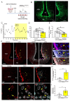
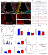
Figure 1
Figure 1. Chemogenetic activation of tanycytes slows down ovariectomy-mediated acceleration of pulsatile LH release and activates arcuate Npy neurons.
(A) Protocol for the iterative tail-blood sampling in mice, (B) Expression of GFP in tanycytes as a reflection of hM3DGq receptor expression in hM3DGqTanycytes mice. Right panel, higher magnification of the framed region in the left panel. (C) Representative pulsatile LH release in bilaterally ovariectomized (OVX) hM3DGqTanycytes mice 1h before and after i.p. clozapine-N-oxide (CNO; 1mg/Kg) administration, (D) Area under the curve of LH and (H) LH pulse frequency before (grey bars) and after (yellow bars) 1mg/kg CNO i.p. injection in OVX hM3DGqTanycytes. (E) Immunofluorescence of OVX hM3DGqTanycytes mice brains 30 min after vehicle (VH) or CNO i.p. administration. White arrows indicate the arcuate nucleus of the hypothalamus (ARH) tanycytes, pink arrows indicate median eminence tanycytes and yellow arrows indicate cells immunoreactive for cFOS in the proximity of tanycytic processes within the ARH. cFos positive immunolabeling is observed in red, GFP (representative of hM3DGq expression) in white and DAPI in blue as nuclear counterstaining. (G) Fluorescent in situ hybridization of OVX hM3DGqTanycytes brain mice for Fos (red dots), Npy (cyan dots), and DAPI (white) 30 min after CNO i.p. injection. White arrows indicate Fos expression in tanycytes and yellow arrows indicate Fos expression in NPY neurons. (H) Number of cells positive for Fos within the ARH nucleus, (I) Percentage of Npy neurons positive for Fos within the ARH nucleus. VH N=4 and CNO N=3. Graphs are plotted as Mean±SEM. Statistical differences were determined by a paired two-sided Student’s t-test (D and E) or an unpaired two-sided Student’s t-test (H and I). * p<0.05, **p<0.01.
Figure 2
Figure 2. Esr1 transcripts are expressed in tanycytes in female mice.
(A) Fluorescent in situ hybridization for Esr1 in the brain of Esr1loxP/loxP mice. White dots show Esr1 transcripts and the red labelling shows immunoreactivity for vimentin, which is a marker for tanycytes. (B) Representative diagram of the design of the fluorescent activated cell sorting (FACS) experiment and dot-plots showing the tissue autofluorescence control and the gating strategy for cell sorting in the median eminence (ME)/arcuate nucleus of the hypothalamus (ARH) region. (C) Real-time qPCR for Esr1 coding ERα and Esr2 coding ERβ in tanycytes isolated by FACS from estrus tdTomatoTanycytes female mice, N=5. (D) Real-time qPCR for Esr1 in FACS-isolated tanycytes at different stages of the estrous cycle; Proestrus (N=6), Estrus (N=6) and Diestrus (N=5). (E) Representative diagram of the design of the FACS experiment and dot-plots showing the tissue autofluorescence and antibody isotype controls and the gating strategy for cell sorting in the ME/ARH. (F) Percentage of tomato-positive tanycytes expressing ERα-immunoreactivity during the different stages of the estrous cycle; Proestrus (N=4), Estrus (N=5) and Diestrus (N=5). B Bars are shown as Mean±SEM. Statistical differences were determined by an unpaired two-sided Student’s t-test (C) or a one-way ANOVA (normal data and homogeneity of variances) followed by test Fisher’s LSD test (D and F). * p<0,05 and ** p<0,01.
Figure 3
Figure 3. Selective impairment of ERα production in adult tanycytes alters female fertility.
(A) Immunoreactivity for ERα (shown in red) in Esr1loxP/loxP and Esr1TanKO mouse littermates. Nuclei were labeled with DAPI (white). Esr1loxP/loxP;RosamTmG mice were injected into the lateral ventricle with an AAV1/2 expressing Dio2::gfp (Esr1loxP/loxP) or Dio2::Cre (Esr1TanKO). White arrowheads indicate the cell bodies of the tanycytic layer bordering the floor and the ventral wall of the third ventricle. Vimentin immunoreactivity is shown in yellow and cre-mediated membrane GFP expression in blue. a-d subpanels depict higher magnification of the framed regions in the corresponding left panel. (B) Estrous cyclicity in females before and after stereotaxic surgery in Esr1loxP/loxP and Esr1TanKO mice. (C) Percentage of permanency in proestrus, estrus, or diestrus of each mouse before and after mock (Esr1loxP/loxP) or Cre-mediated genetic recombination in the Esr1 gene (Esr1TanKO) in tanycytes. Esr1loxP/loxP N=11 and Esr1TanKO N=12. (D) Fertility index. (D-E), ovarian weight (E) and uterine weight (F) at estrous in Esr1loxP/loxP and Esr1TanKO littermates one month after genetic recombination. (G) Plasmatic levels of 17β-estradiol in Esr1loxP/loxP and Esr1TanKO mice at different time points after mock or cre-mediated genetic recombination. (H-J) Mean pulse frequency (H), pulse amplitude (I), and basal levels of LH release (J). N=12 for Esr1Wt and N=13 for Esr1TanKO mice. (K) Representative figure of LH secretion observed for 2h in intact Esr1Wt (blue, K) and Esr1TanKO (red, K) littermates. Bars are shown as Mean±SEM. Statistical differences were determined by paired (C) and unpaired two-sided Student’s t-tests (D-F and H-J) or a two-way ANOVA (normal data and homogeneity of variances) followed by test Fisher’s LSD test (G). * p<0,05, ** p<0,01 and **p<0.001.
Figure 4
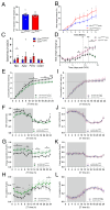
Figure 4. ERα deletion in tanycytes produce hypothalamic insensitivity.
(A) Body weight distribution of Esr1loxP/loxP and Esr1TanKO mice before the stereotaxic injection. (B) Percentage of body weight change after a vehicle (Esr1loxP/loxP, N=6) or Tat-Cre (Esr1TanKO, N=7) injection into the third ventricle of intact female mice. (C) Relative transcript levels in the mediobasal hypothalamus of estrous Esr1loxP/loxP and Esr1TanKO female mice, N=5 for each group. (D) Body weight change after OVX before (day 0-9) and after a daily subcutaneous injection of sesame oil (day 10-11) or estradiol benzoate (day 12-14) (N=5 for Esr1loxP/loxP and N=6 for Esr1TanKO). (E,I) 24h cumulative food intake for Esr1loxP/loxP (E) and Esr1TanKO (I) mice. (F, J) 24-h respiratory exchange ratio, RER Esr1loxP/loxP (F) and Esr1TanKO (J) littermates. (G, K) 24-h Energy expenditure for Esr1loxP/loxP (G) and Esr1TanKO (K) animals. (H, L) 24-h Fatty acid oxidation for Esr1loxP/loxP (H) and Esr1TanKO (L) mice. (E-H) Esr1loxP/loxP N=5 and (I-L) Esr1TanKO N=6. Bars are shown as Mean±SEM. Statistical differences were determined by an unpaired two-sided Student’s t-tests (A, C) and a one-way ANOVA (normal data and homogeneity of variances) followed by test Fisher’s LSD test (B, D-L). * p<0,05, ** p<0,01 and *** p<0,001.
Figure 5
Figure 5. Estrogen receptor α expression in female tanycytes is required for estrogens to exert its anorexigenic effect and fasting-induced cFos expression in NPY neurons.
(A) Food intake after overnight ad libitum feeding in female Esr1loxP/loxP and Esr1tanKO littermates treated or not with the ERα agonist PPT at lights off (N=4 per group). (B) Hourly food intake at refeeding after overnight fasting (12h) in female Esr1loxP/loxP (N=5) and Esr1TanKO (N=6) littermates treated or not with the ERα agonist PPT at lights off. (C) cFos expression in the tuberal region of the hypothalamus after overnight fasting in Npy::Gfp;Esr1loxP/loxP and Npy::Gfp;Esr1TanKO littermates treated with DMSO (VH) or PPT. White arrows show GFP-fluorescent NPY neurons expressing cFOS (D) Total number of cFOS-positive cells in the arcuate nucleus of the hypothalamus (ARH), and (E) Number of cFOS-positive GFP-expressing NPY neurons in the ARH after overnight fasting in Npy::Gfp;Esr1loxP/loxP and Npy::Gfp;Esr1TanKO littermates treated with DMSO or PPT. (F) LH pulsatility in diestrous mice fed ad libitum or after overnight fasting (12h) in the absence or presence of PPT treatment. Bars are shown as Mean±SEM. Statistical differences were determined by a one-way ANOVA (normal data and homogeneity of variances) followed by test Fisher’s LSD test (A,B, D and E). * p<0,05, ** p<0,01 and *** p<0,001.
Figure 6
Figure 6. ERα expression in female tanycytes in the brain of three post-menopausal women.
Immunoreactivity for ERα (shown in white) and vimentin (shown in green) in 20-µm thick sections of the hypothalamus from women aged 64, 69 and 86 years. Nuclei were counterstained with DAPI (blue). The panels in the second column depict a higher magnification of the framed region in the corresponding left panel. Empty arrows indicate DAPI-positive nuclei which do not show ERα immunoreactivity. Inf: infundibulum or arcuate nucleus.
Figure 7
Figure 7. Tanycyte-neuron Interactions in the arcuate nucleus of the hypothalamus (ARH) and the median eminence (ME) at the crossroads of fertility and metabolism.
Schematic diagram illustrating tanycyte interactions with NPY neurons in the ARH and GnRH neurons in the median eminence (ME) in ovariectomized (left panel) and intact (right panel) mice in which tanycytes are selectively expressing the activator DREADD (left panel) or deficient in ERα signaling (right panel) in fed or fasting (framed) conditions. Tanycytes play a crucial role in translating the estrogen signal into meaningful information for NPY and GnRH neurons, which rely on this signal for their function but cannot directly detect it. Overall, tanycytes serve as a linchpin in coordinating reproduction and metabolism by perceiving the estrogenic signals and regulating the function of both GnRH and NPY neurons, though additional mechanisms may also be involved. The red circles symbolize cFos expression in cells. CNO, clozapine-N-oxide.

References

    1. Hunzicker-Dunn M, Mayo K. In: Knobil and Neill’s Physiology of Reproduction. Fourth Edition. Plant TM, Zeleznik J, editors. Elsevier; New York: 2015. Gonadotropinsignaling in the ovary; pp. 895–946.
    1. Sarkar DK, Minami S. Diurnal variation in luteinizing hormone-releasing hormone and beta-endorphin release in pituitary portal plasma during the rat estrous cycle. Biol Reprod. 1995;53:38–45. - PubMed
    1. Frost SI, Keen KL, Levine JE, Terasawa E. Microdialysis methods for in vivo neuropeptide measurement in the stalk-median eminence in the Rhesus monkey. J Neurosci Methods. 2008;168:26–34. - PMC - PubMed
    1. Caraty A, Orgeur P, Thiery JC. Demonstration of the pulsatile secretion of LH-RH into hypophysial portal blood of ewes using an original technic for multiple samples. C R Seances Acad Sci III. 1982;295:103–6. - PubMed
    1. Clarke IJ, Cummins JT. The temporal relationship between gonadotropin releasing hormone (GnRH) and luteinizing hormone (LH) secretion in ovariectomized ewes. Endocrinology. 1982;111:1737–9. - PubMed