. 2024 Jul 17;7(1):872.
doi: 10.1038/s42003-024-06572-0.

Selenoprotein W modulates tau homeostasis in an Alzheimer's disease mouse model

Affiliations

Selenoprotein W modulates tau homeostasis in an Alzheimer's disease mouse model

Bingyu Ren et al. Commun Biol. .

Abstract

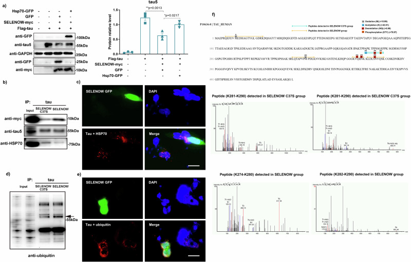
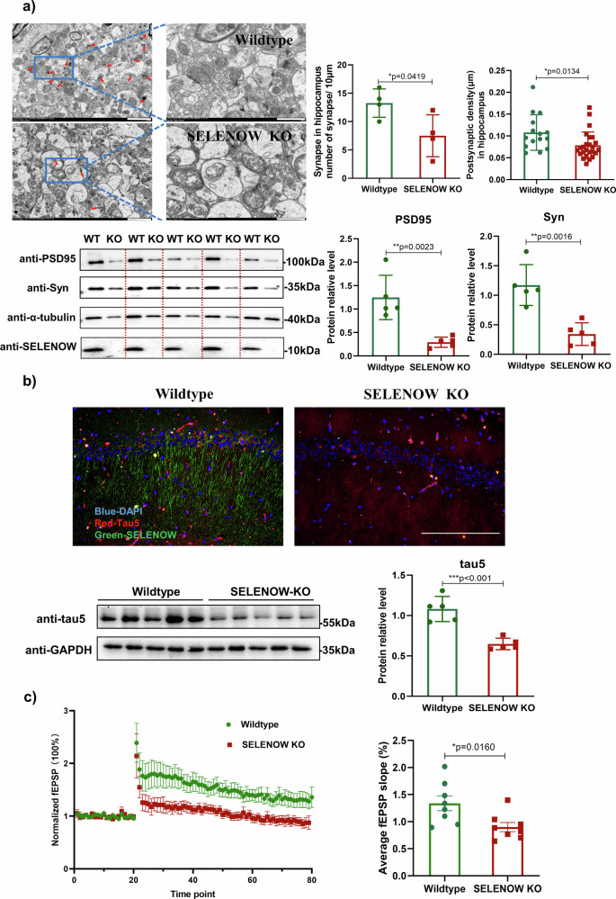
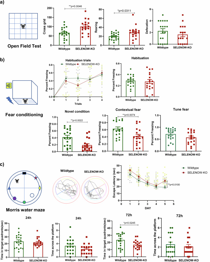
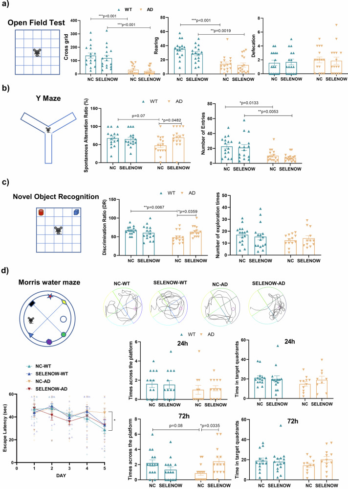
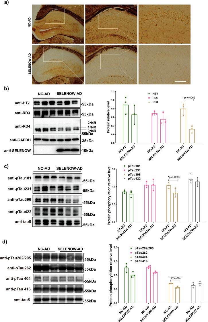
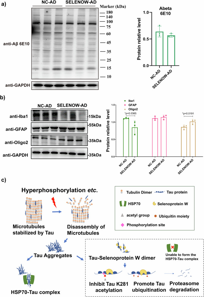
Lower selenium levels are observed in Alzheimer's disease (AD) brains, while supplementation shows multiple benefits. Selenoprotein W (SELENOW) is sensitive to selenium changes and binds to tau, reducing tau accumulation. However, whether restoration of SELENOW has any protective effect in AD models and its underlying mechanism remain unknown. Here, we confirm the association between SELENOW downregulation and tau pathology, revealing SELENOW's role in promoting tau degradation through the ubiquitin‒proteasome system. SELENOW competes with Hsp70 to interact with tau, promoting its ubiquitination and inhibiting tau acetylation at K281. SELENOW deficiency leads to synaptic defects, tau dysregulation and impaired long-term potentiation, resulting in memory deficits in mice. Conversely, SELENOW overexpression in the triple transgenic AD mice ameliorates memory impairment and tau-related pathologies, featuring decreased 4-repeat tau isoform, phosphorylation at Ser396 and Ser404, neurofibrillary tangles and neuroinflammation. Thus, SELENOW contributes to the regulation of tau homeostasis and synaptic maintenance, implicating its potential role in AD.


Conflict of interest statement

The authors declare no competing interests.

Figures

Fig. 1
Fig. 1. SELENOW levels were negatively correlated with tau expression levels.
a Representative western blots of the 8-month-old WT and 3×Tg AD mouse hippocampus, showing the typical overexpression of tau protein and downregulation of SELENOW in AD. b Representative immunostaining of SELENOW (green), tau5 (red) and cell nuclei (blue) in the hippocampal CA1 areas of 8-month-old WT and 3×Tg AD mice. Scale bar = 200 μm. c HEK293TAU cells were transiently transfected with scramble or SELENOW siRNA. Representative immunoblot showing that the knockdown of SELENOW by siRNA upregulated the expression of tau. d HEK293TAU cells were transiently transfected with scramble or SELENOW siRNA and then treated with selenate for 24 h. Representative western blot showed that selenium supplementation by selenate induced the upregulation of SELENOW and reduced the expression of tau. Knockdown of SELENOW blocked selenate-induced tau downregulation. e HEK293TAU cells were transiently transfected with SELENOV or SELENOW. Representative western blot showed that the overexpression of SELENOW reduced the expression of tau while overexpression of SELENOV had no effect on tau expression. f HEK293TAU cells were transiently transfected with SELENOW or its C37S mutant, the same amounts of tau were immunoprecipitated, and the samples were subjected to western blot analysis. Representative results showed that SELENOW immunoprecipitated with tau, while C37 mutated to S in SELENOW diminished their interaction. Data are presented as the mean ± SD. N = 3 in each group, *p < 0.05, **p < 0.01 and ***p < 0.001 by one-way ANOVA.
Fig. 2
Fig. 2. SELENOW induced tau protein clearance through the ubiquitin‒proteasome system.
a HEK293TAU cells were transiently transfected with empty plasmids or SELENOW and then treated with the protein synthesis inhibitor CHX. The samples were collected at several time intervals and subjected to western blot analysis. Protein levels showed accelerated half-life of tau protein by SELENOW overexpression. Protein levels of β-actin were used as a control for normalization. b HEK293TAU cells were transiently transfected with empty plasmids or SELENOW and then treated with the 20 S proteasome inhibitor MG-132 for 6 h. Representative western blot showing that MG-132 blocked SELENOW-induced tau clearance. c HEK293TAU cells were transiently transfected with empty plasmids or SELENOW and then treated with the autophagy inhibitor CQ for 6 h. Representative immunoblot showing that CQ had no effect on SELENOW-induced tau clearance. The transcription levels of MAPT and heat shock proteins were quantified by Q-PCR in HEK293TAU cells treated with (d) MG-132 and (e) the heat shock protein inducer TRC051384. ACTB was used as an endogenous reference gene. f HEK293TAU cells were treated with TRC051384, and the samples were collected for western blotting. Representative immunoblots showed that TRC051384 reversed the effect of SELENOW on tau protein degradation. Data are presented as the mean ± SD. N = 3 in each group, *p < 0.05, **p < 0.01 and ***p < 0.001 by t tests between two groups, and by one-way ANOVA for groups that had more than two.
Fig. 3
Fig. 3. SELENOW competed with Hsp70 for tau binding, inhibited tau acetylation and promoted tau polyubiquitination.
a HEK293TAU cells were transiently transfected with GFP or Hsp70-GFP, and the samples were collected for western blotting. Representative immunoblots showed that HSP70 overexpression partially antagonized the effect of SELENOW on tau protein degradation. HEK293TAU cells were transiently transfected with SELENOW or its C37S mutant, and the same amounts of tau were immunoprecipitated. Representative Western blots of immunoprecipitated samples showing (b) Hsp70 was released from tau while SELENOW binding increased, and (c) the SELENOW overexpression group exhibited increased ubiquitination of tau compared to its C37S mutant. The arrow indicates the predicted band size of tau. HEK293TAU cells were transiently transfected with SELENOW GFP for 48 h, and then PLA fluorescence detection was performed. Representative images showing that (d) SELENOW GFP potently decreased the Hsp70-tau complex and (e) promoted tau ubiquitination compared with adjacent nontransfected cells. Scale bar = 20 μm. f LC/MS-MS detection of soluble tau proteins immunoprecipitated from HEK293TAU cells that overexpressed SELENOW or the C37S mutant. Upper panel: Schematic diagram of tau sequence and modifications detected specifically in the SELENOW C37S or SELENOW group. Lower panel: peptide fragments from K274 to K290 detected by mass spectrometric analysis in SELENOW C37S group and SELENOW group. Data are presented as the mean ± SD. N = 3 in each group, *p < 0.05, **p < 0.01 and ***p < 0.001 by one-way ANOVA.
Fig. 4
Fig. 4. SELENOW deficiency induced synaptic deficits and tau downregulation in the mouse hippocampus.
a Reduced number of synapses and postsynaptic density in the hippocampus of 6-month-old SELENOW KO mice. Representative and quantification of transmission electron microscope (TEM) images showing the synapse distribution (left panels, as indicated by the red arrows) and ultrastructure (right panels, magnified images) in the cornu ammonis 1 (CA1) region of WT and SELENOW KO mice. Scale bar = 2.0 μm. Representative immunoblots showed a significant decrease in PSD95 and the synaptic vesicle marker Syn in the hippocampus of SELENOW KO mice. b Downregulation of tau protein levels in the hippocampus of 6-month-old SELENOW KO mice. Representative immunostaining images of SELENOW (green), tau5 (red) and cell nuclei (blue) in the hippocampal CA1 areas of 6-month-old WT and SELENOW KO mice. Scale bar = 200 μm. Representative immunoblots showed significant downregulation of tau in the hippocampus of SELENOW KO mice. c Impaired LTP recordings in the hippocampus of 6-month-old SELENOW KO mice. Slope of fEPSPs in response to 100-Hz stimulation in the Schaffer collateral CA1 region of WT and SELENOW KO mice (left panel). Quantification of the averaged fEPSP slope (right panel). Representative traces 20 min before and 60 min after θ-burst stimulation are shown. Data are presented as the mean ± SD. N = 4 slices in each group for synapse number analysis, N = 15 synapse from 3 mice in each group for postsynaptic density analysis, N = 5 from 5 mice in each group for immunoblots, and N = 8 slices from 6 mice for LTP recording, *p < 0.05, **p < 0.01 and ***p < 0.001 by t test.
Fig. 5
Fig. 5. SELENOW KO mice exhibited less anxiety-like behavior and impaired memory.
a Locomotion ability and anxiety of the 12-month-old WT and SELENOW KO mice evaluated by the crossing grid numbers, rearing and defecation times in the open field test. b Fear memory of the 12-month-old WT and SELENOW KO mice evaluated by the percent freezing in habituation, contextual fear, novel condition, and tune fear stages in the contextual fear memory task. Representative track trails of WT and KO mice in the contextual fear stage are also provided. c Spatial and working memory of the 12-month-old WT and SELENOW KO mice evaluated by the escape latency, the time the mice spent in the target in 24 h and 72 h trials, and the number of times the mice crossed the platform quadrant in 24 h and 72 h trials of the Morris water maze. The representative track trails of WT and KO mice in the 72 h trials are also provided. Data are presented as the mean ± SD. N = 18 in each group, outliers were detected and excluded using the Grubbs’ test, *p < 0.05, **p < 0.01 and ***p < 0.001 by t test.
Fig. 6
Fig. 6. Brain SELENOW overexpression rescued behavioral deficits in 3×Tg AD mice.
a Locomotion ability and anxiety of 12-month-old WT and 3×Tg AD mice receiving empty vector (NC-WT and NC-AD) or AAV-SELENOW (SELENOW-WT and SELENOW-AD) in their hippocampal CA3 area, evaluated by the number of grid crossings and rearing and defecation times in the open field test. b Spatial memory of the 12-month-old NC-WT, SELENOW-WT, NC-AD and SELENOW-AD mice evaluated by the spontaneous alternation ratio and number of total arm entries in the Y-maze task. c Declarative memory of the 12-month-old NC-WT, SELENOW-WT, NC-AD and SELENOW-AD mice evaluated by the discrimination ratio and total times in the novel object recognition task. d Spatial and working memory of the 12-month-old NC-WT, SELENOW-WT, NC-AD and SELENOW-AD mice evaluated by the escape latency, the time the mice spent in the target in 24 h and 72 h trials, and the number of times the mice crossed the platform quadrant in 24 h and 72 h trials of the Morris water maze. Representative track trials in the 72 h trials are also provided. Data are presented as the mean ± SD. N = 18 in each group, outliers were detected and excluded using the Grubbs’ test, *p < 0.05, **p < 0.01 and ***p < 0.001 by two-way ANOVA.
Fig. 7
Fig. 7. Brain SELENOW overexpression reduced tau pathologies in 3×Tg AD mice.
a Bielschowsky silver staining of neurofibrillary tangles in the hippocampus of 12-month-old NC-AD and SELENOW-AD mice. Scale bar = 500 μm. b Representative western blot results and analysis of soluble total tau (HT7), 3-repeat isoform tau (RD3), and 4-repeat isoform tau (RD4) in brain hippocampus homogenates from 12-month-old NC-AD and SELENOW-AD mice. c Representative western blot results and analysis of tau phosphorylation at different sites, including Thr181, Thr231, Ser396 and Ser422 in brain hippocampus homogenates from 12-month-old NC-AD and SELENOW-AD mice. d Representative western blot results and analysis of tau phosphorylation at different sites, including Ser202/Thr205, Ser262, Ser404 and Ser416 in brain hippocampus homogenates from 12-month-old NC-AD and SELENOW-AD mice. The relative protein phosphorylation levels were defined as phosphorylated tau normalized against total tau (tau5). Data are presented as the mean ± SD. N = 3 in each group, *p < 0.05, **p < 0.01 and ***p < 0.001 by t tests.
Fig. 8
Fig. 8. Brain SELENOW overexpression alleviate neuroinflammation in 3×Tg AD mice.
a Representative western blot results and analysis of Aβ oligomers detected with 6E10 antibody in brain hippocampus homogenates from 12-month-old NC-AD and SELENOW-AD mice. b Representative western blot results and analysis of neuroglial cell markers, including microglia (Iba-1), astrocyte (GFAP) and oligodendrocyte (Oligo2), in brain hippocampus homogenates from 12-month-old NC-AD and SELENOW-AD mice. c A model for SELENOW overexpression in AD promoted the degradation of abnormal tau. SELENOW formed heterodimers with tau via C322, which also mediated tau’s interaction with HSP70 and its auto-acetylation. The interaction of SELENOW and tau affected the formation of the HSP70-tau complex, inhibited tau acetylation at K281, and influences phosphorylation at adjacent sites of Ser396 and Ser404. Non-acetylated lysine sites of tau were more likely to undergo polyubiquitination, leading to proteasomal degradation of abnormal tau. Data are presented as the mean ± SD. N = 3 in each group, *p < 0.05, **p < 0.01 and ***p < 0.001 by t tests.

References

    1. Young ZT, Mok SA, Gestwicki JE. Therapeutic strategies for restoring Tau homeostasis. Cold Spring Harb. Perspect. Med. 2018;8:a024612. doi: 10.1101/cshperspect.a024612. - DOI - PMC - PubMed
    1. Chen J, Berry MJ. Selenium and selenoproteins in the brain and brain diseases. J. Neurochem. 2003;86:1–12. doi: 10.1046/j.1471-4159.2003.01854.x. - DOI - PubMed
    1. Varikasuvu SR, Prasad VS, Kothapalli J, Manne M. Brain Selenium in Alzheimer’s Disease (BRAIN SEAD Study): a systematic review and meta-analysis. Biol. Trace Elem. Res. 2019;189:361–369. doi: 10.1007/s12011-018-1492-x. - DOI - PubMed
    1. Nascimento CQD, et al. Selenium concentrations in elderly people with Alzheimer’s disease: a cross-sectional study with control group. Rev. Brasileira de. Enferm. 2021;74:e20200984. doi: 10.1590/0034-7167-2020-0984. - DOI - PubMed
    1. R. Cardoso B, et al. The APOE ε4 allele is associated with lower selenium levels in the brain: implications for Alzheimer’s Disease. ACS Chem. Neurosci. 2017;8:1459–1464. doi: 10.1021/acschemneuro.7b00014. - DOI - PubMed

Publication types