. 2024 Jul 18;22(1):295.
doi: 10.1186/s12916-024-03495-9.

Foetal gluten immunogenic peptides during pregnancy: a new determinant on the coeliac exposome

Affiliations

Foetal gluten immunogenic peptides during pregnancy: a new determinant on the coeliac exposome

María de Lourdes Moreno et al. BMC Med. .

Abstract

Background: The increasing incidence of coeliac disease is leading to a growing interest in active search for associated factors, even the intrauterine and early life. The exposome approach to disease encompasses a life course perspective from conception onwards has recently been highlighted. Knowledge of early exposure to gluten immunogenic peptides (GIP) in utero could challenge the chronology of early prenatal tolerance or inflammation, rather than after the infant's solid diet after birth.

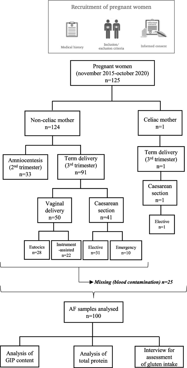
Methods: We developed an accurate and specific immunoassay to detect GIP in amniotic fluid (AF) and studied their accumulates, excretion dynamics and foetal exposure resulting from AF swallowing. One hundred twenty-five pregnant women with different gluten diets and gestational ages were recruited.

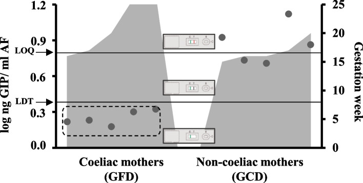
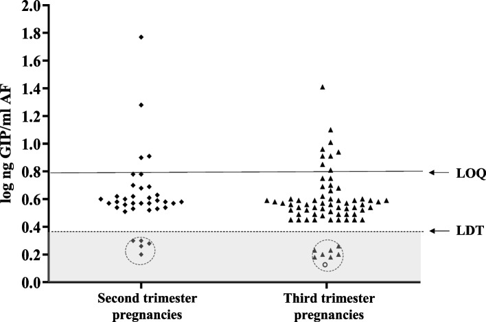
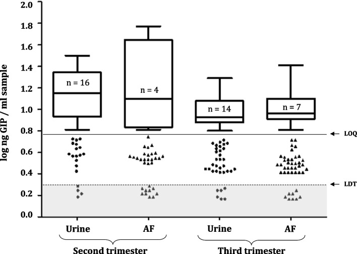
Results: GIP were detectable in AF from at least the 16th gestational week in gluten-consuming women. Although no significant differences in GIP levels were observed during gestation, amniotic GIP late pregnancy was not altered by maternal fasting, suggesting closed-loop entailing foetal swallowing of GIP-containing AF and subsequent excretion via the foetal kidneys.

Conclusions: The study shows evidence, for the first time, of the foetal exposure to gluten immunogenic peptides and establishes a positive correlation with maternal gluten intake. The results obtained point to a novel physiological concept as they describe a plausible closed-loop circuit entailing foetal swallowing of GIP contained in AF and its subsequent excretion through the foetal kidneys. The study adds important new information to understanding the coeliac exposome.

Keywords: Coeliac disease; Exposome; Gluten; Gluten immunogenic peptides.


Conflict of interest statement

The authors declare no competing interests.

Figures

Fig. 1
Fig. 1
Flowchart of study design and pregnancy outcome. AF, amniotic fluid; GIP, gluten immunogenic peptides
Fig. 2
Fig. 2
Workflow for gluten immunogenic peptides detection in AF samples. AF, amniotic fluid; GIP, gluten immunogenic peptides; IC, immunochromatographic; LFT, lateral flow test; LFIA, lateral flow immunoassay; PBS, phosphate-buffered saline; TFA, trifluoroacetic acid
Fig. 3
Fig. 3
Detection and quantification of GIP content in AF samples from amniocentesis of coeliac and non-coeliac pregnant women according to the gestational age. Presence of GIP visible and quantifiable in AF (> LOQ); visual presence of GIP not quantifiable in AF (> LDT, 2.25 ng GIP/mL AF and < LOQ, 6.25 ng GIP/mL AF); absence of GIP in AF (< LDT). Differences in GIP levels in amniotic fluids between the 2 groups were tested with a Mann–Whitney U test. LDT, the limit of technique detection; LOQ, the limit of quantification; AF, amniotic fluid; GIP, gluten immunogenic peptides; GFD, gluten-free diet; GCD, gluten containing diet
Fig. 4
Fig. 4
Comparison of GIP content in AF samples of healthy pregnant women on diverse periods of pregnancy. Visual presence of GIP not quantifiable in AF (> LDT and < QL); presence of GIP visible and quantifiable in AF (> QL); absence of GIP in AF (< LDT). AF from deliveries in the third trimester (at term) (n = 69) and in the second trimester (amniocentesis) (n = 36). Two mothers with monochorionic diamniotic twins were included. Coeliac mother is represented by a circle; non-coeliac mothers in second trimester are indicated by rhombuses; non-coeliac mothers in third trimester are indicated by triangles. Differences in GIP levels in amniotic fluids between the 2 groups were tested with a Mann–Whitney U test. LDT, the limit of technique detection; LOQ, the limit of quantification; GIP, gluten immunogenic peptides; AF, amniotic fluid
Fig. 5
Fig. 5
Amniotic and urinary gluten peptide content and gestational age. Visual presence of GIP not quantifiable (> LDT and < LOQ, scatter plot in white area); absence of GIP (< LDT, scatter plot in grey area) and presence of GIP visible and quantifiable (> LOQ, box and whiskers). Differences between groups were tested with a Wilcoxon signed-rank test in dependent groups and Mann–Whitney U test in independent groups. LDT, the limit of technique detection; LOQ, the limit of quantification; GIP, gluten immunogenic peptides; AF, amniotic fluid
Fig. 6
Fig. 6
Concordance between the punctual measurement of GIP content in the AF and urine samples of pregnant women. According to the QL of technique, individuals with a higher or equal GIP value than LOQ were considered positive (GIP + +) for the presence of GIP, while those with lower GIP content but higher than LDT were considered positive not quantifiable (GIP +) and those with lower GIP content than LDT were considered negative (GIP −). LDT, the limit of technique detection; LOQ, the limit of quantification; GIP, gluten immunogenic peptides
Fig. 7
Fig. 7
Influence of fasting food withdrawal in elective caesarean section in comparison with emergency mode. LDT, the limit of technique detection; LOQ, the limit of quantification; GIP, gluten immunogenic peptides; AF, amniotic fluid
Fig. 8
Fig. 8
Maternal and foetal immune system exposure to gluten immunogenic peptides and immunological detection. GIP, gluten immunogenic peptides
Fig. 9
Fig. 9
Quantitative comparison of normal versus Down syndrome protein and gluten content in AF samples. Black points represent values of total protein content each sample. Grey bars show the GIP content. Visual presence of GIP not quantifiable (> LDT and < LOQ, scatter plot in white area); absence of GIP (< LDT, scatter plot in grey area) and presence of GIP visible and quantifiable (> LOQ, box and whiskers). LDT, the limit of technique detection; LOQ, the limit of quantification; GIP, gluten immunogenic peptides; AF, amniotic fluid

References

    1. Drozdowski LA, Clandinin T, Thomson AB. Ontogeny, growth and development of the small intestine: understanding pediatric gastroenterology. World J Gastroenterol. 2010;16:787–799. doi: 10.3748/wjg.v16.i7.787. - DOI - PMC - PubMed
    1. Martín-Masot R, Diaz-Castro J, Moreno-Fernández J, et al. The role of early programming and early nutrition on the development and progression of celiac disease: a review. Nutrients. 2020;12:3427. doi: 10.3390/nu12113427. - DOI - PMC - PubMed
    1. Semmes EC, Chen JL, Goswami R, et al. Understanding early-life adaptive immunity to guide interventions for pediatric health. Front Immunol. 2021;11:595297. doi: 10.3389/fimmu.2020.595297. - DOI - PMC - PubMed
    1. Nunez N, Réot L, Menu E. Neonatal immune system ontogeny: the role of maternal microbiota and associated factors. How might the non-human primate model enlighten the path? Vaccines (Basel). 2021;9:584. doi: 10.3390/vaccines9060584. - DOI - PMC - PubMed
    1. Park JE, Jardine L, Gottgens B, et al. Prenatal development of human immunity. Science. 2020;368:600–603. doi: 10.1126/science.aaz9330. - DOI - PMC - PubMed

Publication types

LinkOut - more resources