. 2024 Jul 18;15(1):213.
doi: 10.1186/s13287-024-03826-w.

Protein-free media for cardiac differentiation of hPSCs in 2000 mL suspension culture

Affiliations

Protein-free media for cardiac differentiation of hPSCs in 2000 mL suspension culture

Nils Kriedemann et al. Stem Cell Res Ther. .

Abstract

Background: Commonly used media for the differentiation of human pluripotent stem cells into cardiomyocytes (hPSC-CMs) contain high concentrations of proteins, in particular albumin, which is prone to quality variations and presents a substantial cost factor, hampering the clinical translation of in vitro-generated cardiomyocytes for heart repair. To overcome these limitations, we have developed chemically defined, entirely protein-free media based on RPMI, supplemented with L-ascorbic acid 2-phosphate (AA-2P) and either the non-ionic surfactant Pluronic F-68 or a specific polyvinyl alcohol (PVA).

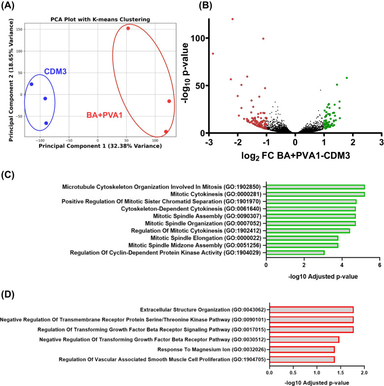
Methods and results: Both media compositions enable the efficient, directed differentiation of embryonic and induced hPSCs, matching the cell yields and cardiomyocyte purity ranging from 85 to 99% achieved with the widely used protein-based CDM3 medium. The protein-free differentiation approach was readily up-scaled to a 2000 mL process scale in a fully controlled stirred tank bioreactor in suspension culture, producing > 1.3 × 10⁹ cardiomyocytes in a single process run. Transcriptome analysis, flow cytometry, electrophysiology, and contractile force measurements revealed that the mass-produced cardiomyocytes differentiated in protein-free medium exhibit the expected ventricular-like properties equivalent to the well-established characteristics of CDM3-control cells.

Conclusions: This study promotes the robustness and upscaling of the cardiomyogenic differentiation process, substantially reduces media costs, and provides an important step toward the clinical translation of hPSC-CMs for heart regeneration.

Keywords: Bioreactor; Cardiomyocytes; Protein-free differentiation media; hPSC.


Conflict of interest statement

F.M. and W.T. are employees of Evotec. The other authors declare no potential competing interests.

Figures

Fig. 1
Fig. 1
BA + PF-68 and BA + PVA1 achieve CM yield and purities comparable to CDM3 in shaken Erlenmeyer flasks. A Schematic of differentiation. hPSC aggregates are formed over three days of expansion in STBRs and transferred to Erlenemeyer flasks, where different differentiation media were used. B Expression of MIXL1-GFP on dd1 for the investigated differentiation media assessed by flow cytometry compared to CDM3 (dashed line; n = 3; results are mean ± s.d.). C Cell yields on dd10 for respective media formulations compared to CDM3 cell yields (dashed line; n = 3; results are mean ± s.d.). D Representative flow cytometry plots revealing the expression of investigated CM markers cardiac troponin T (cTnT), pan-myosin heavy chain (MHC), and sarcomeric actinin (SA) for CDM3, BA + PF-68, and BA + PVA1
Fig. 2
Fig. 2
Protein-free differentiation media closely resemble typical patterns observed for CDM3 at 150 mL process scale. A Schematic of differentiation. hPSC aggregates were formed over two or three days (depending on cell line). The viable cell density was adjusted to 75 × 106 hPSCs per 150 mL bioreactor, and the differentiation was performed. B Viable cell number and viability for the hESC line HES3 MIXL1-GFP for CDM3 (black), BA + PVA1 (blue), BA + PF-68 (green) throughout the 10-day lasting differentiation process (n = 3 for each medium; mean ± s.d.) and C for the hiPSC line Phoenix HSC_ADCF_SeV_iPS2 for CDM3 (black) and BA + PVA1 (blue; n = 3 for each medium; mean ± s.d.). D CM-specific markers cTnT, pan-MHC, and SA for the hESC line MIXL1-GFP for CDM3 (black), BA + PVA1 (blue), BA + PF-68 (green; n = 3 for each medium; mean ± s.d.) and E for the hiPSC line Phoenix for CDM3 (black) and BA + PVA1 (blue; n = 3 for each medium; mean ± s.d.). F Aggregate size development during differentiation for exemplary processes in CDM3 or G BA + PVA1 (cell line Phoenix; each dot represents a single analyzed aggregate, red lines indicate mean ± s.d.)
Fig. 3
Fig. 3
Contraction and electrophysiological analysis revealed no substantial differences between CMs from protein-free differentiation and CDM3. A Bioartificial cardiac tissues (BCTs) were produced from three independent differentiation runs in either BA + PVA1 or CDM3 medium (cell line Phoenix, 3 BCTs per biological replicate for 3 biological replicates; mean ± SEM). The paced and spontaneous contraction forces increase to a similar level (mN/mm2) with increasing preload. The values were normalized to the diameter of the individual BCT. B Individual BCT forces (mN/mm.2) at the maximum stretch (1200 µm; mean ± SEM). C Spontaneous beating frequency (Hz) for BCTs from CMs differentiated in BA + PVA1 or CDM3 at 0 µm and 1200 µm preload (mean ± SEM). D Representative ventricular-like action potentials (APs) for BA + PVA1-derived and E CDM3-derived, seeded CMs were revealed by patch clamp analysis. F Duration of the action potential at 50% of the amplitude (APD50) for CMs derived from BA + PVA1 and CDM3 conditions. G Frequency of APs/min for CMs derived from BA + PVA1 and CDM3 conditions H AP amplitude (in mV) for CMs derived from BA + PVA1 and CDM3 conditions. I Resting membrane potential (in mV) for CMs derived from BA + PVA1 and CDM3 conditions (for BA + PVA1 n = 3 biological replicates, for CDM3 control n = 1 in F-I) J-M Comparison of APD50, AP frequency, AP amplitude, and RMP in three biologically independent CM batches produced in BA + PVA1 (all mean ± SEM)
Fig. 4
Fig. 4
Transcriptomic analysis of CMs from BA + PVA1 and CDM3 revealed significant differences. A Principal component analysis of three biologically independent replicates of CMs from BA + PVA1 (red) and CDM3 (blue) conditions according to K-means clustering. B Volcano plot of transcripts identified in all samples with ≥ 10 counts. Significantly up/downregulated transcripts (p ≥ 0.05) with a fold change ≥ 1/ ≤ − 1 between the two groups are marked in green or red, respectively. Three independent biological samples with comparable cTnT purity (according to flow cytometry analysis) were analyzed for both conditions. C + D Gene Ontology analysis of the most significant results between CMs differentiated in BA + PVA1 or CDM3 to be upregulated in BA + PVA1 (C) or downregulated in BA + PVA1 (D). Only genes of transcripts with a fold change ≥ 1/ ≤ − 1 and p ≥ 0.05 were considered
Fig. 5
Fig. 5
Successful upscaling of differentiation to 2000 mL bioreactor (Bioflo 320). A Viable cell number and viability for the hiPSC line hHSC_1285T_iPS2 in a 2000 mL bioreactor differentiated in BA + PVA1 throughout the 10-day differentiation process (n = 3; mean ± s.d.). B CM-specific markers cTnT, pan-MHC, and SA for the hiPSC line for differentiation runs in A (n = 3; mean ± s.d.). C Microscopic analysis of aggregate diameters during the 10-day differentiation process are comparable between an exemplary 150 mL process and a 2000 mL process when utilizing the same cell line hHSC_1285T_iPS2 (each dot represents a single aggregate; mean ± s.d.) D Exemplary depiction of the respective markers (cTnT, pan-MHC, SA) for a 2000 mL differentiation process. E Total biomass generated in one 2000 mL differentiation process

References

    1. Inoue H, Nagata N, Kurokawa H, Yamanaka S. IPS cells: a game changer for future medicine. EMBO J. 2014;33:409–417. doi: 10.1002/embj.201387098. - DOI - PMC - PubMed
    1. Kempf H, Andree B, Zweigerdt R. Large-scale production of human pluripotent stem cell derived cardiomyocytes. Adv Drug Deliv Rev. 2016;96:18–30. doi: 10.1016/j.addr.2015.11.016. - DOI - PubMed
    1. Zweigerdt R. Large scale production of stem cells. Adv Biochem Eng Biotechnol. 2009 doi: 10.1007/10_2008_27. - DOI - PubMed
    1. Laflamme MA, Murry CE. Heart regenartion. Nature. 2011;473:1–7. doi: 10.1038/jid.2014.371. - DOI - PMC - PubMed
    1. Chong JJH, Yang X, Don CW, Minami E, Liu YW, Weyers JJ, Mahoney WM, Van Biber B, Cook SM, Palpant NJ, Gantz JA, Fugate JA, Muskheli V, Gough GM, Vogel KW, Astley CA, Hotchkiss CE, Baldessari A, Pabon L, et al. Human embryonic-stem-cell-derived cardiomyocytes regenerate non-human primate hearts. Nature. 2014;510:273–277. doi: 10.1038/nature13233. - DOI - PMC - PubMed

Publication types

MeSH terms

LinkOut - more resources