

Abrasion and dental pulp morphological changes in occlusal dysfunction
- PMID: 39020543
- PMCID: PMC11384036
- DOI: 10.47162/RJME.65.2.15
Abrasion and dental pulp morphological changes in occlusal dysfunction
Abstract
Aim: The authors set out to assess if the presence and the degree of severity of the abrasion, as a consequence of the occlusal dysfunction, determine further morphological changes in the dental pulp.
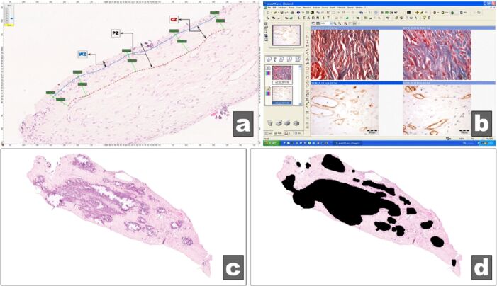
Materials and methods: Study group included teeth dental pulp from 45 cases with occlusal dysfunction, subsequently divided into two subgroups: 24 cases with abrasion (AB) and 21 cases without abrasion (NONAB). The set of morphological parameters of dental pulp were thicknesses of the outer layer, inner layer and entire peripheral pulp zone, the presence of pulpal calcifications and their extent within the dental pulp, the presence of interstitial fibrosis and its extent within the dental pulp and the vascular density (VD) of pulpal capillary network. Tissue samples were fixed in 10% buffered formalin, embedded in paraffin, and sectioned off at 4 μm. Serial slides were stained with Hematoxylin-Eosin (HE), Masson's trichrome (MT) and anti-cluster of differentiation 34 (CD34) antibodies labeled with 3,3'-Diaminobenzidine (DAB) and transformed into virtual slides on which the above-mentioned parameters were studied comparatively with the help of a dedicated in-house software, realized in MATLAB (MathWorks, USA). The numerical values of the assessed parameters were also stratified in classes, thus obtaining score scales for each parameter. Statistical tools used were Lilliefors test, t-test (two-sample assuming equal variances), Mann-Whitney test, Pearson's correlation test, one-way analysis of variance (ANOVA) test and χ² (chi-squared) test.
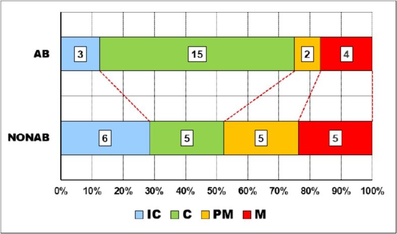
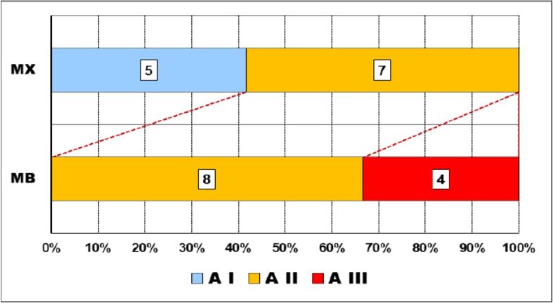
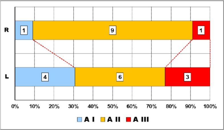
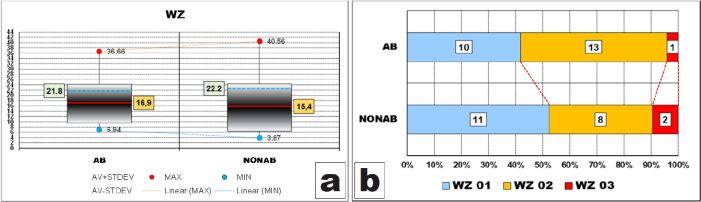
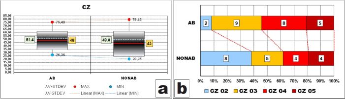
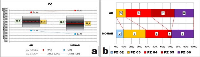
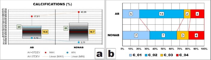
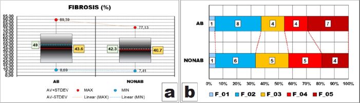
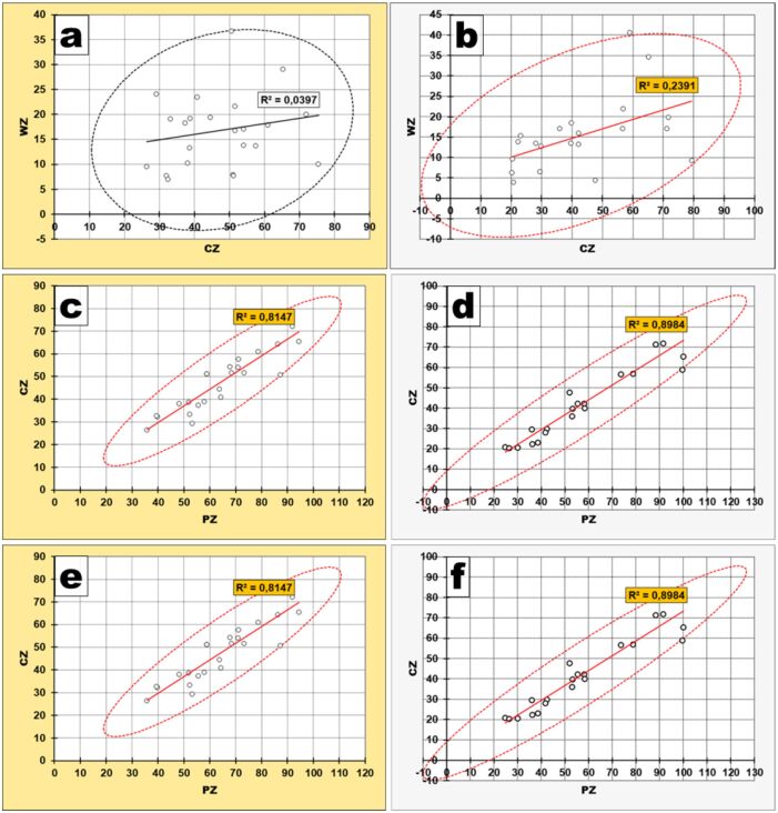
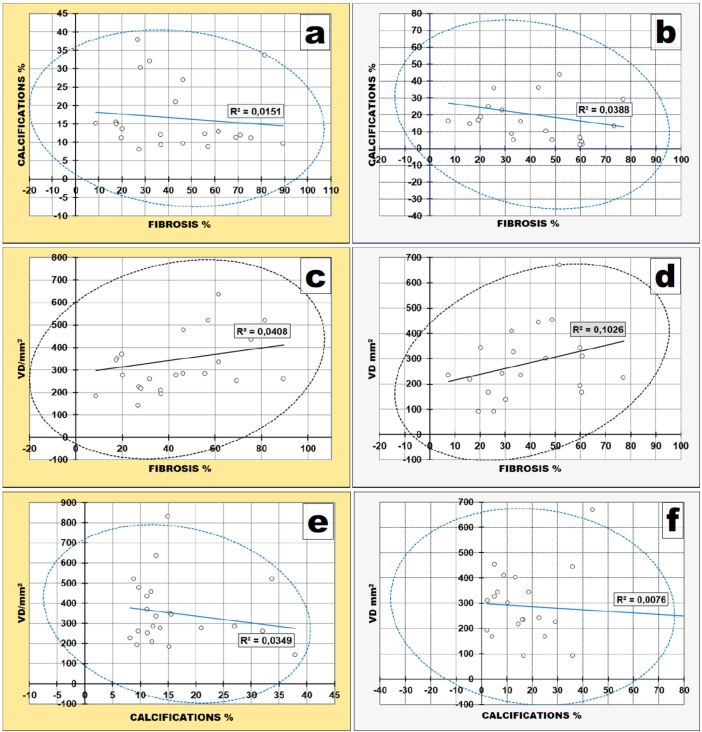
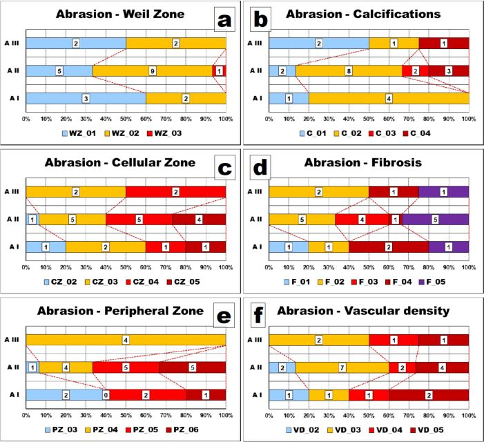
Results and discussions: Both peripheral zone (PZ) and its layers individually tended to be thicker in the teeth with abrasion than in those without abrasion. Also, teeth with abrasion tended to develop larger amounts of calcium deposits in their dental pulp than teeth without abrasion. On the other hand, fibrotic tissue in dental pulp had no relationship with the presence or absence of abrasion. PZ as a whole and its layers evolved together in the same way, with a stronger correlation in the group of teeth without abrasion. Deposits of calcium evolved in the opposite direction to both the amount of fibrous tissue and the capillaries density of the dental pulp. Consequently, the amount of fibrous tissue and VD evolved together in the same way, more pronounced in the teeth without abrasion.
Conclusions: Our preliminary study pointed out that different components of the dental pulp showed slight to moderate changes depending on the degree of abrasion in teeth with occlusal dysfunction.
Conflict of interest statement
The authors declare that they have no conflict of interests.
Figures

References
-
- Burlui V , Morăraşu C . Gnatologie . Iaşi, Romania : Ed. Apollonia ; 2000 . pp. 76 – 89 .
-
- Moreno S, Moreno F. Dental anthropology: clinical importance [Importancia clínica de la antropología dental] Rev Estomatol. 2007;15(2 Suppl 1):42–53.
-
- Ieremia L , Dociu I . Funcţia şi disfuncţia ocluzală . Bucharest, Romania : Ed. Medicală ; 1987 . pp. 24 – 220 .
-
- Addy M, Dowell P. Dentine hypersensitivity - a review. Clinical and in vitro evaluation of treatment agents. J Clin Periodontol. 1983;10(4):351–363. - PubMed
MeSH terms
LinkOut - more resources
Full Text Sources
