. 2025 Feb 3;148(2):564-579.
doi: 10.1093/brain/awae243.

Combined clinical, structural and cellular studies discriminate pathogenic and benign TRPV4 variants

Affiliations

Combined clinical, structural and cellular studies discriminate pathogenic and benign TRPV4 variants

Sarah H Berth et al. Brain. .

Abstract

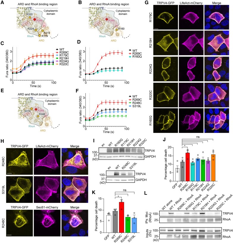
Dominant mutations in the calcium-permeable ion channel TRPV4 (transient receptor potential vanilloid 4) cause diverse and largely distinct channelopathies, including inherited forms of neuromuscular disease, skeletal dysplasias and arthropathy. Pathogenic TRPV4 mutations cause gain of ion channel function and toxicity that can be rescued by small molecule TRPV4 antagonists in cellular and animal models, suggesting that TRPV4 antagonism could be therapeutic for patients. Numerous variants in TRPV4 have been detected with targeted and whole exome/genome sequencing, but for the vast majority, their pathogenicity remains unclear. Here, we used a combination of clinical information and experimental structure-function analyses to evaluate 30 TRPV4 variants across various functional protein domains. We report clinical features of seven patients with TRPV4 variants of unknown significance and provide extensive functional characterization of these and an additional 17 variants, including structural position, ion channel function, subcellular localization, expression level, cytotoxicity and protein-protein interactions. We find that gain-of-function mutations within the TRPV4 intracellular ankyrin repeat domain target charged amino acid residues important for RhoA interaction, whereas ankyrin repeat domain residues outside of the RhoA interface have normal or reduced ion channel activity. We further identify a cluster of gain-of-function variants within the intracellular intrinsically disordered region that may cause toxicity via altered interactions with membrane lipids. In contrast, assessed variants in the transmembrane domain and other regions of the intrinsically disordered region do not cause gain of function and are likely benign. Clinical features associated with gain of function and cytotoxicity include congenital onset of disease, vocal cord weakness and motor-predominant disease, whereas patients with likely benign variants often demonstrated late-onset and sensory-predominant disease. These results provide a framework for assessing additional TRPV4 variants with respect to likely pathogenicity, which will yield critical information to inform patient selection for future clinical trials for TRPV4 channelopathies.

Keywords: Charcot-Marie-Tooth disease; RhoA; TRPV4; calcium signalling; neuropathy; skeletal dysplasia.


Conflict of interest statement

M.H., S.T., T.O.C., C.J.S and B.A.M. are participating in an investigator-initiated natural history study for TRPV4 neuropathy (NCT05600764) that is supported by Actio Biosciences. C.J.S. is supported by Actio Biosciences for laboratory-based studies of TRPV4 channelopathy.

Figures

Figure 1
Figure 1
Evaluation of transmembrane domain variant I545N does not show gain-of-function toxicity. (A) Schematic of the assessments performed for each TRPV4 variant with cell types used for each assay indicated within parentheses. (B) Schematic showing the structure of the TRPV4 homotetramer with the locations of known neuropathy mutations (R186, R232, R237, R269, R315, R316) highlighted in red. RhoA is outlined and shaded green. (C) Averaged calcium imaging traces in MN-1 cells transfected with known pathogenic TRPV4 mutants within the ARD. MN-1 cells were transfected with TRPV4-GFP constructs, loaded with the calcium indicator Fura-2 AM and treated with hypotonic saline (30 mM NaCl) at time = 20 s, n = 9 wells per condition with 20–40 cells per well. Scale bar = 50 µm. (D) Lactate dehydrogenase (LDH) cytotoxicity assays in HEK293T cells transiently transfected with TRPV4 wild-type (WT) or known neuropathy mutants show increased cell death with pathogenic TRPV4 mutants. One-way ANOVA with Dunnett’s post hoc test, n = 4 independent experiments per condition, **P < 0.01, ****P < 0.0001. (E) Location of I545 within the transmembrane domain (TMD) in the TRPV4 protein. (F) Averaged calcium imaging traces in MN-1 cells before and after hypotonic stimulation, n = 9 wells per condition with 20–40 cells per well. (G) Representative images from ratiometric calcium imaging experiments from cells expressing TRPV4 WT, the known pathogenic R269C mutant, or the I545N variant. The pathogenic variant R269C causes elevated basal calcium levels and increased hypotonic-stimulated calcium responses, whereas the I545N variant shows similar calcium responses to WT TRPV4. (H) Fluorescence microscopy of canine kidney epithelial (MDCK) cells co-transfected with GFP-tagged TRPV4 and Life-Act mCherry shows normal trafficking of TRPV4 to the cell membrane and co-localization with cortical actin. Scale bar = 10 µm. (I) Immunoblot from HEK293T cells transiently transfected with WT or mutant TRPV4-GFP shows similar amounts of TRPV4 protein. (J) LDH cytotoxicity assays in HEK293T cells transiently transfected with TRPV4 WT, I545N, or R269C show increased cytotoxicity with expression of the R269C mutant compared to WT TRPV4, but no change in the I545N mutant. One-way ANOVA with Dunnett’s post hoc test, n = 4 independent experiments per condition, *P < 0.05. Error bars indicate standard error of the mean. IDR = intrinsically disordered region.
Figure 2
Figure 2
The M680V channel pore mutant causes loss of function. (A) Schematic of the TRPV4 homotetramer shows M680 within the TRPV4 channel pore and C480 and V515 within the transmembrane domain (TMD). (B) Averaged calcium tracings in MN-1 cells before and after hypotonic stimulation demonstrate a reduction in basal and stimulated intracellular calcium in the M680V variant compared to wild-type (WT) TRPV4, n = 9–16 wells per condition with 20–40 cells per well. (C) Two additional TMD variants (C480F and V515) show normal basal and stimulated intracellular calcium in transiently transfected MN-1 cells, n = 10–12 wells per condition with 20–40 cells per well. (D) Fluorescence microscopy of canine kidney epithelial (MDCK) cells co-transfected with GFP-tagged TRPV4 and Life-Act mCherry shows normal trafficking of TRPV4 M680V, M680K, C480F and V515F variants to the cell membrane and co-localization with cortical actin. (E) Calcium imaging of MN-1 cells shows reduced calcium levels with the M680K variant and dominant-negative effects when the M680K variant is co-transfected with WT TRPV4, n = 9–13 wells per condition with 20–40 cells per well. Scale bar = 10 µm. (F) Calcium imaging of MN-1 cells co-transfected with the M680V variant and WT TRPV4 shows no effect of mutant TRPV4 on the typical WT TRPV4 responses, indicating that M680V does not act as a dominant negative, n = 7–11 wells per condition with 20–40 cells per well. (G) Immunoblot from HEK293T cells transiently transfected with WT or mutant TRPV4-GFP protein show similar amounts of TRPV4 protein. (H) Lactate dehydrogenase (LDH) cytotoxicity assays in HEK293T cells transiently transfected with TRPV4 WT, C480F, V515F, M680V or R269C show no change in cytotoxicity in the TMD variants. One-way ANOVA with Dunnett’s post hoc test, n = 3 independent experiments for M680V, n = 4 for all other conditions. (I) Co-immunoprecipitation experiments from HEK293T cells transfected with Myc-tagged RhoA and GFP-tagged TRPV4 and subjected to immunoprecipitation with anti-Myc antibody demonstrates normal TRPV4-RhoA interaction with all mutants. Error bars indicate standard error of the mean.
Figure 3
Figure 3
Two ankyrin repeat domain mutations outside of the TRPV4-RhoA interface, G321V and V246D, cause loss of function. (A) Schematic of the TRPV4 ankyrin repeat domain (ARD) with the G321 and V264 residues highlighted in yellow and the neuropathy-associated R269 residue highlighted in red. G321 and V264D are located on the surface of the ARD, but not in the RhoA binding region where known neuropathy-causing mutations are located. (B) Representative images from ratiometric calcium imaging experiments in MN-1 cells. Scale bar = 50 µm. (C) Averaged calcium tracings in MN-1 cells before and after hypotonic stimulation show a reduction of basal intracellular calcium and lack of calcium influx in response to hypotonic stress in MN-1 cells transiently transfected with G321V and V246D mutants compared to wild-type (WT) TRPV4 and known neuropathy-causing mutation R269C, n = 9 wells per condition with 20–40 cells per well. (D) Expression of V264D and G321V in canine kidney epithelial (MDCK) cells shows altered cellular localization of these TRPV4 variants. Mutants fail to localize at the plasma membrane but instead co-localize with the endoplasmic reticulum (ER) marker Sec61-mCherry, demonstrating likely misfolding and retention in the ER. Scale bar = 10 µm. (E) Immunoblotting from transiently transfected HEK293T cells shows reduced expressed protein levels in the V264D and G321V mutants compared to WT TRPV4. (F) Co-expression of WT TRPV4 with G321V or with V264D does not alter TRPV4 basal calcium or response to hypotonic stress, indicating that the G321V and V264D mutants do not act in a dominant-negative fashion, n = 9–13 wells per condition with 20–40 cells per well. (G) Measurement of cytotoxicity via lactate dehydrogenase (LDH) assays in HEK293T cells shows reduced toxicity by both G321V and V264D mutants compared to expression of WT TRPV4. One-way ANOVA with Dunnett’s post hoc test, n = 3–5 independent experiments per condition, *P < 0.05. Error bars indicate standard error of the mean.
Figure 4
Figure 4
Functional properties of selected ankyrin repeat domain variants. (A) Schematic of the TRPV4 ankyrin repeat domain (ARD) with the R179, R219, R224 and R320 residues highlighted in yellow and the neuropathy-associated R269 residue highlighted in red. (B) Schematic of TRPV4 ARD with the R160Q variant highlighted in yellow. (C and D) Graphs show averaged calcium tracings in MN-1 cells transfected with ARD variants and stimulated with hypotonic saline. No significant difference was observed between wild-type (WT) TRPV4 and the R179C, R219H and R224Q variants, but the R320C variant shows both increased basal calcium and stimulated calcium levels (C), n = 9–22 wells per condition with 20–40 cells per well. The R160Q variant shows normal basal calcium, but a mild reduction of stimulated calcium response (D), n = 10–13 wells per condition with 20–40 cells per well. (E) Schematic of TRPV4 ARD showing the location of the ARD variants R248C and S319 highlighted in yellow. (F) Averaged calcium tracings in transiently transfected MN-1 cells demonstrate a reduction of stimulated calcium levels in MN-1 cells expressing R248C and S319L compared to WT TRPV4, n = 11–15 wells per condition with 20–40 cells per well. (G and H) Fluorescence microscopy of canine kidney epithelial (MDCK) cells co-transfected with GFP-tagged TRPV4 and Life-Act mCherry shows that R179C, R219H, R224Q, R320C, R160Q (G) and S319L (H) show normal localization at the plasma membrane and co-localization with LifeAct-mCherry. However, the R248C TRPV4 mutant shows reduced plasma membrane targeting and marked co-localization with the endoplasmic reticulum (ER) marker Sec61-mCherry, demonstrating likely misfolding and retention in the ER. Scale bar = 10 µm. (I) Immunoblots from HEK293T cells transiently transfected with WT or mutant TRPV4-GFP demonstrate normal levels of R160Q, R179C, R219H, R224Q and R320C protein compared to WT TRPV4, but reduced R248C and R319L expression. (J and K) Lactate dehydrogenase (LDH) cytotoxicity assays in transiently transfected HEK293T cells show normal levels of toxicity in R160Q, R179C, R219H, R224Q and R320C variants (J) and normal levels of toxicity in R248C and R319L variants (K). One-way ANOVA with Dunnett’s post hoc test, n = 4 (J) and n = 3–4 (K) independent experiments per condition, *P < 0.05. (L) Co-immunoprecipitation experiments from HEK293T cells transfected with Myc-tagged RhoA and GFP-tagged TRPV4 and subjected to immunoprecipitation with anti-Myc antibody demonstrates markedly reduced interaction of TRPV4-R224Q with RhoA and mildly reduced interaction of TRPV4-R320 with RhoA. Error bars indicate standard error of the mean.
Figure 5
Figure 5
Variants at position R271 have differential effects on TRPV4 function. (A) Schematic of the TRPV4 homotetramer shows that residue R271, highlighted in yellow, is located on the ankyrin repeat domain (ARD) surface, adjacent to known TRPV4 neuropathy-causing mutations at the RhoA interface. (B and C) Graphs show averaged calcium tracings in MN-1 cells transfected with R271 variants and stimulated with hypotonic saline. The R271P variant shows increased basal calcium but had minimal calcium response to hypotonic stimulation (B), whereas the R271H (B) and R271C (C) variants show reduced basal calcium and minimal responses to hypotonic stimulation, n = 10–11 wells per condition (B) and n = 9–16 wells per condition (C) with 20–40 cells per well. (D and E) Fluorescence microscopy of canine kidney epithelial (MDCK) cells co-transfected with GFP-tagged TRPV4 and Life-Act mCherry shows that R217H and R271C variants localize normally to the plasma membrane and co-localize with LifeAct-mCherry. However, the R271P variant is predominantly mislocalized to the endoplasmic reticulum (ER). Scale bar = 10 µm. (F) Immunoblots from HEK293T cells transiently transfected with wild-type (WT) or mutant TRPV4-GFP demonstrate normal levels of protein expression. (G) Co-immunoprecipitation studies using Myc-tagged RhoA and GFP-tagged TRPV4 in HEK293T cells demonstrates that the R271P variant, but not R271C or R271H variants, disrupts RhoA binding. (H) Lactate dehydrogenase (LDH) cytotoxicity assays in transiently transfected HEK293T cells show normal levels of toxicity in R271P, R271H and R271C variants. One-way ANOVA with Dunnett’s post hoc test, n = 4 independent experiments per condition, ***P < 0.001. Error bars indicate standard error of the mean.
Figure 6
Figure 6
Specific intrinsically disordered region variants cause gain-of-function of the TRPV4 channel in a manner that is dependent on PIP2 binding. (A) Schematic of the TRPV4 homotetramer and PIP2 binding domain (PBD) within the intrinsically disordered region (IDR). Bottom: The location of the analysed IDR variants and the average number of membrane contacts of each amino acid residue from molecular dynamics simulation on a lipid bilayer composed of POPC (69%), CHOL (20%), DOPS (10%) and PIP2 (1%). (B and C) Graphs show averaged calcium tracings in MN-1 cells transiently transfected with IDR variants and stimulated with hypotonic saline. Expression of the S93F variant leads to increased basal and stimulated calcium levels (B), n = 10–11 wells per condition with 20–40 cells per well. Basal and stimulated calcium levels in the G20R variant are similar to wild-type (WT) (C). The D62N and S94L variants demonstrate mildly elevated basal and partially enhanced stimulated calcium levels (C). The P97R variant shows elevated basal calcium and augmented calcium response to hypotonic stress (C), n = 16–17 wells per condition with 20–40 cells per well. Scale bar = 10 µm. (D) Fluorescence microscopy of canine kidney epithelial (MDCK) cells co-transfected with GFP-tagged TRPV4 and Life-Act mCherry shows normal localization of all IDR variants at the plasma membrane and co-localization with LifeAct-mCherry. (E) Immunoblots from HEK293T cells transiently transfected with WT or mutant TRPV4-GFP demonstrate normal levels of TRPV4 in all IDR variants. (F) Lactate dehydrogenase (LDH) cytotoxicity assays in transiently transfected HEK293T cells show increased cytotoxicity in D62N, S93F, S94L and P79R variants. One-way ANOVA with Dunnett’s post hoc test, n = 4 independent experiments per condition, **P < 0.01, ***P < 0.001, ****P < 0.0001. (G and H) Co-immunoprecipitation using GFP-tagged TRPV4 and either Myc-tagged RhoA (G) or Myc-tagged PACSIN3 (H) in HEK293T cells shows all IDR variants had unchanged binding to RhoA and PACSIN3 compared to WT TRPV4. (I and J) Graphs show averaged calcium tracings in MN-1 cells transiently transfected with TRPV4 variants and PIP2 binding-deficient mutants. Mutating the PIP2 binding domain KRWKR to AAWAA (AWA) in the known neuropathy-causing mutant R269C or in WT TRPV leads to a reduction in basal calcium and stimulated responses, but not in the R616G variant, which causes constitutive activity of the TRPV4 channel (I), n = 12–14 wells per condition with 20–40 cells per well. Introducing the AWA mutation in the P97R variant reduces basal and stimulated calcium levels, negating the gain-of-function effect of the P97R variants (J), n = 9–14 wells per condition with 20–40 cells per well. Error bars indicate standard error of the mean. ARD = ankyrin repeat domain.

References

    1. McCray BA, Schindler A, Hoover-Fong JE, Sumner CJ. Autosomal Dominant TRPV4 Disorders. In: Adam MP, Feldman J, Mirzaa GM, Pagon RA, Wallace SE, Amemiya A, eds. GeneReviews®. University of Washington; 2014. - PubMed
    1. White JP, Cibelli M, Urban L, Nilius B, McGeown JG, Nagy I. TRPV4: Molecular conductor of a diverse orchestra. Physiol Rev. 2016;96:911–973. - PubMed
    1. Taga A, Peyton MA, Goretzki B, et al. TRPV4 mutations causing mixed neuropathy and skeletal phenotypes result in severe gain of function. Ann Clin Transl Neurol. 2022;9:375–391. - PMC - PubMed
    1. Ragamin A, Gomes CC, Bindels-de Heus K, et al. De novo TRPV4 Leu619Pro variant causes a new channelopathy characterised by giant cell lesions of the jaws and skull, skeletal abnormalities and polyneuropathy. J Med Genet. 2022;59:305–312. - PMC - PubMed
    1. Echaniz-Laguna A, Dubourg O, Carlier P, et al. Phenotypic spectrum and incidence of TRPV4 mutations in patients with inherited axonal neuropathy. Neurology. 2014;82:1919–1926. - PubMed

LinkOut - more resources