. 2024 Jul 3:12:1435627.
doi: 10.3389/fchem.2024.1435627. eCollection 2024.

Hyaluronic acid modified indocyanine green nanoparticles: a novel targeted strategy for NIR-II fluorescence lymphatic imaging

Affiliations

Hyaluronic acid modified indocyanine green nanoparticles: a novel targeted strategy for NIR-II fluorescence lymphatic imaging

Haiyan Zhang et al. Front Chem. .

Abstract

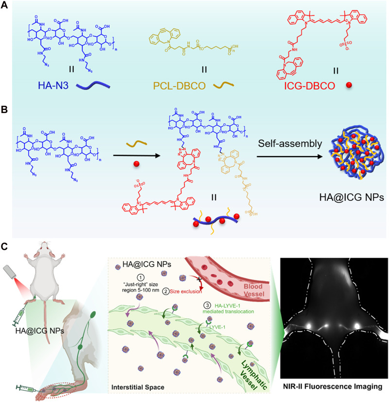
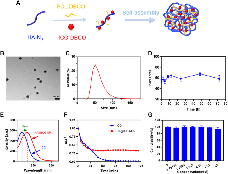
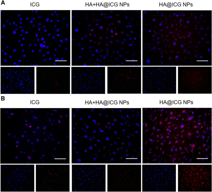
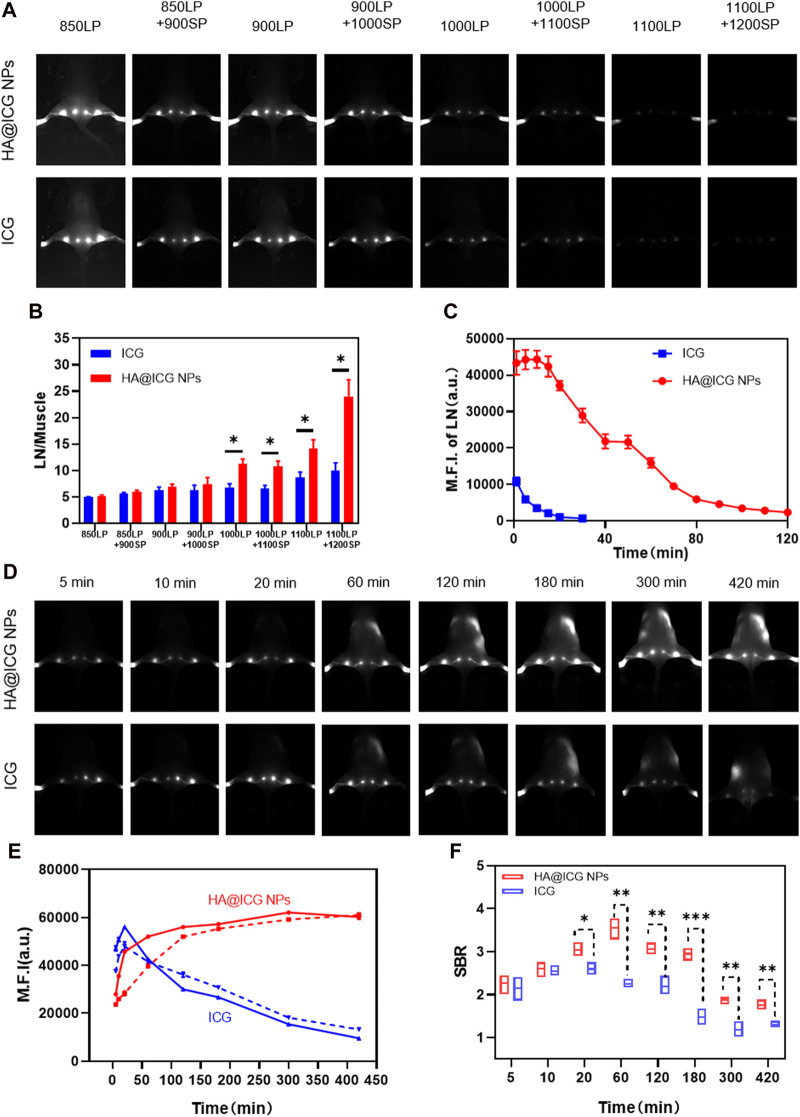
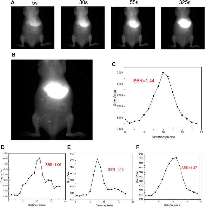
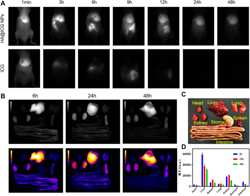
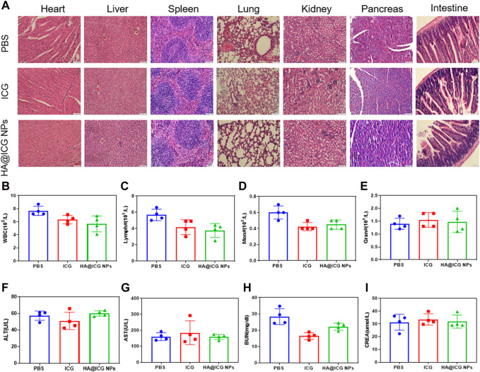
The lymphatic system, alongside blood circulation, is crucial for maintaining bodily equilibrium and immune surveillance. Despite its importance, lymphatic imaging techniques lag behind those for blood circulation. Fluorescence imaging, particularly in the near-infrared-II (NIR-II) region, offers promising capabilities with centimeter-scale tissue penetration and micron-scale spatial resolution, sparking interest in visualizing the lymphatic system. Although indocyanine green (ICG) has been approved by the Food and Drug Administration (FDA) for use as a near-infrared-I (NIR-I) region fluorescent dye, its limitations include shallow penetration depth and low signal-to-noise ratio. Research suggests that ICG's fluorescence emission tail in the second near-infrared window holds potential for high-quality NIR-II imaging. However, challenges like short circulation half-life and concentration-dependent aggregation hinder its wider application. Here we developed HA@ICG nanoparticles (NPs), a superior ICG-based NIR-II fluorescent probe with excellent biocompatibility, prolonging in vivo imaging, and enhancing photostability compared to ICG alone. Leveraging LYVE-1, a prominent lymphatic endothelial cell receptor that binds specifically to hyaluronic acid (HA), our nanoprobes exhibit exceptional performance in targeting lymphatic system imaging. Moreover, our findings demonstrate the capability of HA@ICG NPs for capillary imaging, offering a means to assess local microcirculatory blood supply. These compelling results underscore the promising potential of HA@ICG NPs for achieving high-resolution bioimaging of nanomedicines in the NIR-II window.

Keywords: LYVE-1; NIR-II imaging; fluorescence imaging; hyaluronic acid; indocyanine green; lymphatic imaging.


Conflict of interest statement

The authors declare that the research was conducted in the absence of any commercial or financial relationships that could be construed as a potential conflict of interest.

Figures

FIGURE 1
FIGURE 1
(A,B) Illustration of the preparation process for HA@ICG NPs; (C) Path of the nanoparticles from the interstitial space into the lymphatic vessels:①② “Size effect”: Nanoparticles of just 5–100 nm in size are excluded from the capillaries and diffuse into the lymphatic lumen via the endothelial space of the lymphatic vessels; ③HA-LYVE-1:Translocation of HA@ICG NPs into lymphatic vessels by the high specific affinity of HA for LYVE-1.
FIGURE 2
FIGURE 2
Preparation and characterization of HA@ICG NPs. (A) Schematic illustration of the preparation process for HA@ICG NPs. (B) TEM images of HA@ICG NPs (Scale bar = 100 nm). (C,D) Size distribution and the particle size stability of HA@ICG NPs in PBS over 72 h (E) The fluorescence spectra of ICG and HA@ICG NPs. (F) Fluorescence changes of ICG and HA@ICG NPs under continuous laser radiation (808 nm, 78 mW cm−2) over a period of time in a long-pass filter above 1,000 nm. (G) Cell viability of HLECs treated with different concentrations of HA@ICG NPs (n = 4).
FIGURE 3
FIGURE 3
Images of HLECs incubated with different nanoparticle groups respectively for (A) 2 h and (B) 6 h. Scale bar = 100 µm. HA+HA@ICG NPs Group: HA (10 mg mL−1) pre-treatment for 1 h before adding HA@ICG NPs.
FIGURE 4
FIGURE 4
(A) Fluorescence images of popliteal and sacral LNs at different NIR long pass filters of HA@ICG NPs and free ICG. (B) The ratio of fluorescence intensity of LN to muscle at different wavelengths. (C) Fluorescence changes of the mouse hindlimb limb lymphatic system under continuous laser radiation over a period of time. (D) NIR-II fluorescence imaging of LNs and lymphatic vessels at different time points after s.c. injection of HA@ICG NPs and free ICG, respectively. (E) Time dependent fluorescence intensity of popliteal (solid line) and sacral (imaginal line) LNs. (F) A comparison of the signal-to-background ratio between the HA@ICG NPs group and free ICG for lymphatic vessel imaging. Data expressed as Mean ± SD (n = 3). *p < 0.05, **p < 0.01, and ***p < 0.001.
FIGURE 5
FIGURE 5
(A)The NIR-II vascular imaging in normal mice after intravenous injection under a 1,000 nm long-pass filter at different times. (B) ROI regions in NIR-II fluorescence images of blood vessels post i.v. injection of HA@ICG NPs at 325 s. (C–F) The signal-to-background ratio of four ROI regions (1, 2, 3, 4) in (B). Injection dosage: 300 μM ICG, 150 μL; laser: 808 nm, 10 ms, 78 mW cm−2.
FIGURE 6
FIGURE 6
(A) Near-infrared II fluorescence images of mice at different time points after injection of HA@ICG NPs and ICG via caudal vein. (B–D) The NIR-II fluorescence imaging and quantitative analysis of isolated organs removed at 6, 24 and 48 h after tail vein injection of HA@ICG NPs.
FIGURE 7
FIGURE 7
(A) H&E staining of the main organs from the mice including heart, liver, spleen, lung, kidney, pancreas and intestine after different treatments for 7 days. Scale bar = 100 μm. (B–I) Haemato-biochemical parameters of the mice after treatment with PBS, ICG, HA@ICG NPs for 7 days (n = 4).

Similar articles

Cited by

References

    1. Cao J., Zhu B., Zheng K., He S., Meng L., Song J., et al. (2020). Recent progress in NIR-II contrast agent for biological imaging. Front. Bioeng. Biotechnol. 7, 487. 10.3389/fbioe.2019.00487 - DOI - PMC - PubMed
    1. Carr J. A., Franke D., Caram J. R., Perkinson C. F., Saif M., Askoxylakis V., et al. (2018). Shortwave infrared fluorescence imaging with the clinically approved near-infrared dye indocyanine green. Proc. Natl. Acad. Sci. 115, 4465–4470. 10.1073/pnas.1718917115 - DOI - PMC - PubMed
    1. Chen Y., Xue L., Zhu Q., Feng Y., Wu M. (2021). Recent advances in second near-infrared region (NIR-II) fluorophores and biomedical applications. Front. Chem. 9, 750404. 10.3389/fchem.2021.750404 - DOI - PMC - PubMed
    1. Crawford T., Moshnikova A., Roles S., Weerakkody D., DuPont M., Carter L. M., et al. (2020). pHLIP ICG for delineation of tumors and blood flow during fluorescence-guided surgery. Sci. Rep. 10, 18356. 10.1038/s41598-020-75443-5 - DOI - PMC - PubMed
    1. Hu Z., Fang C., Li B., Zhang Z., Cao C., Cai M., et al. (2019). First-in-human liver-tumour surgery guided by multispectral fluorescence imaging in the visible and near-infrared-I/II windows. Nat. Biomed. Eng. 4, 259–271. 10.1038/s41551-019-0494-0 - DOI - PubMed

LinkOut - more resources