. 2024 Jun 15;16(3):28-54.
doi: 10.62347/RTMH4490. eCollection 2024.

Association of connexin36 with adherens junctions at mixed synapses and distinguishing electrophysiological features of those at mossy fiber terminals in rat ventral hippocampus

Affiliations

Association of connexin36 with adherens junctions at mixed synapses and distinguishing electrophysiological features of those at mossy fiber terminals in rat ventral hippocampus

Deepthi Thomas et al. Int J Physiol Pathophysiol Pharmacol. .

Abstract

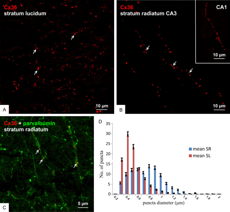
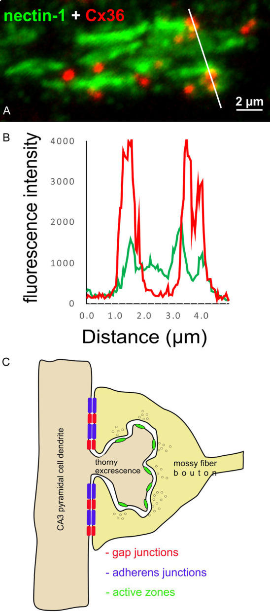
Background: Granule cells in the hippocampus project axons to hippocampal CA3 pyramidal cells where they form large mossy fiber terminals. We have reported that these terminals contain the gap junction protein connexin36 (Cx36) specifically in the stratum lucidum of rat ventral hippocampus, thus creating morphologically mixed synapses that have the potential for dual chemical/electrical transmission.

Methodology: Here, we used various approaches to characterize molecular and electrophysiological relationships between the Cx36-containing gap junctions at mossy fiber terminals and their postsynaptic elements and to examine molecular relationships at mixed synapses in the brainstem.

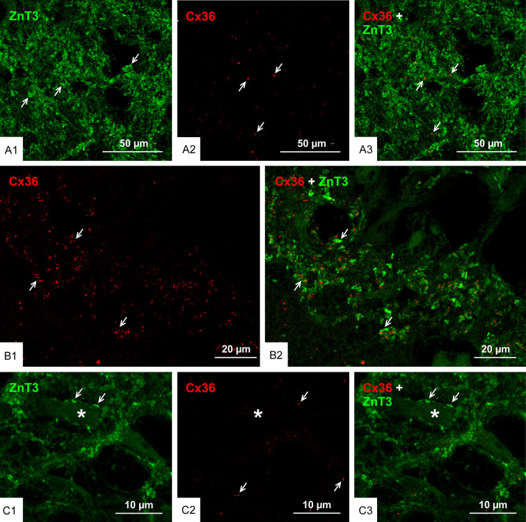
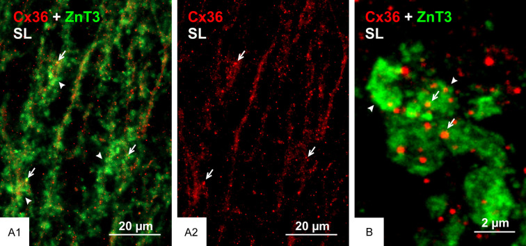
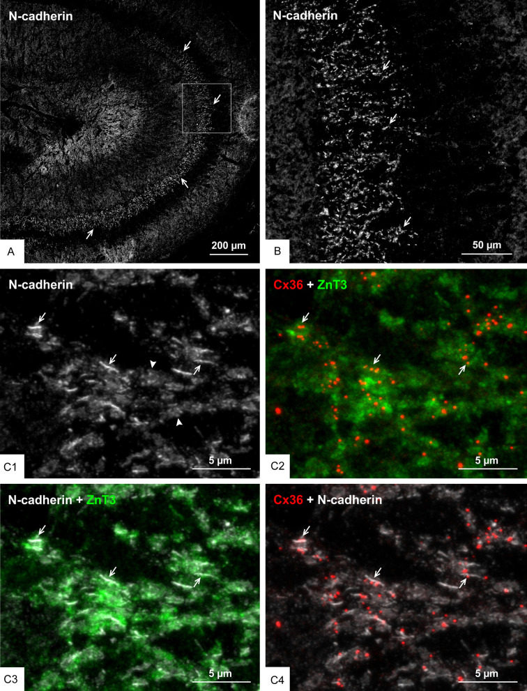
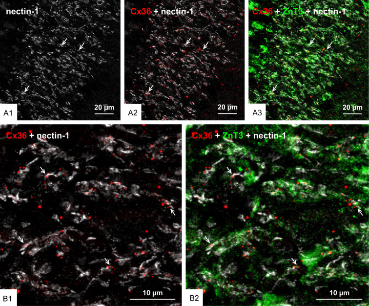
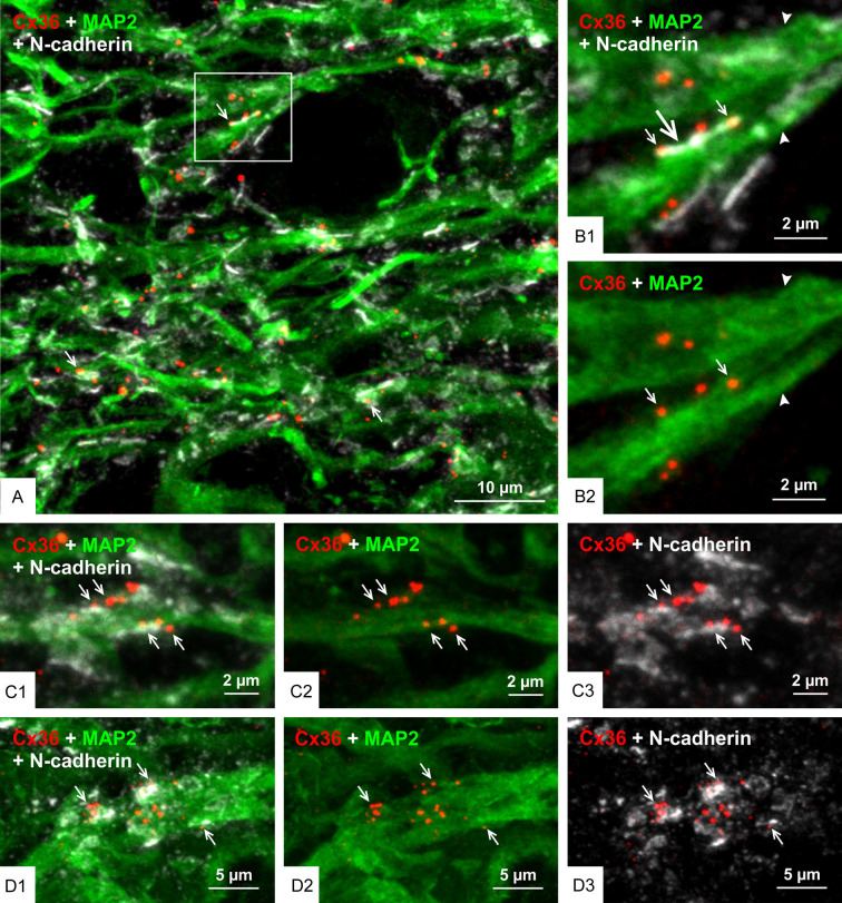
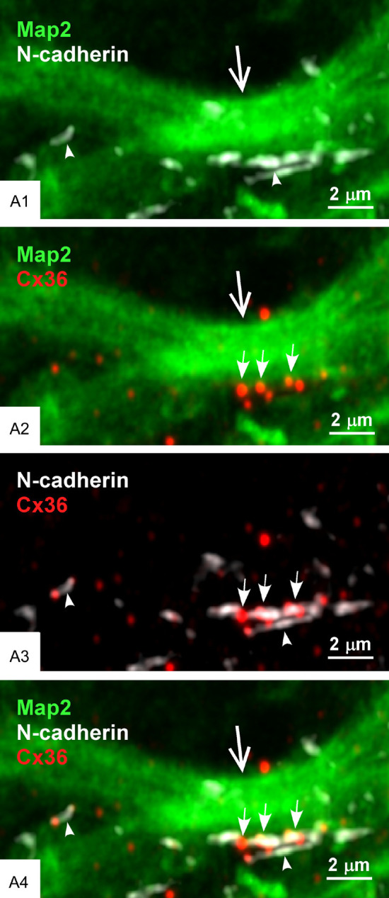
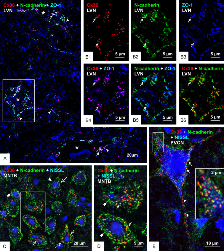
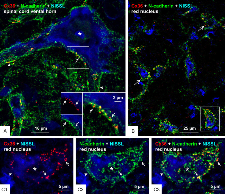
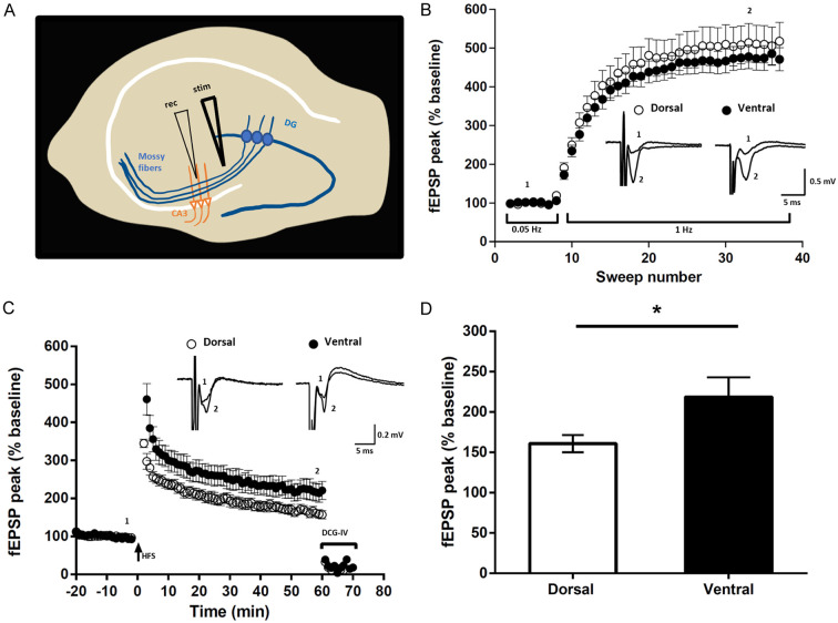
Results: In rat and human ventral hippocampus, many of these terminals, identified by their selective expression of vesicular zinc transporter-3 (ZnT3), displayed multiple, immunofluorescent Cx36-puncta representing gap junctions, which were absent at mossy fiber terminals in the dorsal hippocampus. In rat, these were found in close proximity to the protein constituents of adherens junctions (i.e., N-cadherin and nectin-1) that are structural hallmarks of mossy fiber terminals, linking these terminals to the dendritic shafts of CA3 pyramidal cells, thus indicating the loci of gap junctions at these contacts. Cx36-puncta were also associated with adherens junctions at mixed synapses in the brainstem, supporting emerging views of the structural organization of the adherens junction-neuronal gap junction complex. Electrophysiologically induced long-term potentiation (LTP) of field responses evoked by mossy fiber stimulation was greater in the ventral than dorsal hippocampus.

Conclusions: The electrical component of transmission at mossy fiber terminals may contribute to enhanced LTP responses in the ventral hippocampus.

Keywords: Gap junctions; brainstem; electrical coupling; hippocampus; immunofluorescence; long-term potentiation; mixed chemical/electrical synapses; neurotransmission.


Conflict of interest statement

None.

Figures

Figure 1
Figure 1
Association of Cx36 with ZnT3+ mossy fiber terminals in adult rat ventral hippocampus. (A) The ventral hippocampus sectioned transversely along its longitudinal axis showing low magnification overview of immunofluorescence labelling for ZnT3 in the hippocampal stratum lucidum (arrows) and hilus (arrowheads). (B) Higher magnification of the boxed area in (A), showing a high density of ZnT3+ mossy fiber terminals (arrows) in the stratum lucidum. (C) Double immunofluorescence labelling showing a moderate density of Cx36-puncta in the stratum lucidum of the ventral hippocampus (C1) and their association with mossy fiber terminals in this region (C2, arrows). (D) Confocal magnification of double labelling in the stratum lucidum showing clusters of Cx36-puncta (D1, arrows) localized to ZnT3+ mossy fiber terminals (D2, arrows).
Figure 2
Figure 2
Distribution of Cx36-puncta in regions of adult rat ventral hippocampus. (A-C) Comparison of the appearance of immunofluorescent Cx36-puncta in the ventral stratum lucidum showing their moderate density and occurrence often in clusters (A, arrows) vs. those in the ventral stratum radiatum of the CA3 region (B) and CA1 region (B, inset) showing Cx36-puncta of generally large size and organized in linear arrays (B, arrows) along parvalbumin+ dendrites (C, arrows). (D) Distribution of Cx36-puncta size in the stratum lucidum (SL) vs. stratum radiatum (SR), showing a shift to a larger diameter of those in the stratum radiatum.
Figure 3
Figure 3
Paucity of Cx36-puncta among axon collaterals of mossy fibers in the ventral hippocampal hilus. (A) Double immunofluorescence labelling in a central region of the hilus showing a high concentration of ZnT3+ terminals formed by axon collaterals of mossy fibers (A1, arrows) and the same field showing sparse Cx36-puncta (A2, arrows), with a lack of Cx36-puncta association with ZnT3+ terminals (A3, arrows). (B) A field in the same section as imaged in (A) and serving as positive control for labelling of Cx36 with the typically moderate density of Cx36-puncta (B1, arrows) in the CA3c region of the stratum lucidum and the localization of these puncta to ZnT3+ mossy fiber terminals (B2, arrows). (C) Image of a presumptive mossy cell (C1, asterisk) lying at the border between the ventral blade of the granule cell layer and the hilus, showing ZnT3+ terminals on the mossy cell somata (C1, arrows) and very sparse labelling for Cx36 in the same field (C2, arrows), where ZnT3+ terminals on the mossy cell lack association with Cx36-puncta (C3, arrows).
Figure 4
Figure 4
Immunofluorescence localization of Cx36 in the stratum lucidum of human hippocampus. (A) Overlay image showing a high density of labelling for ZnT3 at mossy fiber terminals in the stratum lucidum of ventral hippocampus and co-distribution of labelling for Cx36 (A1), which appears as Cx36-puncta of moderate density (A2). (B) Higher magnification of ZnT3+ mossy fiber terminals (arrowheads) with their overlapping or closely associated Cx36-puncta (arrows).
Figure 5
Figure 5
Association of Cx36 with N-cadherin+ adherens junctions at mossy fiber terminals in adult rat ventral hippocampus. (A, B) Low magnification overview of the distribution of N-cadherin+ adherens junctions in regions of the ventral hippocampus including the stratum lucidum (A, arrows), and magnification of the boxed area in (A) showing a high density of N-cadherin+ adherens junctions in the stratum lucidum (B, arrows). (C) Confocal magnification of the stratum lucidum showing N-cadherin+ adherens junctions seen as linear strands in on edge views (C1, arrows) and as discs in en face views (C1, arrowheads). Cx36-puncta in the same field is localized to ZnT3+ mossy fiber terminals as seen in red/green overlay (C2, arrows), and N-cadherin+ adherens junctions are also localized to those terminals as shown in white/green overlay (C3, arrows), with Cx36-puncta often localized at or adjacent to those junctions as shown in white/red overlay (C4, arrows).
Figure 6
Figure 6
Association of Cx36 with nectin-1+ adherens junctions at mossy fiber terminals in adult rat ventral hippocampus. (A) Low magnification overview of the distribution of nectin-1+ adherens junctions in the ventral hippocampus, showing immunofluorescence labelling of nectin-1 highly concentrated in the stratum lucidum (A1, arrows), where it is co-distributed with Cx36-puncta (A2, arrows) and ZnT3+ mossy fiber terminals in this region (A3, arrows). (B) Confocal magnification of a region in stratum lucidum similar to that in (A), showing the vast majority of Cx36-puncta overlapping with or immediately adjacent to nectin-1+ adherens junctions (B1, arrows) localized to ZnT3+ mossy fiber terminals (B2, arrows).
Figure 7
Figure 7
Configuration of Cx36-puncta and adherens junction in relation to CA3 pyramidal cell dendrites in the stratum lucidum of rat ventral hippocampus. (A) Triple immunofluorescence labelling in the CA3b region of the stratum lucidum showing numerous Cx36-puncta associated with N-cadherin+ adherens junctions localized to CA3 pyramidal cell dendrites labelled for the microtubule marker MAP2 (arrows). (B) Higher magnification of the boxed area in (A), showing large MAP2+ pyramidal cell dendrites (arrowheads) with a N-cadherin+ adherens junction viewed on-edge along a dendrite (B1, large arrows) and several Cx36-puncta localized to the junction (B1, small arrows), where the Cx36-puncta are shown in red/green channel overlay (B2, arrows). (C) Triple labelling in the CA3c region of the stratum lucidum showing multiple Cx36-puncta co-distributed with N-cadherin+ adherens junctions localized to both the upper and lower edges of a large MAP2+ pyramidal cell dendrite (C1, arrows). The same image is shown with overlay of only the red/green channels and overlay of only the red/white channels to visualize Cx36-puncta association with the dendrite (C2, arrows) and with N-cadherin (C3, arrows). (D) Triple labelling in the CA3c stratum lucidum showing N-cadherin+ adherens junctions viewed en-face and overlaying a MAP2+ dendrite (D1, arrows), with clusters of Cx36-puncta (D2, arrows) closely associated with the adherens junctions that in en-face view appear to consist of multiple N-cadherin+ puncta (D3, arrows).
Figure 8
Figure 8
Higher resolution confocal airyscan imaging of the AJ-nGJ complex at CA3 pyramidal cell dendrites in stratum lucidum of rat ventral hippocampus. (A1-A4) Adherens junctions along MAP2+ CA3 pyramidal cells dendrite (A1, large arrow) are shown to consist of multiple intermittent small junctions (A1, arrowheads), with the same region displaying a cluster of Cx36-puncta (A2, small arrows). Overlay of N-cadherin/Cx36 labelling shows Cx36-puncta abutted to the ends of individual junctional components of the N-cadherin+ adherens junctions (A3, arrows), all of which is closely adjacent to the MAP2+ CA3 pyramidal cell dendrite (A4).
Figure 9
Figure 9
Immunofluorescence intensity profiling and loci of the AJ-nGJ complex at mossy fiber terminals on CA3 pyramidal cell dendrites. (A) Line profile of immunofluorescence labelling of Cx36 and nectin-1 at mossy fiber terminals in the ventral hippocampus of adult rat, with line tracking through several Cx36-puncta. (B) Histogram of immunofluorescence intensity along the white line in (A), showing overlap or close spatial proximity of peak labelling intensity for Cx36-puncta and nectin-1. (C) Schematic diagram illustrating locations of the components for mixed synaptic neurotransmission from mossy fiber terminals to CA3 pyramidal cell dendrites. The chemical synapse component is localized on the spine head at active zones and the electrical component consisting of Cx36-containing gap junctions closely associated with adherens junctions at the AJ-nGJ complex is localized at contacts between the mossy fiber terminal and dendritic shaft of the CA3 pyramidal cell.
Figure 10
Figure 10
Relationships of Cx36, N-cadherin and ZO-1 at morphologically mixed synapses in the LVN, MNTB and PVCN of adult mouse. (A) Low magnification of the LVN, showing immunolabelling associated with three neuronal somata (not counterstained, but marked by asterisks) and their initial dendrites (arrowheads) decorated with Cx36-puncta that are co-localized with N-cadherin and ZO-1, seen as white in overlay of triple labelling (arrows). (B) Higher magnification of the boxed area at a neuronal somata in (A), showing individual labelling of Cx36-puncta (B1, arrows), N-cadherin (B2, arrows) and ZO-1 (B3, arrows), and with overlays showing near total co-localization of Cx36-puncta with ZO-1 (B4, arrows), substantial but not total co-localization of N-cadherin with ZO-1 (B5, arrows) and Cx36-puncta partially overlapping with or adjacent to labelling of N-cadherin (B6, arrows). (C, D) Overlay image of double labelling for Cx36 and N-cadherin with blue fluorescence counterstain in the MNTB, showing neuronal somata (C, large arrows; boxed area in C magnified in D) displaying Cx36-puncta associated with N-cadherin (small arrows) and patches of labelling for N-cadherin lacking association with Cx36-puncta (double arrowheads). (E) Overlay image showing Cx36-puncta association with N-cadherin on the somata (arrows) and initial dendrites (arrowheads) of a blue fluorescence counterstained octopus neuron in the PVCN, with the boxed area shown at higher magnification in the inset.
Figure 11
Figure 11
Relationships of Cx36 and N-cadherin at morphologically mixed synapses on motoneurons in the spinal cord and on large neurons in the red nucleus. (A) Double immunofluorescence labelling of Cx36 and N-cadherin with blue fluorescence Nissl counterstain in the lumbar spinal cord ventral horn of adult rat, showing overlay image of Cx36-puncta associated with labelling of N-cadherin on the somata (arrow) and initial dendrites (arrowheads) of a large motoneuron (asterisk), with inset of boxed area showing labelling of Cx36 (red) and N-cadherin (green) (arrows) shown magnified in overlay (arrow). (B) Double immunofluorescence labelling of Cx36 and N-cadherin with blue fluorescence Nissl counterstain in the red nucleus of adult mouse, with overlay image showing distribution of Cx36-puncta with N-cadherin on the surface of large neuronal somata (large arrows). (C) Magnification of the single neuronal somata (asterisk) in the boxed area of (B) (rotated clockwise by 90 degrees), showing nearly all somal Cx36-puncta (C1, arrows) associated with labelling of N-cadherin (C2, arrows), as seen in overlay (C3, arrows), and separate labelling of N-cadherin devoid of association with Cx36 (arrowhead).
Figure 12
Figure 12
Properties of mossy fiber field EPSPs examined in transverse slices of rat dorsal vs. ventral hippocampus. A. Schematic of a hippocampal slice showing the positioning of stimulating (stim) and recording (rec) electrodes. B. Plot showing FF at mossy fiber synapses, where increased stimulation frequency from low (0.05 Hz) to moderate (1 Hz) stimulation produced a pronounced increase in fEPSP amplitude. Inset traces represent averaged response when stimulated at 0.05 Hz (1) and at 1 Hz (2) as indicated. C. Induction of LTP at mossy fiber synapses is independent of NMDAR activation, as shown by the persistence of mossy fiber LTP recorded in the presence of the NMDAR antagonist APV. Inset traces represent averaged response recorded before (1) and 60 min after high-frequency stimulation (HFS) (2) as indicated. At the end of the LTP induction protocol, the large degree of inhibition (75-80%) of fEPSPs by the mGluR2 agonist DCG-IV verified that stimulation evoked responses originated from activation of mossy fiber synapses. D. Averaged fEPSP amplitude measured from the last 5 min of LTP in dorsal and ventral hippocampal slices, showing significantly greater potentiation in the ventral slices. *P < 0.05, unpaired Student’s t test with Welch’s correction and error bars representing s.e.m.

Similar articles

References

    1. Connors BW, Long MA. Electrical synapses in the mammalian brain. Annu Rev Neurosci. 2004;27:393–418. - PubMed
    1. Connors BW. Electrical signaling with neuronal gap junctions. In: Harris A, Locke D, editors. Connexins: A Guide. Springer: Humana Press; 2009. pp. 143–164.
    1. Pereda AE, Curti S, Hoge G, Cachope R, Flores CE, Rash JE. Gap junction-mediated electrical transmission: regulatory mechanisms and plasticity. Biochim Biophys Acta. 2013;1828:134–146. - PMC - PubMed
    1. Pereda AE. Electrical synapses and their functional interactions with chemical synapses. Nat Rev Neurosci. 2014;15:250–263. - PMC - PubMed
    1. Nagy JI, Pereda AE, Rash JE. Electrical synapses in mammalian CNS: past eras, present focus and future directions. Biochim Biophys Acta Biomembr. 2018;1860:102–123. - PMC - PubMed

LinkOut - more resources