. 2024 Jul 3:11:1356986.
doi: 10.3389/fnut.2024.1356986. eCollection 2024.

Effect of FADS1 SNPs rs174546, rs174547 and rs174550 on blood fatty acid profiles and plasma free oxylipins

Affiliations

Effect of FADS1 SNPs rs174546, rs174547 and rs174550 on blood fatty acid profiles and plasma free oxylipins

Miriam Rabehl et al. Front Nutr. .

Abstract

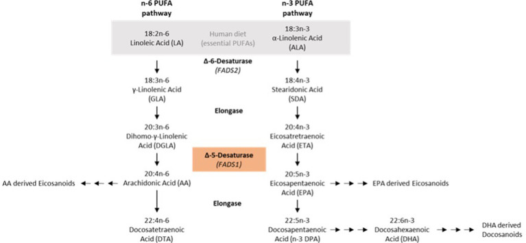
Introduction: Previous studies have indicated that activity of fatty acid desaturase 1 (FADS1), is involved in cardiometabolic risk. Recent experimental data have shown that FADS1 knockdown can promote lipid accumulation and lipid droplet formation in liver cells. In this study, we aimed to characterize whether different FADS1 genotypes affect liver fat content, essential fatty acid content and free oxylipin mediators in the blood.

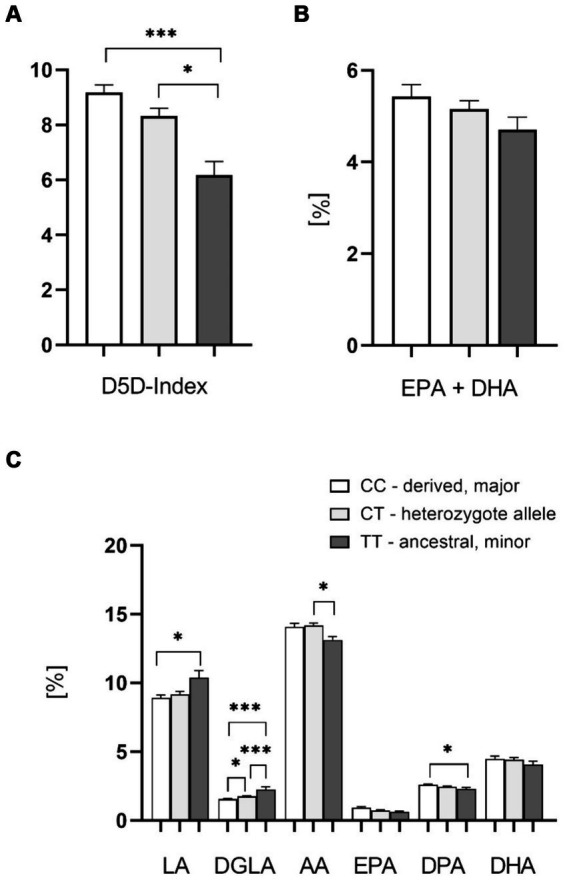
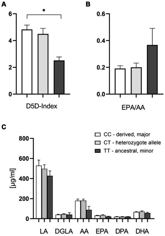
Methods: We analyzed the impact of FADS1 single-nucleotide polymorphisms (SNPs) rs174546, rs174547, and rs174550 on blood fatty acids and free oxylipins in a cohort of 85 patients from an academic metabolic medicine outpatient center. Patients were grouped based on their genotype into the homozygous major (derived) allele group, the heterozygous allele group, and the homozygous minor (ancestral) allele group. Omega-3 polyunsaturated fatty acids (n-3 PUFA) and omega-6 polyunsaturated fatty acids (n-6 PUFA) in the blood cell and plasma samples were analyzed by gas chromatography. Free Oxylipins in plasma samples were analyzed using HPLC-MS/MS. Liver fat content and fibrosis were evaluated using Fibroscan technology.

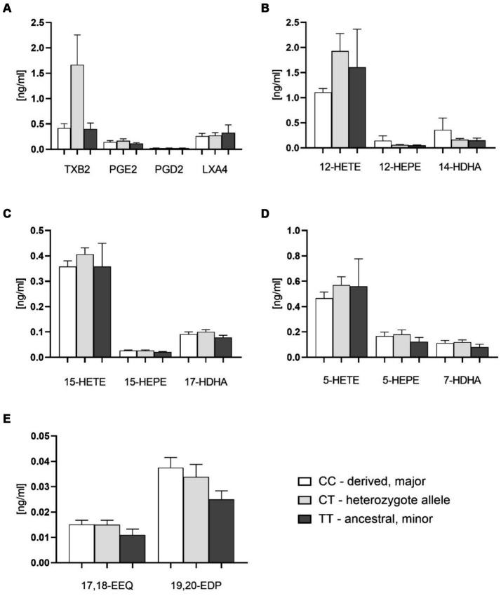
Results: Patients with the homozygous ancestral (minor) FADS1 genotype exhibited significantly lower blood levels of the n-6 PUFA arachidonic acid (AA), but no significant differences in the n-3 PUFAs eicosapentaenoic acid (EPA) and docosahexaenoic acid (DHA). There were no significant differences in liver fat content or arachidonic acid-derived lipid mediators, such as thromboxane B2 (TXB2), although there was a trend toward lower levels in the homozygous ancestral genotype group.

Discussion: Our findings suggest that FADS1 genotypes influence the blood levels of n-6 PUFAs, while not significantly affecting the n-3 PUFAs EPA and DHA. The lack of significant differences in liver fat content and arachidonic acid-derived lipid mediators suggests that the genotype-related variations in fatty acid levels may not directly translate to differences in liver fat or inflammatory lipid mediators in this cohort. However, the trend towards lower levels of certain lipid mediators in the homozygous ancestral genotype group warrants further investigation to elucidate the underlying mechanisms of different FADS1 genotypes and potential implications for cardiometabolic risk.

Keywords: FADS1; MAFLD; Oxylipins; PUFA; steatosis hepatis.


Conflict of interest statement

MRo is owner of Lipidomix GmbH. The remaining authors declare that the research was conducted in the absence of any commercial or financial relationships that could be construed as a potential conflict of interest.

Figures

Figure 1
Figure 1
N-6/n-3 PUFA synthesis pathways.
Figure 2
Figure 2
Effect of the FADS1 rs174546 genotype on PUFAs in the blood cell fraction. (A) D5D index is significantly higher with the C allele carriers (CC > CT > TT). (B) Slightly lower EPA + DHA content (determined in analogy to the Omega-3-Index) in the blood cell fraction from patients with the homozygous ancestral FADS1 genotype (TT). (C) Significantly higher levels of n-6 PUFAs linoleic acid (LA) and dihomo-gamma-linolenic acid (DGLA), and lower levels of arachidonic acid (AA) and n-3 PUFA docosapentaenoic acid (DPA) with the minor TT genotype. n = 37 for the homozygous derived genotype CC, n = 37 for the heterozygous genotype CT, n = 11 for the homozygous ancestral genotype TT, *p < 0.05, **p < 0.01, ***p < 0.001, one-way ANOVA with subsequent Tukey’s HSD testing.
Figure 3
Figure 3
Effect of the FADS1 rs174546 genotype on plasma PUFAs content. (A) The D5D index is significantly higher with the CC alleles in comparison to TT alleles. (B) Slightly higher EPA/AA plasma ratio with the homozygous ancestral FADS1 genotype (ns, p = 0.067). (C) Slightly lower AA (20:4 n-6) levels with the homozygous ancestral FADS1 genotype. n = 19 for the homozygous derived genotype, n = 19 for the heterozygous genotype, n = 4 for the homozygous ancestral genotype, *p < 0.05, Kruskal-Wallis and subsequent Dunn’s testing.
Figure 4
Figure 4
Plasma free oxylipins in groups with FADS1 rs174546 genotypes. (A) Thromboxane B2 (TXB2), prostaglandin D2 (PGD2), prostaglandin E2 (PGE2) and lipoxin A4 (LXA4) contents in plasma of different FADS1 genotypes. (B) 12-Lipoxygenase-derived oxylipin contents from AA (12-hydroxyeicosatetraenoic acid, 12-HETE), EPA (12-hydroxyeicosapentaenoic acid, 12-HEPE) and DHA (14-hydroxydocosahexaenoic acid, 14-HDHA) in plasma of different FADS1 genotypes. (C) 15-Lipoxygenase-derived oxylipin contents from AA (15-HETE), EPA (15-HEPE) and DHA (17-HDHA). (D) 5-Lipoxygenase-derived oxylipins from AA (5-HETE), EPA (5-HEPE) and DHA (7-HDHA) in plasma of different FADS1 genotypes. (E) Cytochrome P450-derived epoxy metabolites from EPA (17,18-epoxyeicosatetraenoic acid 17,18-EEQ) and DHA (19,20-epoxydocosapentaenoic acid, 19,20-EDP) in plasma of different FADS1 genotypes. n = 36 for the homozygous derived genotype, n = 36 for the heteozygous genotype, n = 10 for the homozygous ancestral genotype. There were no significant differences between groups from Kruskal-Wallis testing.

Similar articles

Cited by

References

    1. Sangro P, de la Torre Alaez M, Sangro B, D'Avola D. Metabolic dysfunction-associated fatty liver disease (MAFLD): an update of the recent advances in pharmacological treatment. J Physiol Biochem. (2023) 79:869–79. doi: 10.1007/s13105-023-00954-4, PMID: - DOI - PMC - PubMed
    1. Younossi ZM, Koenig AB, Abdelatif D, Fazel Y, Henry L, Wymer M. Global epidemiology of nonalcoholic fatty liver disease-Meta-analytic assessment of prevalence, incidence, and outcomes. Hepatology. (2016) 64:73–84. doi: 10.1002/hep.28431, PMID: - DOI - PubMed
    1. Cornier MA, Dabelea D, Hernandez TL, Lindstrom RC, Steig AJ, Stob NR, et al. The metabolic syndrome. Endocr Rev. (2008) 29:777–822. doi: 10.1210/er.2008-0024, PMID: - DOI - PMC - PubMed
    1. Godoy-Matos AF, Silva Junior WS, Valerio CM. NAFLD as a continuum: from obesity to metabolic syndrome and diabetes. Diabetol Metab Syndr. (2020) 12:60. doi: 10.1186/s13098-020-00570-y, PMID: - DOI - PMC - PubMed
    1. Gu W, Hortlik H, Erasmus HP, Schaaf L, Zeleke Y, Uschner FE, et al. Trends and the course of liver cirrhosis and its complications in Germany: Nationwide population-based study (2005 to 2018). Lancet Reg Health Eur. (2022) 12:100240. doi: 10.1016/j.lanepe.2021.100240, PMID: - DOI - PMC - PubMed

LinkOut - more resources