

A case of lipoblastoma in a pediatric patient
- PMID: 39021668
- PMCID: PMC11253145
- DOI: 10.1016/j.radcr.2024.05.054
A case of lipoblastoma in a pediatric patient
Abstract
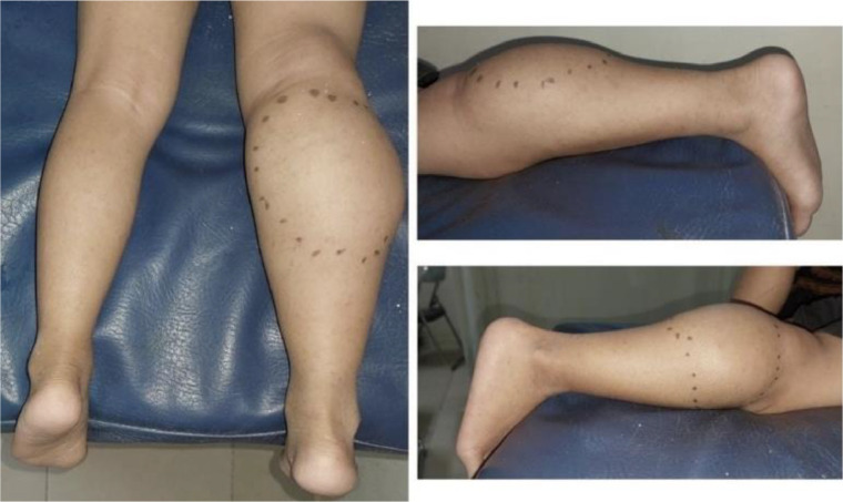
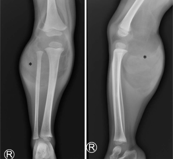
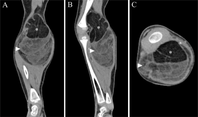
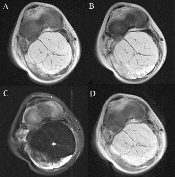
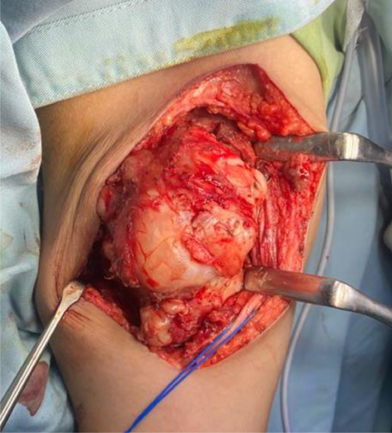
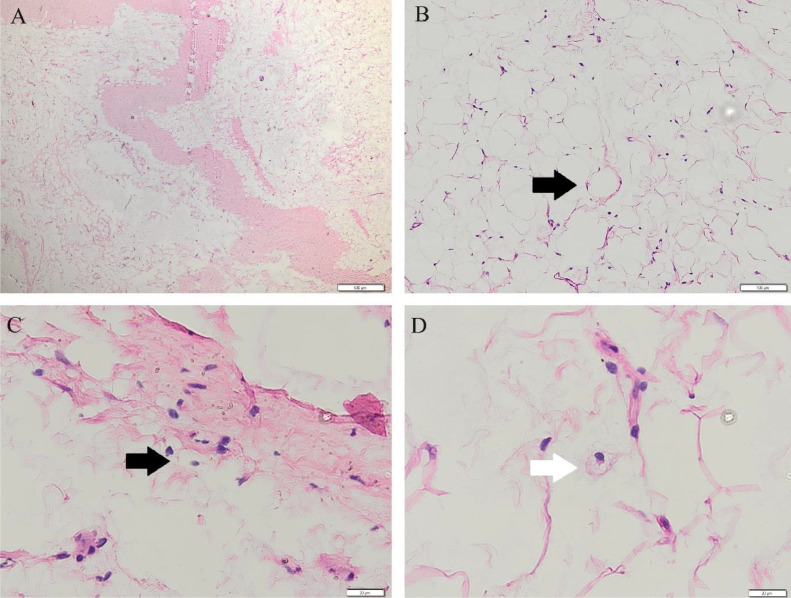
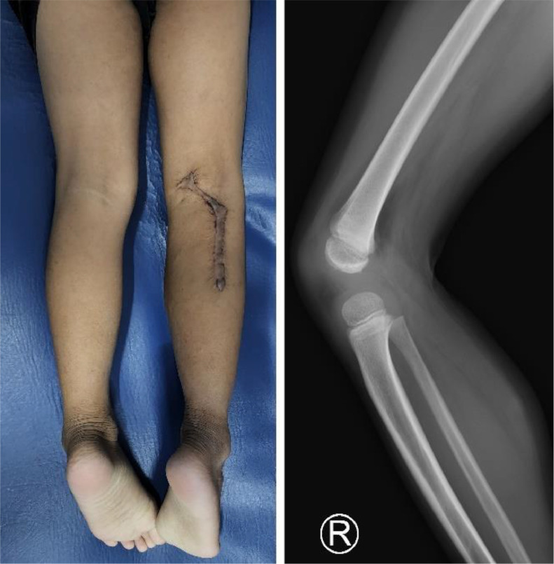
Lipoblastoma is a benign soft tissue tumor that originates from embryonic white fat. Lipoblastoma presents as a slow-growing mass that commonly occurs in the extremities of young children. Histological examination remains the gold standard in confirming lipoblastoma; however, radiology examination can help identify and evaluate the extent and characterization of the mass prior to the excision. Here, we report a 7-year-old male patient who presented with a painless mass in the right popliteal extending to the proximal cruris areas, and the imaging modalities suggested the presence of fat within the mass. The patient then underwent complete excision, and histopathology examination revealed lipoblastoma. This study highlights the possibility of lipoblastoma in older children and the role of imaging examinations in the diagnosis.
Keywords: Lipoblastoma; Lipoblastomatosis; MRI; Pediatric.
© 2024 The Authors. Published by Elsevier Inc. on behalf of University of Washington.
Figures
References
-
- Shek K.W., Cheng S.S., Tse K.S., Lai K.C., Chan M.K. Lipoblastoma: different features on magnetic resonance imaging. Hong Kong J Radiol. 2015;18:302–306.
-
- Hicks J, Dilley A, Patel D, Barrish J, Zhu SH, Brandt M. Lipoblastoma and lipoblastomatosis in infancy and childhood: histopathologic, ultrastructural, and cytogenetic features. Ultrastruct Pathol. 2001;25(4):321–333. - PubMed
-
- Speer AL, Schofield DE, Wang KS, Shin CE, Stein JE, Shaul DB, et al. Contemporary management of lipoblastoma. J Pediatr Surg. 2008;43(7):1295–1300. - PubMed
-
- Abraham-Mendoza S, Padilla-Guevara R, Cortés-García C, Morales-Piñón E, García-Salazar J, Téllez-Bernal E. Lipoblastoma: case report and literature review. Gaceta Mexicana de Oncología. 2018;16:348–351.
Publication types
LinkOut - more resources
Full Text Sources
