

Case Reports
doi: 10.14744/SEMB.2023.45578.
eCollection 2024.
Biliary Cystadenoma with High Dysplasia Detected Incidentally in a Young Patient Admitted for Percutaneous Abscess Drainage
Affiliations
- PMID: 39021698
- PMCID: PMC11249983
- DOI: 10.14744/SEMB.2023.45578
Item in Clipboard
Case Reports
Biliary Cystadenoma with High Dysplasia Detected Incidentally in a Young Patient Admitted for Percutaneous Abscess Drainage
Sisli Etfal Hastan Tip Bul.
.
Abstract
Biliary cystadenomas are uncommon lesions with clinical and radiological characteristics that overlap with other cystic liver lesions. Here, we intended to discuss a biliary cystadenoma found in a 37-year-old female patient who had been treated for a liver abscess and had been sent to our clinic with a long-term hydatid cyst diagnosis.
Keywords: Abscess; biliary cystadenocarcinoma; biliary cystadenoma; computed tomography; excision; ultrasonography.
© Copyright 2024 by The Medical Bulletin of Sisli Etfal Hospital.
Conflict of interest statement
The authors declare no conflict of interest.
Figures
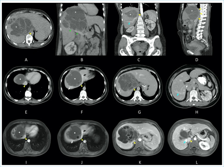
Imaging prior to the treatment. On axial CT (a-h) and MRI (i-l) images with intravenous contrast, the BCAC (asterisk), inferior vena cava (IVC; yellow arrowhead), left hepatic vein (black arrow), middle hepatic vein (white arrow), right hepatic vein (blue arrow) and portal vein (green arrowhead) are shown. Because of the mass effect, there is a deviation in the middle and left hepatic veins as well as IVC compression at the hepatic level (yellow arrowhead).

CT-guided drainage of the lesion with a catheter.

Post-treatment magnetic resonance imaging shows the multiseptal and multiloculated cystic lesion. (a) axial T1 weighted imaging (WI), (b) T2-fat saturated image, (c) diffusion WI, (d) apparent diffusion coefficient, (e) contrast-enhanced axial plan, (f) coronal plan T2-WI.

Intraoperative images show (a) Biliary cyst adenoma (asterisk) compressing the right and left lobes and the caudate lobe in the central sector, (b) the area of previously placed drainage catheter at the junction of segment 4-8 (black arrow), (c) Inferior vena cava (long arrow) with portal vein (arrows head) isolated in the central sector, (d) Inferior view of the vena cava (long arrow) after resection.
Similar articles
-
[Biliary cystic neoplasm: biliary cystadenoma and biliary cystadenocarcinoma].Korean J Gastroenterol. 2006 Jan;47(1):5-14. Korean J Gastroenterol. 2006. PMID: 16434863 Review. Korean.
-
Biliary cystadenoma and other complicated cystic lesions of the liver: diagnostic and therapeutic challenges.World J Surg. 2006 Aug;30(8):1560-6. doi: 10.1007/s00268-005-0461-7. World J Surg. 2006. PMID: 16865321
-
Biliary cystadenoma in an endemic zone of hydatid cyst: A rare surgical surprise.Ann Hepatobiliary Pancreat Surg. 2020 Feb;24(1):85-89. doi: 10.14701/ahbps.2020.24.1.85. Epub 2020 Feb 27. Ann Hepatobiliary Pancreat Surg. 2020. PMID: 32181435 Free PMC article.
-
Cystadenoma and cystadenocarcinoma of the liver: a single center experience.J Am Coll Surg. 2005 May;200(5):727-33. doi: 10.1016/j.jamcollsurg.2005.01.005. J Am Coll Surg. 2005. PMID: 15848365
-
Diagnostic and Therapeutic Challenges of Intrahepatic Biliary Cystadenoma and Cystadenocarcinoma: A Report of 10 Cases and Review of the Literature.Int Surg. 2015 Jul;100(7-8):1212-9. doi: 10.9738/INTSURG-D-15-00025.1. Int Surg. 2015. PMID: 26595495 Review.
References
-
- Brittingham C, Tuma F. StatPearls. Treasure Island (FL): StatPearls Publishing; 2023. Hepatic cystadenoma. - PubMed
Publication types
LinkOut - more resources
Full Text Sources