. 2024 Jun 15;10(12):e33159.
doi: 10.1016/j.heliyon.2024.e33159. eCollection 2024 Jun 30.

Neighbor effect on conformational spaces of alanine residue in azapeptides

Affiliations

Neighbor effect on conformational spaces of alanine residue in azapeptides

Ho-Jin Lee et al. Heliyon. .

Abstract

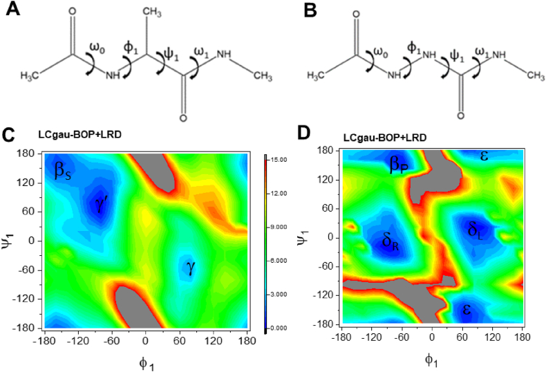
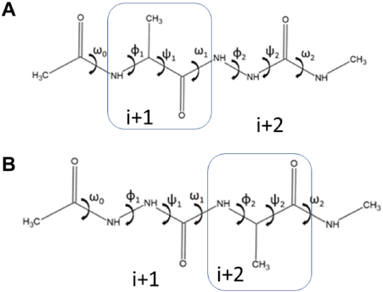
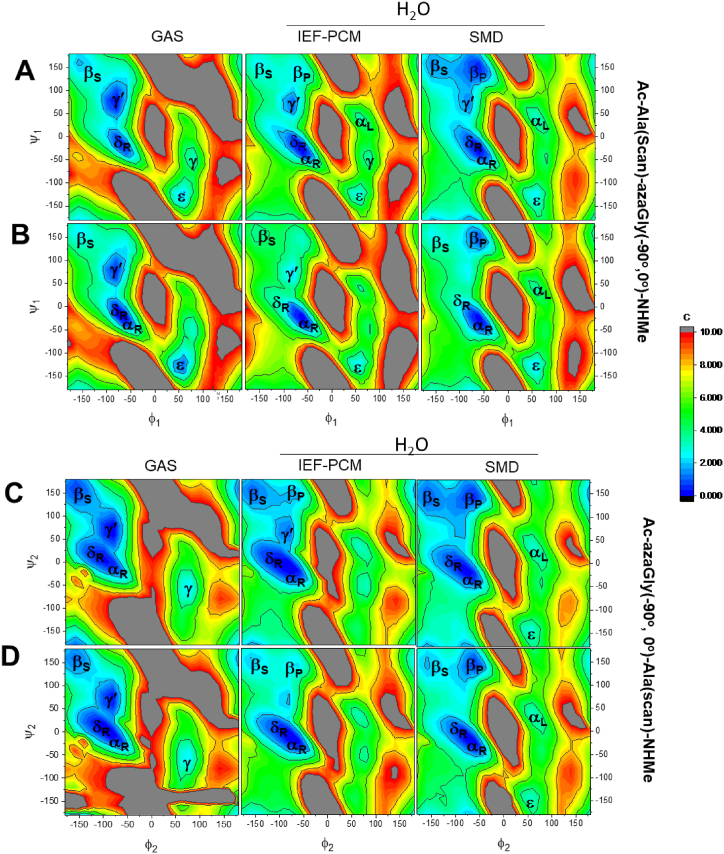
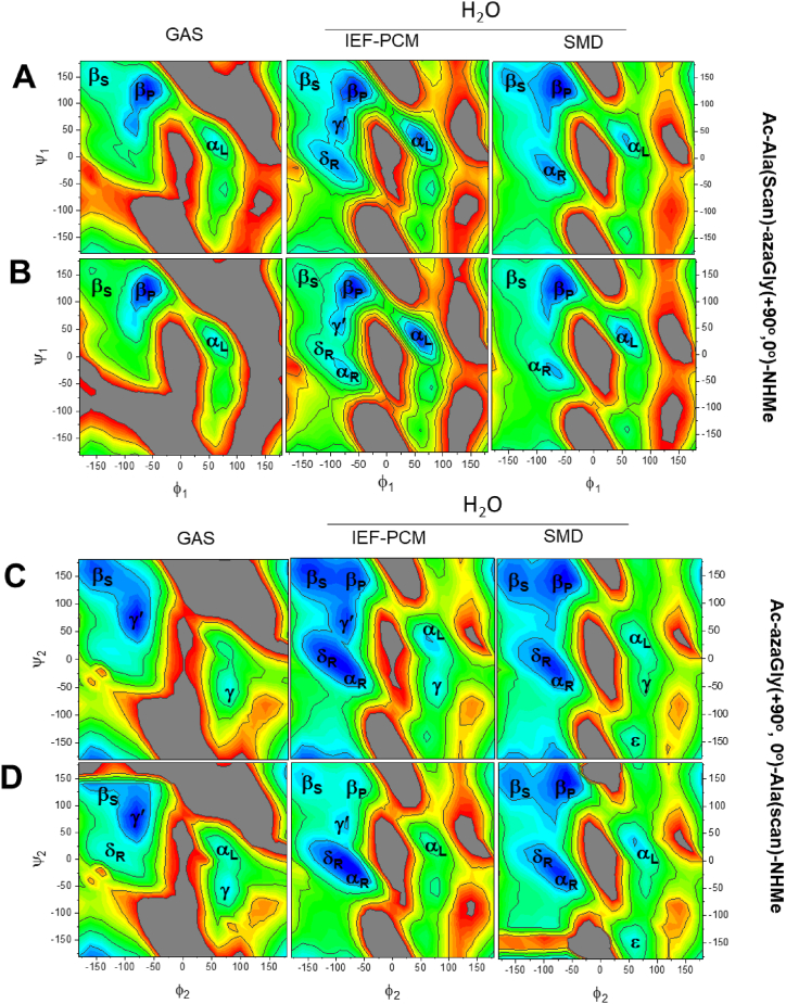
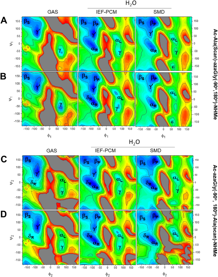
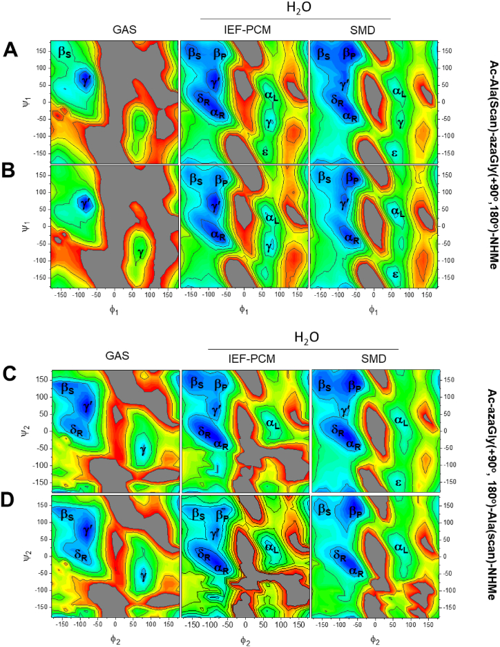
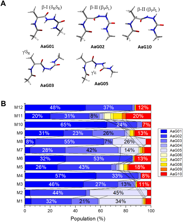
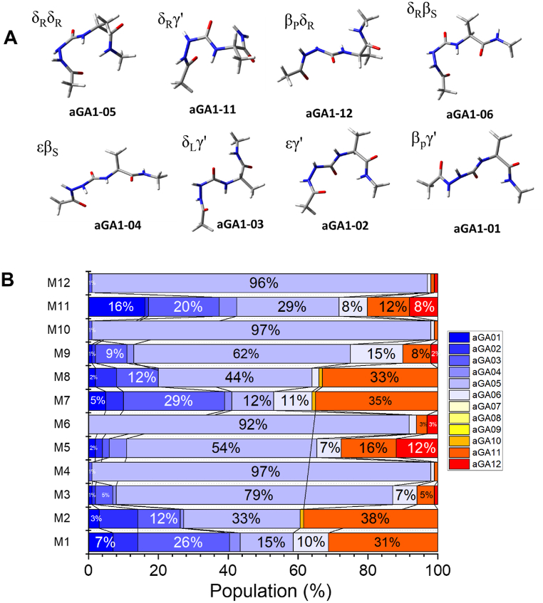
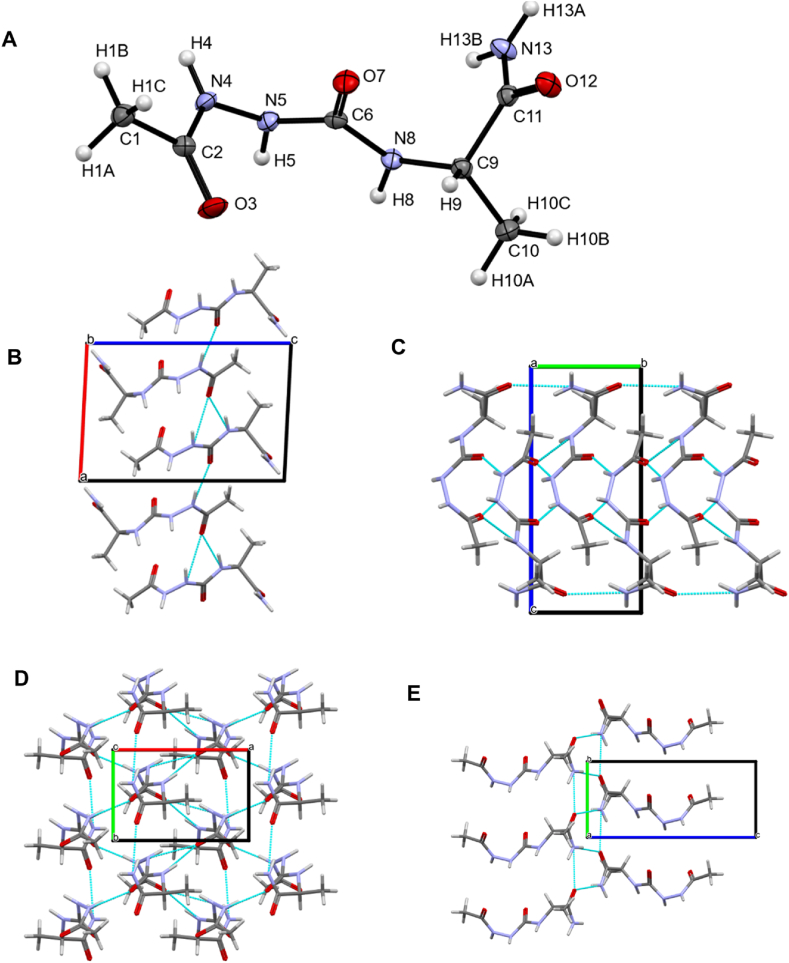
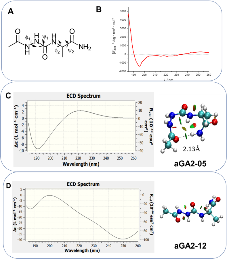
The conformational properties of Alanine (Ala) residue have been investigated to understand protein folding and develop force fields. In this work, we examined the neighbor effect on the conformational spaces of Ala residue using model azapeptides, Ac-Ala-azaGly-NHMe (3, AaG), and Ac-azaGly-Ala-NHMe (4, aGA1). Ramachandran energy maps were generated by scanning (φ, ψ) dihedral angles of the Ala residues in models with the fixed dihedral angles (φ = ±90°, ψ = ±0° or ±180°) of azaGly residue using LCgau-BOP and LCgau-BOP + LRD functionals in the gas and water phases. The integral-equation-formalism polarizable continuum model (IEF-PCM) and a solvation model density (SMD) were employed to mimic the solvation effect. The most favorable conformation of Ala residue in azapeptide models is found as the polyproline II (βP), inverse γ-turn (γ'), β-sheet (βS), right-handed helix (αR), or left-handed helix (αL) depending on the conformation of neighbor azaGly residue in isolated form. Solvation methods exhibit that the Ala residue favors the βP, δR, and αR conformations regardless of its position in azapeptides 3 and 4 in water. Azapeptide 5, Ac-azaGly-Ala-NH2 (aGA2), was synthesized to evaluate the theoretical results. The X-ray structure showed that azaGly residue adopts the polyproline II (βP) and Ala residue adopts the right-handed helical (αR) structure in aGA2. The conformational preferences of aGA2 and the dimer structure of aGA2 based on the X-ray structure were examined to assess the performance of DFT functionals. In addition, the local minima of azapeptide 6, Ac-Phe-azaGly-NH2 (FaG), were compared with the previous experimental results. SMD/LCgau-BOP + LRD methods agreed well with the reported experimental results. The results suggest the importance of weak dispersion interactions, neighbor effect, and solvent influence in the conformational preferences of Ala residue in model azapeptides.

Keywords: And Lcgau-BOP+LRD; Azapeptide; DFT functionals; Foldamer; LCgau-BOP; βI-turn; βII-turn.


Conflict of interest statement

The authors declare that they have no known competing financial interests or personal relationships that could have appeared to influence the work reported in this paper.

Figures

Fig. 1
Fig. 1
Chemical structure and dihedral angle nomenclature of Ac-Ala-NHMe (1) and Ac-azaGly-NHMe (2) peptide models, respectively (a, b). Potential energy maps of (c) Ac-Ala-NHMe (1) and (d) Ac-azaGly-NHMe (2), respectively, at the LCgau-BOP + LRD/6-31 + G(d) level in the gas phase. Geometry optimization of all variables except (φ1, ψ1) was performed on a grid with 15° spacing. The dihedral angle of ω0 and ω1 was set to ∼180°. The energy area from the lowest energy (blue) to 15 kcal/mol (red) is shown. The Ala residue favors βS(C5) and γ′ regions; the azaGly residue favors the δR and δL, βP, or ε regions. The nomenclature of conformers was adopted from Karplus et al. (For interpretation of the references to colour in this figure legend, the reader is referred to the Web version of this article.)
Fig. 2
Fig. 2
Chemical structures and dihedral angle nomenclature of (a) Ac-Ala-azaGly-NHMe (3, AaG) and (b) Ac-azaGly-Ala-NHMe (4, aGA1).
Fig. 3
Fig. 3
Ramachandran energy maps of Ac-Ala(φ11 = scan)-azaGly (φ2 = −90°, ψ2 = 0°)-NHMe (A and B) and Ac-azaGly(φ1 = −90°, ψ1 = 0°)-Ala(φ22 = scan)-NHMe (C, D) at the LCgau-BOP (A and C) and LCgau-BOP + LRD (B and D) with the 6-31 + G(d) basis set. Geometry optimization of all variables except (φ1, ψ1) for AaG or (φ2, ψ2) for aGA1 was performed on a grid with 15° spacing. The dihedral angle of ω0 and ω2 was ∼180°. The energy area from the lowest energy (blue) to 10 kcal/mol (red) is shown. IEF-PCM and SMD solvation methods were used to consider the water environment. (For interpretation of the references to colour in this figure legend, the reader is referred to the Web version of this article.)
Fig. 4
Fig. 4
Ramachandran energy maps of Ac-Ala(φ11 = scan)-azaGly(φ2 = +90°, ψ2 = 0°)-NHMe (A and B) and Ac-azaGly(φ1 = +90°, ψ1 = 0°)-Ala(φ,ψ = scan)-NHMe (C and D) at the LCgau-BOP (A and C) and LCgau-BOP + LRD (B and D) with the 6-31 + G(d) basis set. Geometry optimization of all variables except (φ1, ψ1) for AaG or (φ2, ψ2) for aGA was performed on a grid with 15° spacing. The dihedral angle of ω0 and ω2 was ∼180°. The energy area from the lowest energy (blue) to 10 kcal/mol (red) is shown. IEF-PCM and SMD methods were used to consider the water environment. (For interpretation of the references to colour in this figure legend, the reader is referred to the Web version of this article.)
Fig. 5
Fig. 5
Ramachandran energy maps of Ac-Ala(φ11 = scan)-azaGly(φ2 = −90°, ψ2 = 180°)-NHMe (A and B) and Ac-azaGly(φ1 = +90°, ψ1 = 180°)-Ala(φ22 = scan)-NHMe (C and D) at the LCgau-BOP (A and C) and LCgau-BOP + LRD (B and D) with the 6-31 + G(d) basis set. Geometry optimization of all variables except (φ1, ψ1 = scan) for AaG or (φ2, ψ2 = scan) for aGA1 was performed on a grid with 15° spacing. The dihedral angle of ω0 and ω2 was ∼180°. The energy area from the lowest energy (blue) to 10 kcal/mol (red) is shown. IEF-PCM and SMD solvation methods were used to consider the water effect. (For interpretation of the references to colour in this figure legend, the reader is referred to the Web version of this article.)
Fig. 6
Fig. 6
Ramachandran energy maps of Ac-Ala(φ11 = scan)-azaGly (φ2 = +90°, ψ2 = 180°)-NHMe (A and B) and Ac-azaGly(φ1 = +90°, ψ1 = 180°)-Ala(φ22 = scan)-NHMe (C, D) at the LCgau-BOP (A and C) and LCgau-BOP + LRD (B and D) with the 6-31 + G(d) basis set. Geometry optimization of all variables except (φ1, ψ1 = scan) for AaG or (φ2, ψ2 = scan) for aGA1 was performed on a grid with 15° spacing. The dihedral angle of ω0 and ω2 was set to ∼180°. The energy area from the lowest energy (blue) to 10 kcal/mol (red) is shown. IEF-PCM and SMD solvation methods were used to consider the water effect. (For interpretation of the references to colour in this figure legend, the reader is referred to the Web version of this article.)
Fig. 7
Fig. 7
The most favorable conformation of Ac-Ala-azaGly-NHMe (3, AaG) at the SMD/LCgau-BOP + LRD/6–311++G(2d,2p) (A) and population of conformers in the gas and water (B). For y-axis, M1:LCgau-BOP; M2:LCgau + LRD; M3:IEF-PCM/LCgau-BOP; M4:IEF-PCM/LCgau-BOP + LRD; M5: SMD/LCgau-BOP; M6: SMD/LCgau-BOP + LRD; M7: B3LYP; M8:B3LYP-D3; M9: IEF-PCM/B3LYP; M10: IEF-PCM/B3LYP-D3; M11: SMD/B3LYP; M12: SMD/B3LYP-D3 with the 6–311++G(2d,2p) basis set used.
Fig. 8
Fig. 8
The most favorable conformation of Ac-azaGly-Ala-NHMe (4, aGA1) at the SMD/LCgau-BOP + LRD/6–311++G(2d,2p) (A) and population of conformers for azapeptide 4 in the gas and water (B). For y-axis, M1:LCgau-BOP; M2:LCgau + LRD; M3:IEF-PCM/LCgau-BOP; M4:IEF-PCM/LCgau-BOP + LRD; M5: SMD/LCgau-BOP; M6: SMD/LCgau-BOP + LRD; M7: B3LYP; M8:B3LYP-D3; M9: IEF-PCM/B3LYP; M10: IEF-PCM/B3LYP-D3; M11: SMD/B3LYP; M12: SMD/B3LYP-D3 with the 6–311++G(2d,2p) basis set used.
Fig. 9
Fig. 9
(A) Crystal structure of Ac-azaGly-Ala-NH2 (azapeptide 5, aGA2). Atomic displacement ellipsoids are contoured at the 0.5 probability level. Crystal packing with H-bonds is shown as cyan dotted lines: (B) H-bonded chains of molecules along the diagonal of the ab plane of the unit cell (view along the b axis). (C and D) H-bonded 2-D network of molecules in the ab plane(view along unit cell axis a and c, respectively). (E) H-bonded ladder of molecules along unit cell axis b (view along axis a). (For interpretation of the references to colour in this figure legend, the reader is referred to the Web version of this article.)
Fig. 10
Fig. 10
CD spectrum of azapeptide 5. (A) Chemical structure of azapeptide 5 (B) Experimental CD spectrum of azapeptide 5 in water at 5 °C. (C and D) Calculated electronic circular dichroism (ECD) for aGA2-05 and aGA2-12 at the SMD/B3LYP-D3 functional with a 6–311++G(2d,2p) basis set in water. NCI analysis of aGA2-05 and aGA2-12. NCI plot shows the regions of attractive (blue) and repulsive (red) interactions, as well as regions of van der Waals interactions (green) for the B3LYP-D3 functional (Multiwfn and VMD software were used to generate the figures). (For interpretation of the references to colour in this figure legend, the reader is referred to the Web version of this article.)

Similar articles

References

    1. Perczel A., Angyan J.G., Kajtar M., Viviani W., Rivail J.L., Marcoccia J.F., Csizmadia I.G. Peptide models. 1. Topology of selected peptide conformational potential energy surfaces (glycine and alanine derivatives) J. Am. Chem. Soc. 1991;113(16):6256–6265.
    1. Head-Gordon T., Head-Gordon M., Frisch M.J., Brooks C.L., III Pople JA: theoretical study of blocked glycine and alanine peptide analogs. J. Am. Chem. Soc. 1991;113(16):5989–5997.
    1. Perczel A., Farkas O., Jákli I., Topol I.A., Csizmadia I.G. Peptide models XXXIII. Extrapolation of low-level Hartree-Fock data of peptide conformation to large basis set SCF, MP2, DFT, and CCSD(T) results. The Ramachandran surface of alanine dipeptide computed at various levels of theory. J. Comput. Chem. 2003;24(9):1026–1042. - PubMed
    1. Hudáky I., Hudáky P., Perczel A. Solvation model induced structural changes in peptides. A quantum chemical study on Ramachandran surfaces and conformers of alanine diamide using the polarizable continuum model. J. Comput. Chem. 2004;25(12):1522–1531. - PubMed
    1. Wang Z.X., Duan Y. Solvation effects on alanine dipeptide: a MP2/cc-pVTZ//MP2/6-31G** study of (Phi, Psi) energy maps and conformers in the gas phase, ether, and water. J. Comput. Chem. 2004;25(14):1699–1716. - PubMed

LinkOut - more resources