. 2024 Jul 1;14(7):5084-5098.
doi: 10.21037/qims-24-284. Epub 2024 Jun 27.

A nomogram for risk stratification of central cervical lymph node metastasis in patients with papillary thyroid carcinoma

Affiliations

A nomogram for risk stratification of central cervical lymph node metastasis in patients with papillary thyroid carcinoma

Ying Zou et al. Quant Imaging Med Surg. .

Abstract

Background: Whether to perform prophylactic central lymph node dissection for cN0 papillary thyroid carcinoma (PTC) patients is still controversial. This retrospective study aimed to develop and validate a nomogram based on ultrasound and dual-energy computed tomography (DECT) for the risk stratification of central lymph node metastasis (CLNM) in patients with PTC.

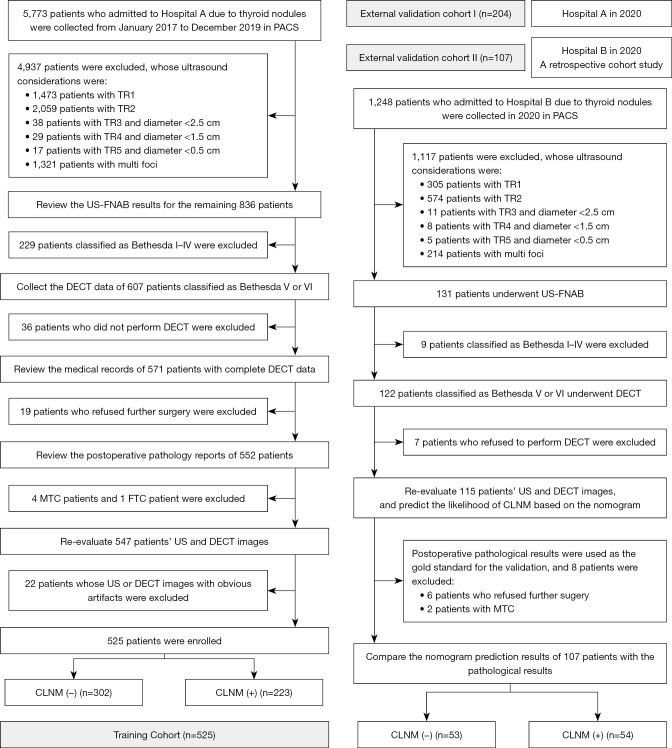
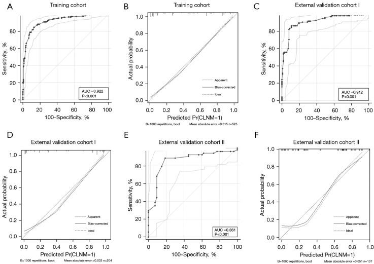
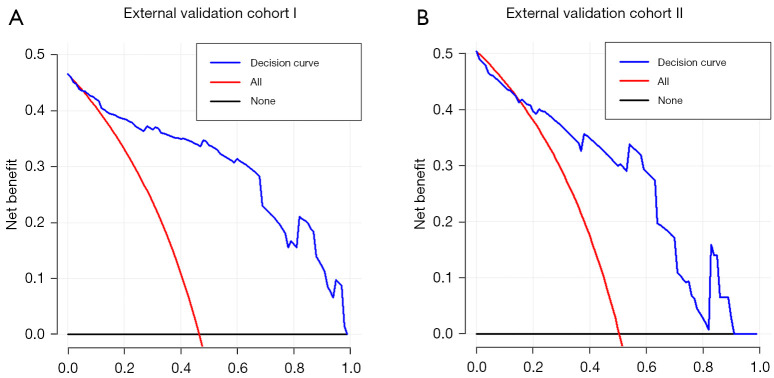
Methods: A total of 525 patients from 2017 to 2019 [Tianjin First Central Hospital (Hospital A)] were retrospectively analyzed to form the training cohort and to conduct internal validation. Another group of 204 patients in 2020 (Hospital A) formed the temporal validation cohort. A total of 107 patients in 2020 [Binzhou Medical University Hospital (Hospital B)] formed the geographic validation cohort, which was a retrospective cohort study. The area under the curve (AUC), calibration curve, and decision curve were used to evaluate the performance of the nomogram. The locally weighted regression curve was used for risk stratification.

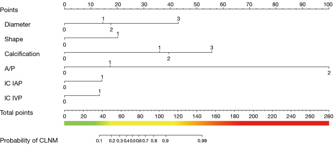
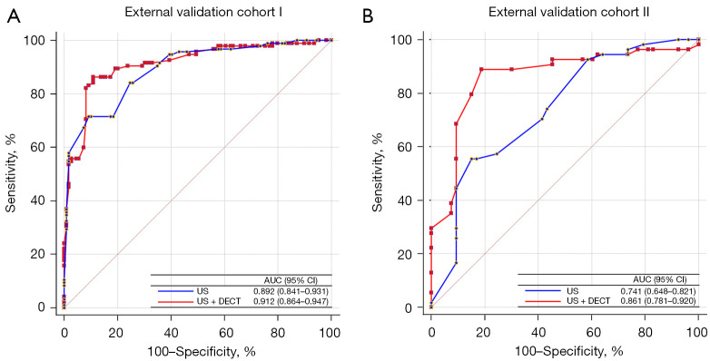
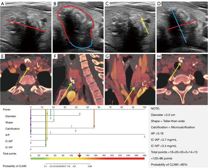
Results: Diameter, taller-than-wide, calcification, capsular invasion, and iodine concentration in the arterial and venous phases were independent risk predictors of CLNM. The AUC of the nomogram was 0.922 (95% confidence interval: 0.895-0.943) in the training cohort. Two external validation cohorts demonstrated the good performance of the nomogram in predicting CLNM, with AUCs of 0.912 and 0.861. The significantly improved net reclassification index and integrated discriminatory improvement index indicated that DECT was a powerful supplement to ultrasound for predicting CLNM. The risk stratification system divided all patients into low-risk (0-50 points), intermediate-risk (51-100 points), and high-risk groups (>100 points).

Conclusions: The nomogram and risk stratification system estimated the utility of CLNM to guide individualized treatment of patients with PTC.

Keywords: Papillary thyroid carcinoma (PTC); dual-energy computed tomography (DECT); lymphatic metastasis; nomogram; risk assessment.


Conflict of interest statement

Conflicts of Interest: All authors have completed the ICMJE uniform disclosure form (available at https://qims.amegroups.com/article/view/10.21037/qims-24-284/coif). The authors have no conflicts of interest to declare.

Figures

Figure 1
Figure 1
Flowchart of inclusion and exclusion in the current study. Hospital A is Tianjin First Central Hospital, School of Medicine, Nankai University; Hospital B is Binzhou Medical University Hospital. CLNM, central lymph node metastasis; DECT, dual-energy computed tomography; FTC, follicular thyroid carcinoma; MTC, medullary thyroid carcinoma; PACS, picture archiving and communication systems; TR, thyroid imaging reporting and data system for ultrasonography; US, ultrasound; US-FNAB, ultrasound-guided fine needle aspiration biopsy.
Figure 2
Figure 2
The nomogram for predicting CLNM in patients with PTC. According to the 8th AJCC staging systems, the diameter was classified into four categories according to the definition of diameter as follows: T1a: ≤1 cm, T1b: 1–2 cm, T2: 2–4 cm, ≥ T3: >4 cm, which corresponded to 0, 1, 2, and 3 in “Diameter” in the nomogram. Refer to American College of Radiology Thyroid Imaging, Reporting, and Data for grouping criteria, the shape of the thyroid nodule included wider-than-tall and taller-than-wide, which corresponded to 0 and 1 in “Shape” in the nomogram. Refer to American College of Radiology Thyroid Imaging, Reporting, and Data for grouping criteria, the calcification of the thyroid nodule included none or large comet-tail, macrocalcification, rim calcification, and microcalcification, which corresponded to 0, 1, 2, and 3 in “Calcification” in the nomogram. A/P was graded by values of <25%, 25–50%, or >50%, proven by a previous study, which corresponded to 0, 1, and 2 in “A/P” in the nomogram. The cutoff value of each DECT quantitative parameter was displayed in Table S5, and these parameters were converted from continuous variables to categorical variables accordingly, which corresponded to 0 and 1 in “IC IAP and IC IVP” in the nomogram. The cutoff value of IC IAP and IC IVP were 2.4 mg/mL and 3.2 mg/mL, respectively. AJCC, American Joint Committee on Cancer; A/P, the ratio of capsular abutment over the lesion perimeter; CLNM, central lymph node metastasis; DECT, dual-energy computed tomography; IAP, in the arterial phase; IC, iodine concentration; IVP, in the venous phase; PTC, papillary thyroid carcinoma.
Figure 3
Figure 3
The ROC curves and calibration curves of the three cohorts. The ROC curves and calibration curves of the nomogram for the probability of CLNM in the training (A,B), external validation cohort I (C,D), and external validation cohort II (E,F). AUC, area under the curve; CLNM, central lymph node metastasis; ROC, receiver operating characteristic.
Figure 4
Figure 4
Compare the AUCs of the two models constructed using US alone and combined US and DECT in the external validation cohorts Ⅰ and Ⅱ. AUC, area under the curve; CI, confidence interval; DECT, dual-energy computed tomography; US, ultrasound.
Figure 5
Figure 5
Decision curve analysis of the nomogram in the two external validation cohorts. The X-axis is the threshold probability, which represents the expected benefit of intervention equal to that of avoiding intervention. If the probability of a patient developing CLNM exceeds the threshold probability, it is considered that the PTC patient should undergo preventive CLND, and vice versa. The Y-axis is the net benefit, which is equal to the proportion of patients with true positives minus the proportion of patients with false positives, that is, weighted by the relative harm of abandoning the intervention versus the adverse effects of unnecessary interventions. The blue line represents the nomogram. The red line and black line represent the assumption of all patients with and without CLNM, respectively. CLND, central lymph node dissection; CLNM, central lymph node metastasis; PTC, papillary thyroid carcinoma.
Figure 6
Figure 6
An example of using the nomogram to illustrate the correct evaluation of individual risk of CLNM in patients with PTC. A 29-year-old female was incidentally found a thyroid lesion in the left lobe during a cervical spine CT examination outside the hospital. Ultrasound manifestation: hypo-echo in the middle of the inferior pole of the thyroid, the diameter was 3.0 cm (A, red line), irregular shape, A/P =0.18 (B), with microcalcification (C, yellow arrow), taller-than-wide (D). On the iodine map of dual-energy CT, the IC in the arterial (E-G) and venous (H) phases of the measured lesion were 3.7 and 3.4 mg/mL, respectively. A vertical line of each variable was drawn. The values on the “Points” scale intersected by the lines were added to obtain total points (18+20+55+0+14+13=120). The total points >100 points, considered as a high-risk patient. The graph revealed that the risk of CLNM was over 82% by drawing a vertical line on the “Total points” scale. Postoperative pathological results showed that (left lobe) PTC, the diameter was 3.0 cm, metastatic carcinoma was found in the central cervical region (3/7). This image is published with the patient’s consent. (B) The blue line represented the capsular abutment, which was defined as a lack of intervening tissue between PTC lesions and normal thyroid capsules; the red line represented the capsular protrusion, which was defined as the disruption of the perithyroidal echogenic line between the primary site of PTC and the normal thyroid capsule on sonography. (D) Taller-than-wide was defined as the anteroposterior diameter of the nodule (blue line) that was larger than its transverse diameter (red line) on a transverse plane. The yellow arrows in (E-H) pointed to the thyroid primary lesions in the iodine maps of the arterial (E-G) and the venous (H) phases. Combining the axial (E), sagittal (F), and coronal (G) images, the primary lesion with the largest cross-sectional area was selected to measure. The region of interest was placed on the substantial part as large as possible, pay attention to avoid cystic degeneration, necrosis, or calcification, and not involve adjacent blood vessels. According to the 8th AJCC staging systems, the diameter was classified into four categories according to the definition of diameter as follows: T1a: ≤1 cm, T1b: 1–2 cm, T2: 2–4 cm, ≥ T3: >4 cm, which corresponded to 0, 1, 2, and 3 in “Diameter” in the nomogram. Refer to American College of Radiology Thyroid Imaging, Reporting, and Data for grouping criteria, the shape of the thyroid nodule included wider-than tall and taller-than wide, which corresponded to 0 and 1 in “Shape” in the nomogram. Refer to American College of Radiology Thyroid Imaging, Reporting, and Data for grouping criteria, the calcification of the thyroid nodule included none or large comet-tail, macrocalcification, rim calcification, and microcalcification, which corresponded to 0, 1, 2, and 3 in “Calcification” in the nomogram. A/P was graded by values of <25%, 25–50%, or >50%, proven by a previous study, which corresponded to 0, 1, and 2 in “A/P” in the nomogram. The cutoff value of each DECT quantitative parameter was displayed in Table S5, and these parameters were converted from continuous variables to categorical variables accordingly, which corresponded to 0 and 1 in “IC IAP and IC IVP” in the nomogram. The cutoff value of IC IAP and IC IVP were 2.4 mg/mL and 3.2 mg/mL, respectively. AJCC, American Joint Committee on Cancer; A/P, the ratio of capsular abutment over the lesion perimeter; CLNM, central lymph node metastasis; CT, computed tomography; DECT, dual-energy computed tomography; IAP, in the arterial phase; IC, iodine concentration; IVP, in the venous phase; PTC, papillary thyroid carcinoma.

Similar articles

Cited by

References

    1. Siegel RL, Miller KD, Wagle NS, Jemal A. Cancer statistics, 2023. CA Cancer J Clin 2023;73:17-48. 10.3322/caac.21763 - DOI - PubMed
    1. Cho SY, Lee TH, Ku YH, Kim HI, Lee GH, Kim MJ. Central lymph node metastasis in papillary thyroid microcarcinoma can be stratified according to the number, the size of metastatic foci, and the presence of desmoplasia. Surgery 2015;157:111-8. 10.1016/j.surg.2014.05.023 - DOI - PubMed
    1. Zhao Q, Ming J, Liu C, Shi L, Xu X, Nie X, Huang T. Multifocality and total tumor diameter predict central neck lymph node metastases in papillary thyroid microcarcinoma. Ann Surg Oncol 2013;20:746-52. 10.1245/s10434-012-2654-2 - DOI - PubMed
    1. Carling T, Carty SE, Ciarleglio MM, Cooper DS, Doherty GM, Kim LT, Kloos RT, Mazzaferri EL, Sr, Peduzzi PN, Roman SA, Sippel RS, Sosa JA, Stack BC, Jr, Steward DL, Tufano RP, Tuttle RM, Udelsman R; . American Thyroid Association design and feasibility of a prospective randomized controlled trial of prophylactic central lymph node dissection for papillary thyroid carcinoma. Thyroid 2012;22:237-44. 10.1089/thy.2011.0317 - DOI - PubMed
    1. So YK, Seo MY, Son YI. Prophylactic central lymph node dissection for clinically node-negative papillary thyroid microcarcinoma: influence on serum thyroglobulin level, recurrence rate, and postoperative complications. Surgery 2012;151:192-8. 10.1016/j.surg.2011.02.004 - DOI - PubMed

LinkOut - more resources