. 2023 Dec;8(12):783-798.
doi: 10.1038/s41578-023-00581-x. Epub 2023 Jul 26.

New Opportunities and Old Challenges in the Clinical translation of Nanotheranostics

Affiliations

New Opportunities and Old Challenges in the Clinical translation of Nanotheranostics

Peter J Gawne et al. Nat Rev Mater. 2023 Dec.

Abstract

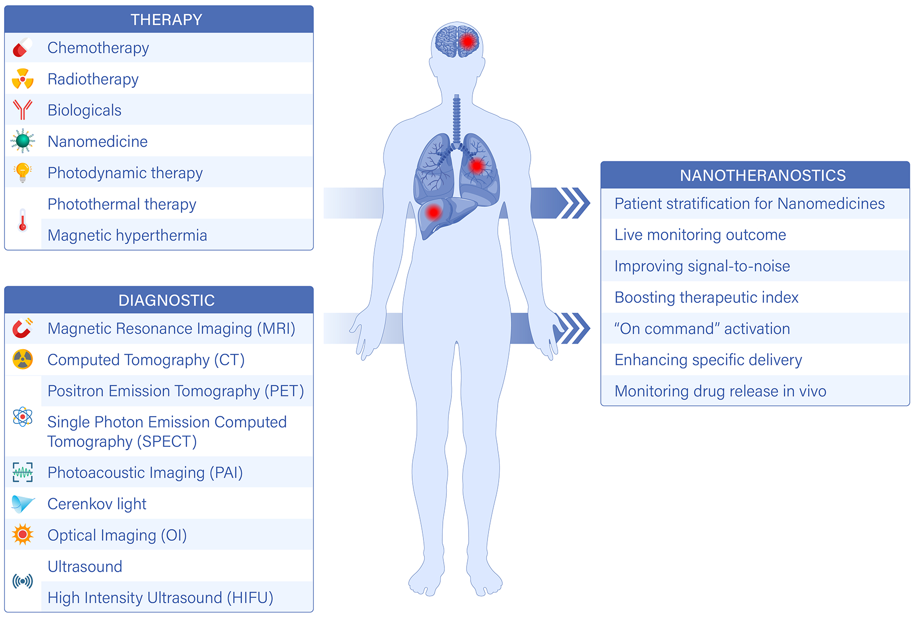
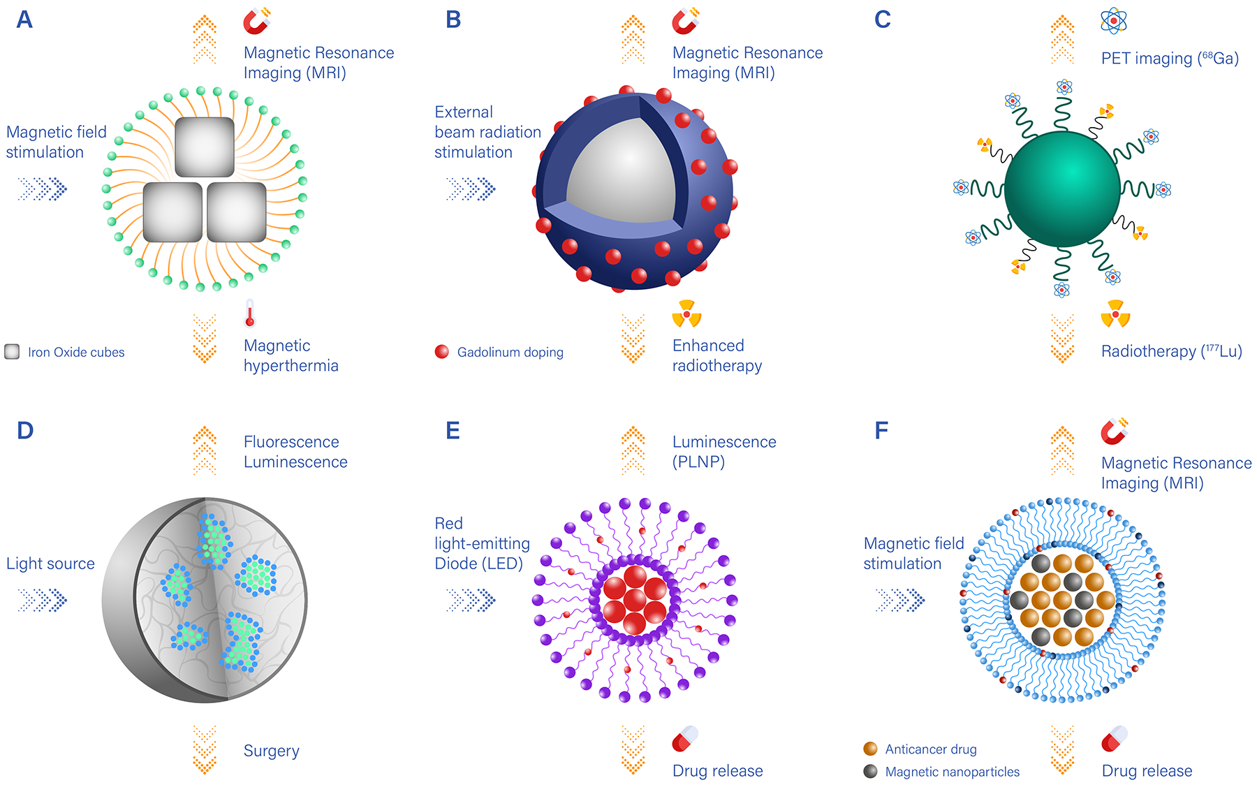
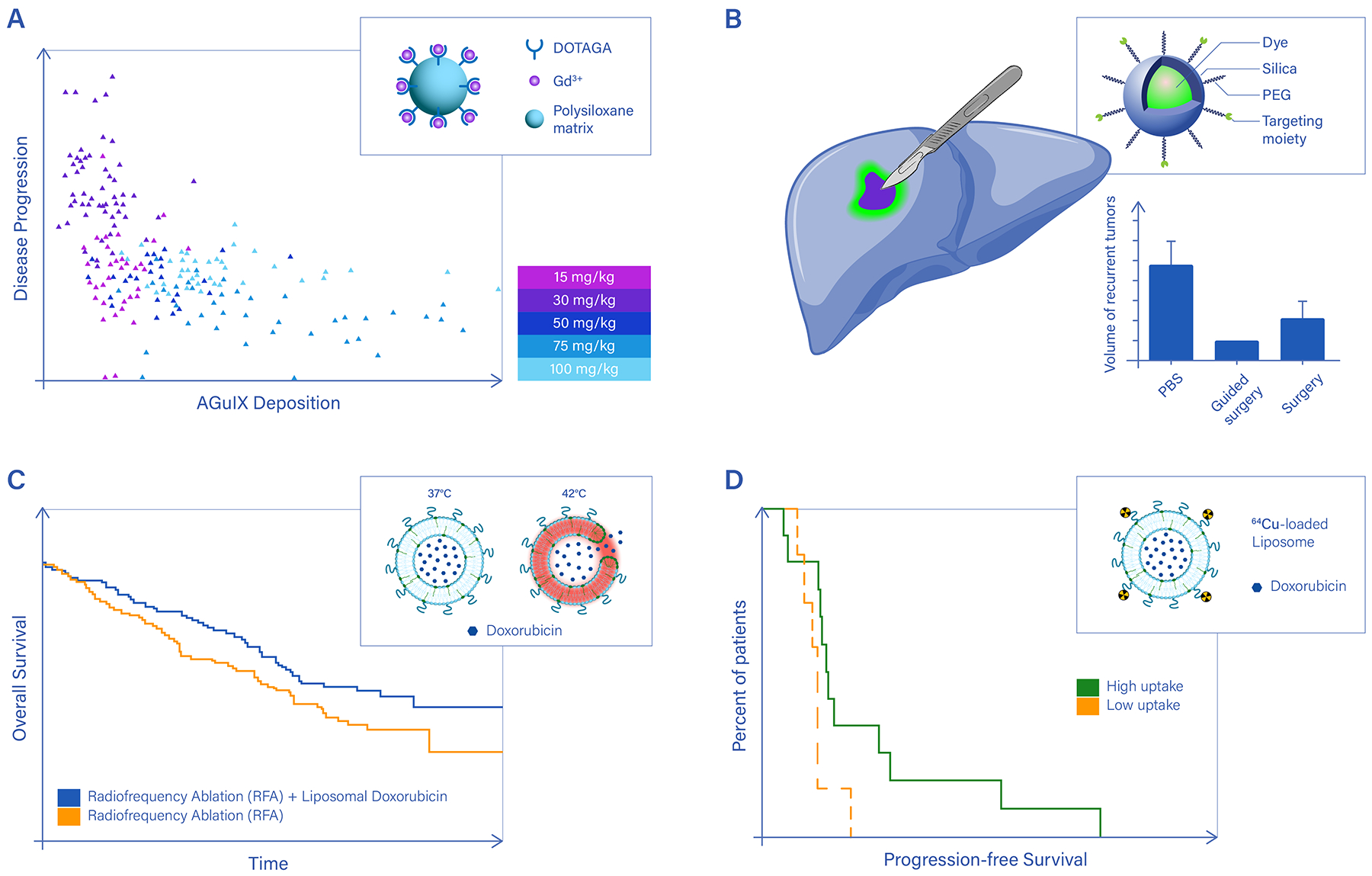
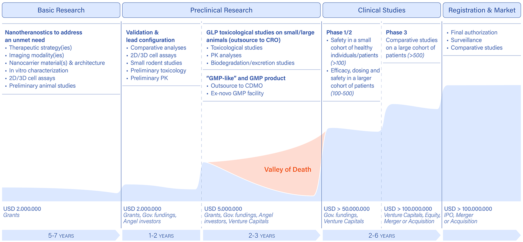
Nanoparticle-based systems imbued with both diagnostic and therapeutic functions, known as nanotheranostics, have enabled remarkable progress in guiding focal therapy, inducing active responses to endogenous and exogenous biophysical stimuli, and stratifying patients for optimal treatment. However, although in recent years more nanotechnological platforms and techniques have been implemented in the clinic, several important challenges remain that are specific to nanotheranostics. In this Review, we first discuss some of the many ways of 'constructing' nanotheranostics, focusing on the different imaging modalities and therapeutic strategies. We then outline nanotheranostics that are currently used in humans at different stages of clinical development, identifying specific advantages and opportunities. Finally, we define critical steps along the winding road of preclinical and clinical development and suggest actions to overcome technical, manufacturing, regulatory and economical challenges for the safe and effective clinical translation of nanotheranostics.


Conflict of interest statement

Competing interests The authors declare no competing interests.

Figures

Figure 1 |
Figure 1 |. Theranostic nanoparticles.
Nanotheranostics combine therapeutic and imaging functions in a nanocarrier. This can be achieved by the incorporation of specific therapeutic and imaging agents into a nanoparticle, or can be a result of the innate properties of the material, or a combination of the two. Targeting molecules (such as small ligands, peptides, aptamers, antibodies and fragments of antibodies) can be incorporated into a nanoparticle to enhance the recognition of specific cellular and subcellular targets. It is also possible to use materials with the ability to specifically recognize the diseased tissue. Moreover, nanotheranostics can be designed to be activated by endogenous and exogenous stimuli. Compared to molecular agents and nanomedicines, nanotheranostics offer several advantages, including patient stratification, ‘on command’ activation and enhanced therapeutic efficacy.
Figure 2 |
Figure 2 |. Examples of nanotheranostics with different imaging, therapeutic and targeting components.
A. Clusters of iron oxide nanocubes coated by polymeric or lipid chains (yellow) and presenting surface-targeting moieties (blue). Upon magnetic stimulation, iron oxide nanocubes generate thermal energy, which can be used for magnetic hyperthermia, and can be visualized via magnetic resonance imaging (MRI); B. Nanocarriers carrying heavy metals (such as Gd) locally enhance the therapeutic efficacy of external beam radiation and can be imaged via MRI; C. Nanocarriers conjugated to two different radionuclides can be used for positron emission tomography (PET) imaging (using 89Zr) and radiotherapy (using 177Lu); D. Nanoparticles encapsulating luminescent/fluorescent molecules, which under external light stimulation can be visualised and guide surgical resection; E. Nanocarriers loaded with persistent luminescence nanoparticles (PLNP) and chemotherapeutic drugs that are released by passive diffusion over time; F. Nanocarriers loaded with iron oxide particles for MR imaging guidance, which under high-intensity focused ultrasound stimulation trigger the release of anti-cancer molecules.
Figure 3 |
Figure 3 |. Clinical demonstrations of nanotheranostics.
A. Enhancing the efficacy of external beam radiation therapy using Gd-based nanoparticles (AGuIX) under magnetic resonance imaging (MRI). Clinical data on cancer metastases to the brain show a positive correlation between AGuIX deposition within the lesions, as detected via MRI, and disease regression. Upon external beam radiation therapy, brain metastases receiving higher amounts of AGuIX grow less or even reduce in diameter (relative lesion size < 1) over a 28-day observation period. DOTAGA: 1,4,7,10-tetraazacyclododecane,1-glutaric acid-4,7,10-triacetic acid ligands. The legend shows the dose of AGuIX injected in the patients. Each symbol in the plot corresponds to a different injected dose and patient. B. Multimodal imaging nanoparticles carrying targeting moieities to specifically recognize cancer cells help visualize tumor margins, inspect lymph nodes, and accurately guide surgical resection of the bulk malignancy. However, to fully unveil the advantage of this approach, future clinical studies will have to address patient survival and disease recurrence, as was done for indocyanine. The Kaplan-Meier plot compares the overall survival for patients with rectal cancer following a laparoscopic lateral pelvic lymph node dissection performed with or without indocyanine green fluorescence imaging. Image-guided surgery with indocyanine positively correlates with improved survival and reduced risk of local disease relapse. C. Boosting the spatio-temporal specificity of conventional therapies using thermoresponsive nanomedicines. The Kaplan-Meier plot shows a significant improvement in overall survival for hepatocellular carcinoma patients treated with radiofrequency ablation plus thermosensitive doxorubicin liposomes. The chemotherapeutic agent is released preferentially at the tumor site, following the localized temperature increase and consequent destabilization of the liposomes. Drug release from thermosensitive nanomedicines has been achieved in clinical settings using radiofrequency ablation and high-intensity focused ultrasound; D. 89Zr-labeled, docetaxel loaded polymeric nanoparticles have been used to treat patients with various cancers (primary and metastatic) and image particle deposition in each single lesion for 7 patients. Although at the overall patient level no statistically significant correlation was found between nanoparticle accumulation in the cancer tissue and response to therapy, individual lesions with higher nanoparticle deposition (red lesion in Patient 1 and yellow lesion in Patient 6) underwent a reduction in size of up to 50% within a 96-hour observation period. Panel a adapted from Ref., panel b from Ref., panel c from Ref., panel d from Ref..
Figure 4.
Figure 4.. Milestones in the clinical translation of nanotheranostics.
Early design, development and characterization of novel nanotheranostics entirely relies on research grants, which help demonstrating a new idea and technology all the wat up to small-rodent models. This basic research phase is almost exclusively funded by governmental research grants. More systematic analyses and in-vitro/in-vivo characterizations of the new technology are conducted in the preclinical research phase. This aims at identifying a lead product, with a specific imaging/therapeutic agent combination as well as material and architecture configurations. This phase is typically funded by industrial-like governmental grants, such as small business innovation research (SBIR) and small business technology transfer (STTR) programs in the US; European Innovation Council (EIC) Transition and Accelerator programmes in the EU, and angel investors and foundations. A second portion of the preclinical research phase deals with the manufacturing and toxicological testing of the proposed product, following well-coded procedures (good manufacturing practice, GMP, and good laboratory practice, GLP) in agreement with the relevant regulatory bodies (FDA, EMA and others). This phase can be funded by governmental agencies, angel donors and foundations, as well as venture capital firms investing in early-stage products. Finally, the clinical studies aim to assess the safety, efficacy and economic convenience of the proposed product over different phases, depending on the type of product and disease. These activities typically require significant capitals involving multiple venture capital firms as well as pharmaceutical companies. PK: pharmacokinetics; CRO: contract research organization; CDMO: contract development and manufacturing organization).

References

    1. Wagner FE; Haslbeck S; Stievano L; Calogero S; Pankhurst QA; Martinek KP, Before striking gold in gold-ruby glass. Nature 2000, 407 (6805), 691–2. - PubMed
    1. Nano on reflection. Nature Nanotechnology 2016, 11 (10), 828–834. - PubMed
    1. Weber DO, Nanomedicine. Health Forum J 1999, 42 (4), 32, 36–7. - PubMed
    1. Mitchell MJ; Billingsley MM; Haley RM; Wechsler ME; Peppas NA; Langer R, Engineering precision nanoparticles for drug delivery. Nat Rev Drug Discov 2021, 20 (2), 101–124. - PMC - PubMed
    1. Stater EP; Sonay AY; Hart C; Grimm J, The ancillary effects of nanoparticles and their implications for nanomedicine. Nat Nanotechnol 2021, 16 (11), 1180–1194. - PMC - PubMed

LinkOut - more resources