. 2024 Jun 13:39:100805.
doi: 10.1016/j.bbih.2024.100805. eCollection 2024 Aug.

Changes in neuroinflammatory biomarkers correlate with disease severity and neuroimaging alterations in patients with COVID-19 neurological complications

Affiliations

Changes in neuroinflammatory biomarkers correlate with disease severity and neuroimaging alterations in patients with COVID-19 neurological complications

Fernanda G Q Barros-Aragão et al. Brain Behav Immun Health. .

Abstract

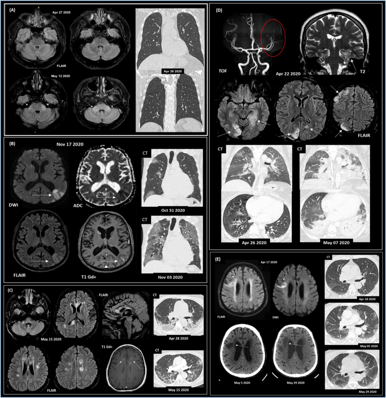
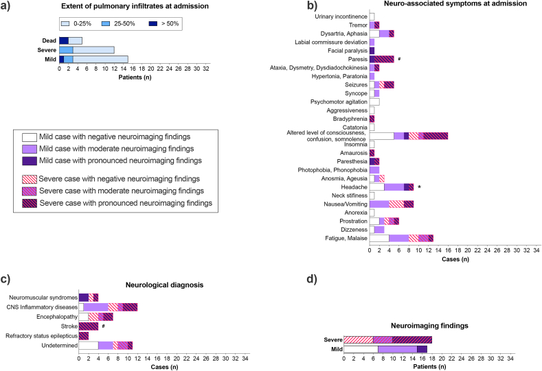
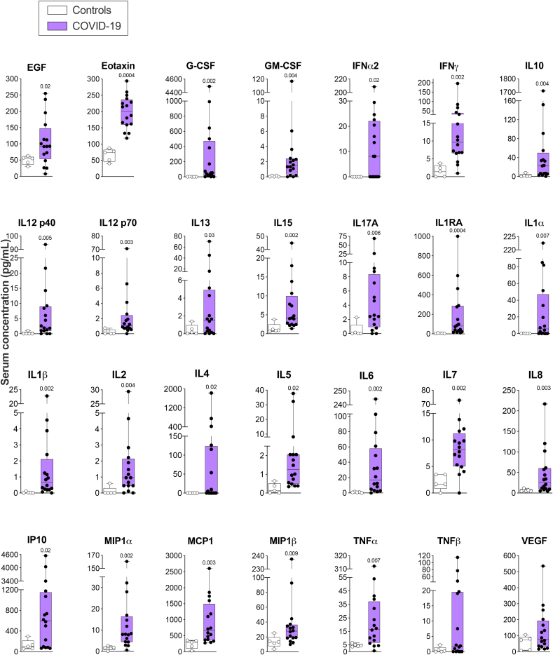
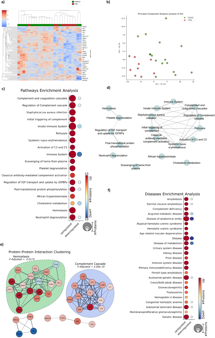
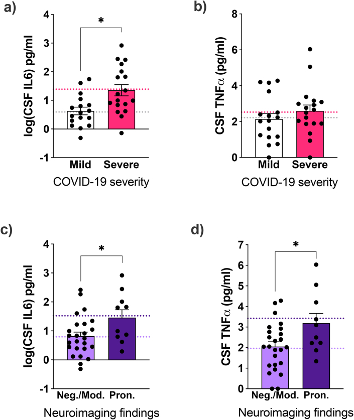
COVID-19 induces acute and persistent neurological symptoms in mild and severe cases. Proposed concomitant mechanisms include direct viral infection and strain, coagulopathy, hypoxia, and neuroinflammation. However, underlying molecular alterations associated with multiple neurological outcomes in both mild and severe cases are majorly unexplored. To illuminate possible mechanisms leading to COVID-19 neurological disease, we retrospectively investigated in detail a cohort of 35 COVID-19 mild and severe hospitalized patients presenting neurological alterations subject to clinically indicated cerebrospinal fluid (CSF) sampling. Clinical and neurological investigation, brain imaging, viral sequencing, and cerebrospinal CSF analyses were carried out. We found that COVID-19 patients presented heterogeneous neurological symptoms dissociated from lung burden. Nasal swab viral sequencing revealed a dominant strain at the time of the study, and we could not detect traces of SARS-CoV-2's spike protein in patients' CSF by multiple reaction monitoring analysis. Patients presented ubiquitous systemic hyper-inflammation and broad alterations in CSF proteomics related to inflammation, innate immunity, and hemostasis, irrespective of COVID-19 severity or neuroimaging alterations. Elevated CSF interleukin-6 (IL6) correlated with disease severity (sex-, age-, and comorbidity-adjusted mean Severe 24.5 pg/ml, 95% confidence interval (CI) 9.62-62.23 vs. Mild 3.91 pg/mL CI 1.5-10.3 patients, p = 0.019). CSF tumor necrosis factor-alpha (TNFα) and IL6 levels were higher in patients presenting pronounced neuroimaging alterations compared to those who did not (sex-, age-, and comorbidity-adjusted mean TNFα Pronounced 3.4, CI 2.4-4.4 vs. Non-Pronounced 2.0, CI 1.4-2.5, p = 0.022; IL6 Pronounced 33.11, CI 8.89-123.31 vs Non-Pronounced 6.22, CI 2.9-13.34, p = 0.046). Collectively, our findings put neuroinflammation as a possible driver of COVID-19 acute neurological disease in mild and severe cases.

Keywords: COVID-19; Coronavirus; Inflammation; Neuro-infectious diseases; Neuroinflammation.


Conflict of interest statement

none.

Figures

Fig. 1
Fig. 1
Representative paired neuroimaging and chest CT findings from COVID-19 patients presenting neurological symptoms. (A) Subject #16 (65 y/o; male) with encephalitis and a positive result for SARS-CoV-2 RT-qPCR in the CSF screening. Initial symptoms of fever and vomiting, evolving with drowsiness, ataxia, acute diplopia and convergent strabismus. No signs of viral pneumonia were detected on the CT scan. Case previously reported (Freitas et al., 2021). (B) Subject #25 (83 y/o; male; deceased) with acute CNS ischemic lesions (vasculitis); viral pneumonia, fever, respiratory distress, and rhabdomyolysis. Coronal chest CT images (lung window) show multiple areas of round ground-glass opacities, mainly on the right lung, with increasing extent after three days. (C) Subject #14 (50 y/o; female) presented acute demyelinating brain lesions and COVID-19 pneumonia. Axial CT images show areas of consolidation distributed predominantly at peripheral and posterior areas of the lower lobes and improvement in lung infiltrates after 17 days. (D) Subject #27 (29 y/o; female; deceased) with a previous diagnosis of Systemic Lupus Erythematosus, presenting CNS vasculitis; refractory epileptic status; and viral pneumonia. Evolution to progressive global systemic failure. Chest CT shows mild round ground-glass opacity on the middle lobe and upper right lung. Eleven days later, abnormalities increased with diffuse consolidation, mainly in the lower lobes. (E) Subject #23 (78 y/o; male; deceased) presented a subacute ischemic stroke with an altered level of consciousness, seizures, and coma; viral pneumonia. Axial CT images show no lung parenchymal findings consistent with viral pneumonia that progresses to mild patchy ground-glass opacity in the lungs' periphery 17 days after. Lung abnormalities are almost entirely resolved after 41 days. Subjects (B–E) with positive RT-qPCR for SARS-CoV-2 in the nasopharyngeal swab. Abbreviations: FLAIR, DWi, ADC, T1, T1 Gd+, T2 refer to MRI sequences, and Gd + represents venous contrast. Time of flight (TOF) arterial angiographic MRI sequence.
Fig. 2
Fig. 2
COVID-19 induces neurological disturbances despite the low extent of pulmonary infiltrates. (A) Pulmonary infiltrates extent in CT scans at hospital admission (n = 33). (B-D) Patients were grouped as presenting negative (white bars), moderate (light purple) or pronounced (dark purple) neuroimaging (CT/MRI) findings related to COVID-19 hospitalization (n = 35). Dashed lines represent severe disease or death. CNS inflammatory diseases include meningitis, encephalitis, meningoencephalitis, encephalomyelitis, myelitis, CNS vasculitis, and acute disseminated encephalomyelitis (ADEM). Neuromuscular syndromes include Guillain-Barré Syndrome (GBS), cranial neuropathy, peripheral facial paralysis, and myopathy. See Suppl. Table 2 for individualized data. *p < 0.017, disease severity as an outcome, Fisher's exact test; #p < 0.017, neuroimaging positivity as an outcome, Chi-Square test, followed by pairwise comparison with Bonferroni correction. (For interpretation of the references to colour in this figure legend, the reader is referred to the Web version of this article.)
Fig. 3
Fig. 3
COVID-19 patients with neurological symptoms present modified systemic inflammatory biomarkers. Data are shown as the median, interquartile range (box), and min-max points (whiskers). Symbols indicate individual patients (n = 5 control, 16 COVID-19). Mann-Whitney test corrected for False Discovery Rate (FDR) using the two-stage step-up (Benjamini, Krieger, and Yekutieli) method. Discoveries are signalized by the above q-values (FDR-adjusted p-values).
Fig. 4
Fig. 4
COVID-19 patients with neurological symptoms display cerebrospinal fluid (CSF)-altered proteomic patterns related to inflammation, innate immunity, and hemostasis. (A) Hierarchical clustering analysis from uninfected normal pressure hydrocephalus (NPH) controls (CTRL, n = 10) and COVID-19 (n = 16) CSF using the measured relative abundance of the proteins. (B) Enrichment analysis using the statistically significant COVID-19 dysregulated proteins searched against a public databank to obtain up and downregulated proteins matched with biological pathways. (C) Disease enrichment analysis of COVID-19 statistically significant dysregulated proteins. (D) The enriched pathways' interaction evidences the connection between the pathways modulated by COVID-19. (E) Protein-Protein interaction clustering analysis of the COVID-19 statistically significant dysregulated proteins.
Fig. 5
Fig. 5
COVID-19 severity and neuroimaging abnormalities are linked to cerebrospinal fluid (CSF) neuroinflammatory biomarkers in COVID-19 patients presenting neurological symptoms. COVID-19 patients were grouped by (a, b) disease severity or by (c, d) presenting negative or moderate (Neg./Mod.) or pronounced (Pron.) neuroimaging alterations. Data are shown as mean and SEM. Symbols indicate individual patients. *p < 0.05, ANCOVA using sex, age, diabetes, and numbers of neurological, cardiovascular, and other comorbidities as covariates. (a) F(1,27) = 6.3, p = 0.019; (b) F(1,27) = 0.25, p = 0.624; (c) F(1,27) = 4.4, p = 0.046; (d) F(1,27) = 5.9, p = 0.022. Dashed lines indicate model-adjusted means.

References

    1. Alvim R.G., Lima T.M., Rodrigues D.A.S., Marsili F.F., Bozza V.B.T., Higa L.M., Monteiro F.L., Leitão I.C., Carvalho R.S., Galliez R.M., Castineiras T.M.P.P., Nobrega A., Travassos L.H., Ferreira Jr OC., Tanuri A., Vale A.M., Castilho L.R. An affordable anti-SARS-COV-2 spike protein ELISA test for early detection of IgG seroconversion suited for large-scale surveillance studies in low-income countries. medRxiv. 2020 doi: 10.1101/2020.07.13.20152884. - DOI
    1. Angriman F., Ferreyo B., Burry L., Fan E. Interleukin-6 receptor blockade in patients with COVID-19: placing clinical trials into context. Lancet Respir. Med. 2021;9:655–664. https://pubmed.ncbi.nlm.nih.gov/33930329/ Available at: - PMC - PubMed
    1. Burton M.D., Johnson R.W. Interleukin-6 trans-signaling in the senescent mouse brain is involved in infection-related deficits in contextual fear conditioning. Brain Behav. Immun. 2012;26:732–738. - PMC - PubMed
    1. Candido D.S., et al. Evolution and epidemic spread of SARS-CoV-2 in Brazil. Science. 2020;369(1979):1255–1260. https://pubmed.ncbi.nlm.nih.gov/32703910/ Available at: - PMC - PubMed
    1. Chou S.H.-Y., Beghi E., Helbok R., Moro E., Sampson J., Altamirano V., Mainali S., Bassetti C., Suarez J.I., McNett M., GCS-NeuroCOVID Consortium and ENERGY Consortium Global Incidence of neurological manifestations among patients hospitalized with COVID-19-A report for the GCS-NeuroCOVID Consortium and the ENERGY Consortium. JAMA Neurol. 2021;4 http://www.ncbi.nlm.nih.gov/pubmed/33974053 Available at: - PMC - PubMed

LinkOut - more resources