. 2024 Aug 20;98(8):e0061824.
doi: 10.1128/jvi.00618-24. Epub 2024 Jul 18.

The Raf kinase inhibitors Dabrafenib and Regorafenib impair Zika virus replication via distinct mechanisms

Affiliations

The Raf kinase inhibitors Dabrafenib and Regorafenib impair Zika virus replication via distinct mechanisms

Lucas Wilken et al. J Virol. .

Abstract

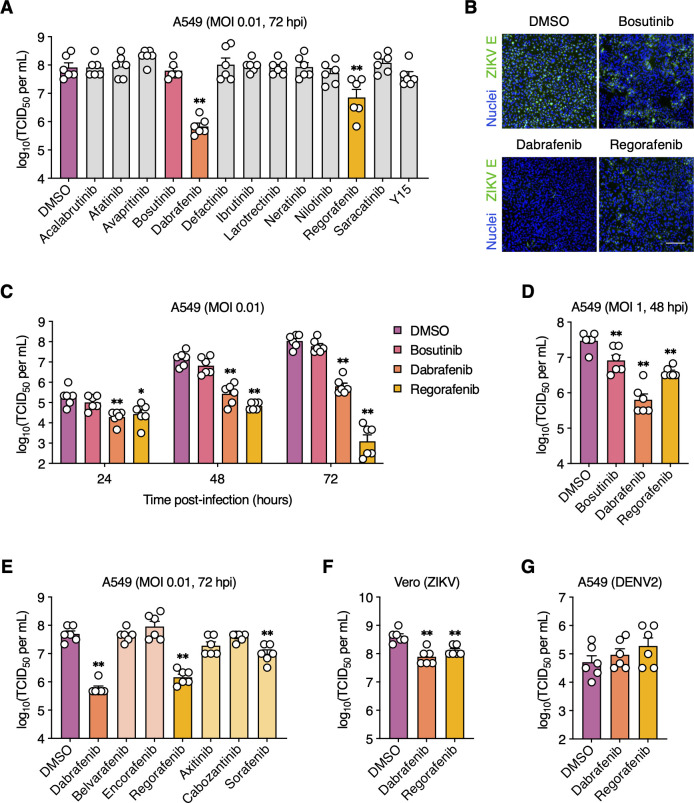
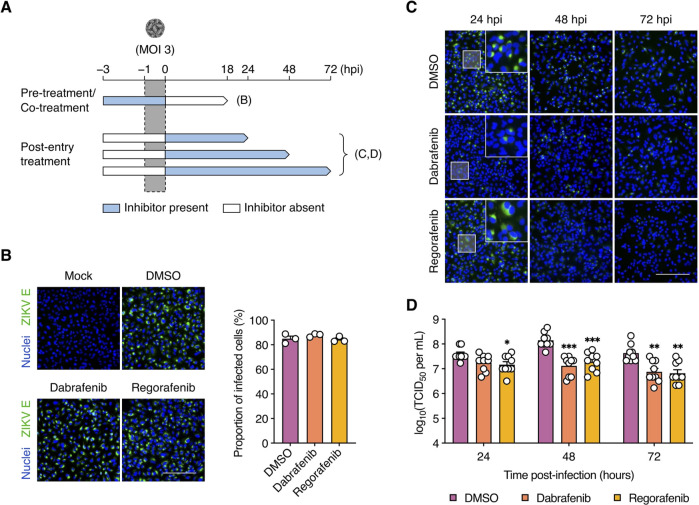
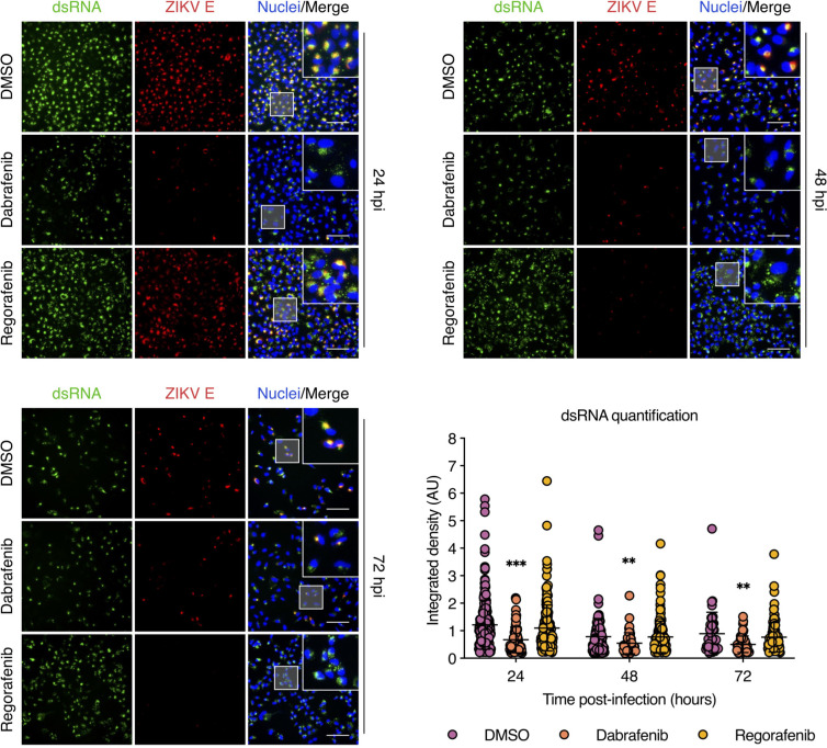
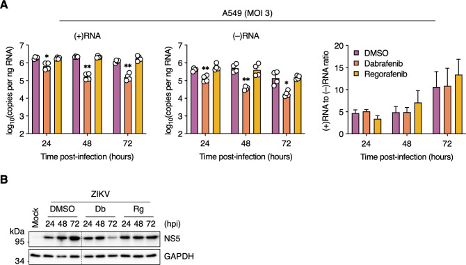
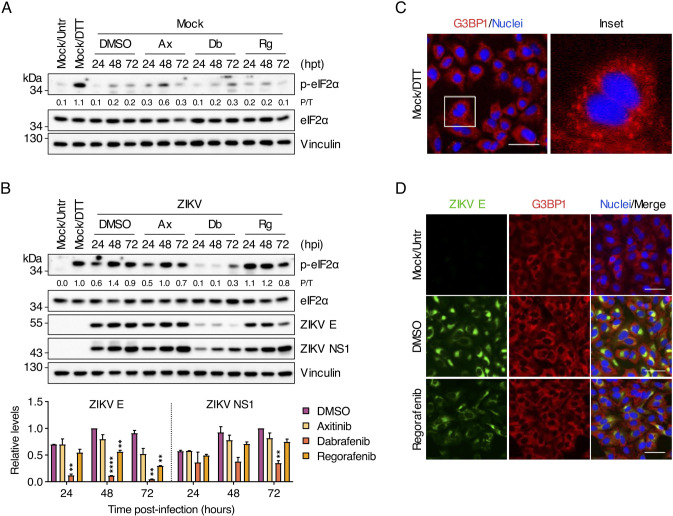
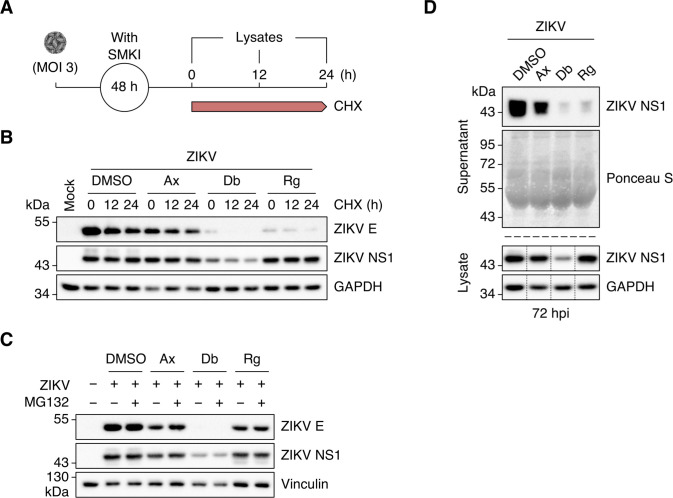
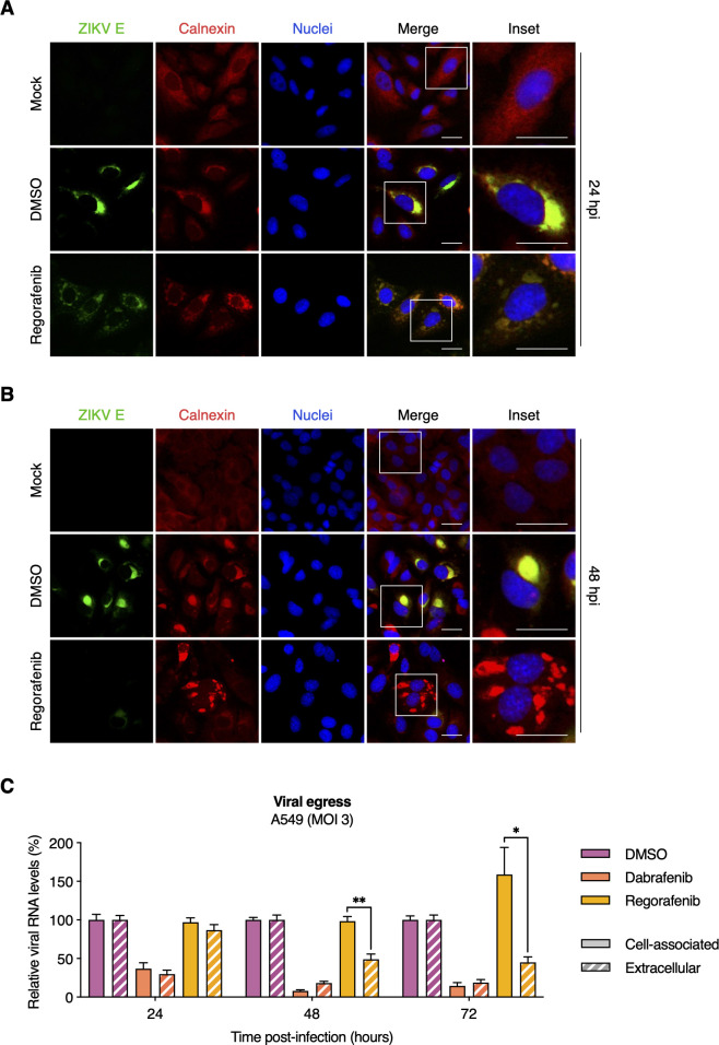
Zika virus (ZIKV) is a re-emerging mosquito-borne flavivirus that has been associated with congenital neurological defects in fetuses born to infected mothers. At present, no vaccine or antiviral therapy is available to combat this devastating disease. Repurposing drugs that target essential host factors exploited by viruses is an attractive therapeutic approach. Here, we screened a panel of clinically approved small-molecule kinase inhibitors for their antiviral effects against a clinical isolate of ZIKV and thoroughly characterized their mechanisms of action. We found that the Raf kinase inhibitors Dabrafenib and Regorafenib potently impair the replication of ZIKV, but not that of its close relative dengue virus. Time-of-addition experiments showed that both inhibitors target ZIKV infection at post-entry steps. We found that Dabrafenib, but not Regorafenib, interfered with ZIKV genome replication by impairing both negative- and positive-strand RNA synthesis. Regorafenib, on the other hand, altered steady-state viral protein levels, viral egress, and blocked NS1 secretion. We also observed Regorafenib-induced ER fragmentation in ZIKV-infected cells, which might contribute to its antiviral effects. Because these inhibitors target different steps of the ZIKV infection cycle, their use in combination therapy may amplify their antiviral effects which could be further explored for future therapeutic strategies against ZIKV and possibly other flaviviruses.

Importance: There is an urgent need to develop effective therapeutics against re-emerging arboviruses associated with neurological disorders like Zika virus (ZIKV). We identified two FDA-approved kinase inhibitors, Dabrafenib and Regorafenib, as potent inhibitors of contemporary ZIKV strains at distinct stages of infection despite overlapping host targets. Both inhibitors reduced viral titers by ~1 to 2 log10 (~10-fold to 100-fold) with minimal cytotoxicity. Furthermore, we show that Dabrafenib inhibits ZIKV RNA replication whereas Regorafenib inhibits ZIKV translation and egress. Regorafenib has the added benefit of limiting NS1 secretion, which contributes to the pathogenesis and disease progression of several flaviviruses. Because these inhibitors affect distinct post-entry steps of ZIKV infection, their therapeutic potential may be amplified by combination therapy and likely does not require prophylactic administration. This study provides further insight into ZIKV-host interactions and has implications for the development of novel antivirals against ZIKV and possibly other flaviviruses.

Keywords: RNA virus; drug repurposing; flavivirus; host-directed antivirals; kinase inhibitors.


Conflict of interest statement

The authors declare no conflicts of interest.

Figures

Fig 1
Fig 1
Dabrafenib and Regorafenib inhibit ZIKV but not DENV2 replication. (A and B) A549 cells were mock-infected or infected with ZIKV (MOI = 0.01 PFU/cell) and subsequently treated with the indicated SMKIs or DMSO (vehicle control). (A) Viral titers in the supernatant were determined at 72 hpi by endpoint dilution assay. (B) ZIKV infection was visualized at 72 hpi by immunofluorescence staining for ZIKV E protein (green). Nuclei were stained with Hoechst 33342 (blue). Scale bar, 200 µm. Representative images of two independent experiments are shown. (C) A549 cells were infected with ZIKV (MOI = 0.01 PFU/cell) and subsequently treated with DMSO, bosutinib (5 µM), Dabrafenib (10 µM), or Regorafenib (2.5 µM). Viral titers in the supernatant were determined at 2, 24, 48, and 72 hpi by endpoint dilution assay. (D) A549 cells were infected with ZIKV (MOI = 1 PFU/cell) and subsequently treated with DMSO, bosutinib (5 µM), Dabrafenib (10 µM), or Regorafenib (2.5 µM). Viral titers in the supernatant were determined at 48 hpi by endpoint dilution assay. (E) A549 cells were infected with ZIKV (MOI = 0.01 PFU/cell) and subsequently treated with the indicated SMKIs or DMSO. Viral titers in the supernatant were determined at 72 hpi by endpoint dilution assay. (F) Vero cells were infected with ZIKV (MOI = 0.01 PFU/cell) and subsequently treated with DMSO, Dabrafenib (2.5 µM), or Regorafenib (1 µM). Viral titers in the supernatant were determined at 48 hpi by endpoint dilution assay. (G) A549 cells were mock-infected or infected with DENV2 (MOI = 0.01 PFU/cell) and subsequently treated with DMSO, Dabrafenib (10 µM), or Regorafenib (2.5 µM). Viral titers in the supernatant were determined at 72 hpi by endpoint dilution assay. Data are expressed as mean ± standard error of the mean (SEM) from two independent experiments with three biological replicates per experiment. *, P < 0.05; **, P < 0.01 (Mann–Whitney U test vs “DMSO”).
Fig 2
Fig 2
Dabrafenib and Regorafenib target ZIKV infection at the post-entry stage. (A) Schematic representation of the time-of-addition assays. (B) A549 cells were pre-treated with DMSO (vehicle control), Dabrafenib (10 µM), or Regorafenib (2.5 µM) for 2 h prior to infection with ZIKV (MOI = 3 PFU/cell) as well as during the adsorption period. ZIKV infection was visualized at 18 hpi by immunofluorescence staining for ZIKV E protein (green). Nuclei were stained with Hoechst 33342 (blue). Scale bar, 200 µm. Images are representative of three independent experiments. Bar graph shows the proportions of infected cells determined from immunofluorescence images (one per experiment) using ImageJ. (C and D) A549 cells were infected with ZIKV (MOI = 3 PFU/cell) and subsequently treated with DMSO, Dabrafenib (10 µM), or Regorafenib (2.5 µM). (C) ZIKV infection was visualized at 24, 48, and 72 hpi by immunofluorescence staining for ZIKV E protein (green). Nuclei were stained with Hoechst 33342 (blue). Scale bar, 200 µm. Images are representative of three independent experiments. (D) Viral titers in the supernatant were determined at 24, 48, and 72 hpi by endpoint dilution assay. Data are expressed as mean ± SEM from three independent experiments with three biological replicates per experiment. *, P < 0.05; **, P < 0.01; ***, P < 0.001 (Mann–Whitney U test vs “DMSO”).
Fig 3
Fig 3
Dabrafenib, but not Regorafenib, reduces viral dsRNA levels. A549 cells were infected with ZIKV (MOI = 3 PFU/cell) and subsequently treated with DMSO (vehicle control), Dabrafenib (10 µM), or Regorafenib (2.5 µM). ZIKV infection was visualized at 24, 48, and 72 hpi by immunofluorescence staining for ZIKV E protein (red) and dsRNA (green). Nuclei were stained with Hoechst 33342 (blue). Scale bars, 100 µm. Dot plot shows image-based quantification of the integrated density of dsRNA in individual cells. Data are expressed as mean ± SEM. Data from one of three independent experiments with similar results are shown. *, P < 0.05; **, P < 0.01; ***, P < 0.001 (unpaired Student’s t test vs “DMSO”).
Fig 4
Fig 4
Dabrafenib, but not Regorafenib, attenuates viral RNA synthesis. (A) A549 cells were infected with ZIKV (MOI = 3 PFU/cell) and subsequently treated with DMSO (vehicle control), Dabrafenib (10 µM), or Regorafenib (2.5 µM). Total cell-associated RNA was isolated at 24, 48, and 72 hpi, and analyzed by positive strand-specific RT-qPCR (upper left panel) and negative strand-specific RT-qPCR (upper right panel). Lower left panel: Results shown in the upper panels plotted as the ratio of positive-strand RNA [(+)RNA] to negative-strand RNA [(−)RNA]. Data are expressed as mean ± SEM from two independent experiments with two biological replicates per experiment. *, P < 0.05; **, P < 0.01 (two-way ANOVA vs “DMSO”). (B) A549 cells were mock-infected or infected with ZIKV (MOI = 3 PFU/cell) and were left untreated (Untr) or were treated with DMSO (vehicle control), Dabrafenib (Db; 10 µM), or Regorafenib (Rg; 2.5 µM) for the indicated durations. Whole-cell lysates were analyzed by Western blotting with antibodies against ZIKV NS5 and GAPDH (loading control). Black dotted lines indicate removal of portions of the blots for clarity.
Fig 5
Fig 5
Regorafenib reduces ZIKV E protein, but not NS1, levels and does not activate the integrated stress response. (A) A549 cells were either left untreated (Untr) or were treated with DMSO (vehicle control), Axitinib (Ax; 10 µM), Dabrafenib (Db; 10 µM), or Regorafenib (Rg; 2.5 µM) for the indicated durations. As a positive control, cells were treated with DTT (2 mM) for 30 min prior to harvesting. Whole-cell lysates were analyzed by Western blotting with antibodies against phospho-eIF2α (Ser51), total eIF2α, and vinculin (loading control). (B) A549 cells were mock-infected or infected with ZIKV (MOI = 3 PFU/cell) and were left untreated (Untr) or were treated with DMSO (vehicle control), Axitinib (Ax; 10 µM), Dabrafenib (Db; 10 µM), or Regorafenib (Rg; 2.5 µM) for the indicated durations. As a positive control, cells were treated with DTT (2 mM) for 30 min prior to harvesting. Whole-cell lysates were analyzed by Western blotting with antibodies against phospho-eIF2α (Ser51), total eIF2α, ZIKV E protein, and vinculin (loading control). Ratios of phospho-eIF2α to total eIF2α (P/T) and the relative levels of ZIKV E protein and ZIKV NS1 were determined by densitometric analysis. The latter are shown as bar graphs and are expressed as mean ± standard deviation (SD). **, P < 0.01; ****, P < 0.0001 (multiple t test with Holm–Šidák correction vs “DMSO”). Blots in (A) and (B) are representative of two independent experiments. (C) A549 cells were treated with DTT (2 mM) for 30 min prior to fixation and SGs were visualized by immunofluorescence staining for G3BP1 (red). Nuclei were stained with Hoechst 33342 (blue). Scale bar, 50 µm. (D) A549 cells were mock-infected or infected with ZIKV (MOI = 3 PFU/cell) and were left untreated (Untr) or treated with DMSO (vehicle control) or Regorafenib (2.5 µM) for 24 h. ZIKV infection and SGs were visualized by immunofluorescence staining for ZIKV E protein (green) and G3BP1 (red), respectively. Scale bars, 50 µm. Images are representative of two independent experiments.
Fig 6
Fig 6
Regorafenib treatment results in inefficient ZIKV NS1 secretion. (A) Schematic representation of the cycloheximide (CHX)–chase experiment. (B) A549 cells were mock-infected or infected with ZIKV (MOI = 3 PFU/cell) and subsequently treated with DMSO (vehicle control), Axitinib (Ax; 10 µM), Dabrafenib (Db; 10 µM), or Regorafenib (Rg; 2.5 µM). At 48 hpi, cells were exposed to cycloheximide (10 µg/mL) and chased for 0, 12, and 24 h. Whole-cell lysates were analyzed by Western blotting with antibodies against ZIKV E protein, ZIKV NS1, and GAPDH (loading control). Blots are representative of two independent experiments. (C) A549 cells were mock-infected or infected with ZIKV (MOI = 3 PFU/cell) and subsequently treated with DMSO (vehicle control), Axitinib (Ax; 10 µM), Dabrafenib (Db; 10 µM), or Regorafenib (Rg; 2.5 µM). At 56 hpi, cells were exposed to MG132 (10 µM) or left untreated. Whole-cell lysates were collected at 72 hpi and analyzed by Western blotting with antibodies against ZIKV E protein, ZIKV NS1, and vinculin (loading control). (D) A549 cells were infected with ZIKV (MOI = 3) and subsequently treated with DMSO (vehicle control), Axitinib (Ax; 10 µM), Dabrafenib (Db; 2.5 µM), or Regorafenib (Rg; 2.5 µM). Culture supernatants were collected at 72 hpi, precipitated with TCA and analyzed by Western blotting with an antibody against ZIKV NS1. Ponceau S staining served as a loading control. Whole-cell lysates were processed in parallel and analyzed by Western blotting with antibodies against ZIKV NS1 and GAPDH (loading control). Black dotted lines indicate removal of portions of the blots for clarity.
Fig 7
Fig 7
Regorafenib alters ER morphology in ZIKV-infected cells and limits viral egress. (A and B) A549 cells were mock-infected or infected with ZIKV (MOI = 3 PFU/cell) and subsequently treated with DMSO (vehicle control) or Regorafenib (2.5 µM). At 24 hpi (A) and 48 hpi (B), ZIKV infection and ER were visualized by immunofluorescence staining for ZIKV E protein (green) and calnexin (red), respectively. Nuclei were stained with Hoechst 33342 (blue). Scale bars, 25 µm. Images are representative of two independent experiments. (C) A549 cells were infected with ZIKV (MOI = 3 PFU/cell) and subsequently treated with DMSO (vehicle control), Dabrafenib (10 µM), or Regorafenib (2.5 µM). At 24, 48, and 72 hpi, total cell-associated RNA and extracellular viral RNA were isolated, and quantified by RT-qPCR. Data were normalized to DMSO control, and are expressed as mean ± SEM from two independent experiments with two biological replicates per experiment. *, P < 0.05; **, P < 0.01 (unpaired Student’s t test, “cell associated” vs “extracellular”).

Similar articles

References

    1. Castanha PMS, Marques ETA. 2020. Vaccine development during global epidemics: the Zika experience. Lancet Infect Dis 20:998–999. doi:10.1016/S1473-3099(20)30360-1 - DOI - PMC - PubMed
    1. Enfissi A, Codrington J, Roosblad J, Kazanji M, Rousset D. 2016. Zika virus genome from the Americas. Lancet 387:227–228. doi:10.1016/S0140-6736(16)00003-9 - DOI - PubMed
    1. Kraemer MUG, Reiner RC, Brady OJ, Messina JP, Gilbert M, Pigott DM, Yi D, Johnson K, Earl L, Marczak LB, et al. . 2019. Past and future spread of the arbovirus vectors Aedes aegypti and Aedes albopictus. Nat Microbiol 4:854–863. doi:10.1038/s41564-019-0376-y - DOI - PMC - PubMed
    1. Petersen LR, Jamieson DJ, Powers AM, Honein MA. 2016. Zika virus. N Engl J Med 374:1552–1563. doi:10.1056/NEJMra1602113 - DOI - PubMed
    1. Mlakar J, Korva M, Tul N, Popović M, Poljšak-Prijatelj M, Mraz J, Kolenc M, Resman Rus K, Vesnaver Vipotnik T, Fabjan Vodušek V, Vizjak A, Pižem J, Petrovec M, Avšič Županc T. 2016. Zika virus associated with microcephaly. N Engl J Med 374:951–958. doi:10.1056/NEJMoa1600651 - DOI - PubMed