Review
. 2025 Mar;41(3):461-485.
doi: 10.1007/s12264-024-01259-2. Epub 2024 Jul 18.

The Principle of Cortical Development and Evolution

Affiliations
Review

The Principle of Cortical Development and Evolution

Zhengang Yang. Neurosci Bull. 2025 Mar.

Erratum in

Abstract

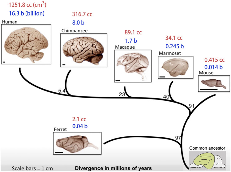
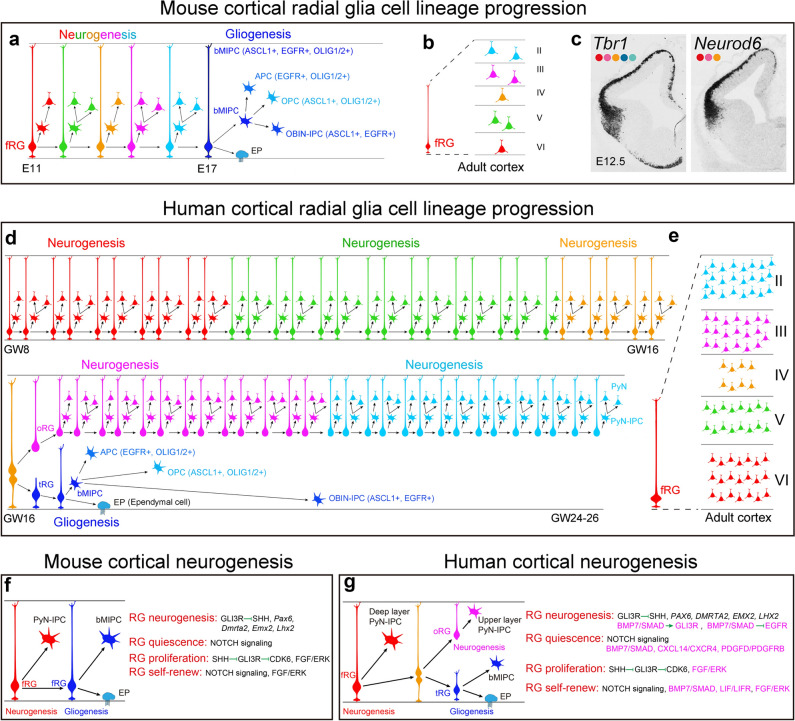
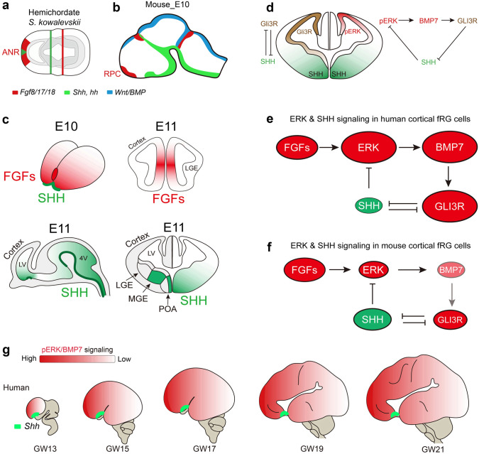
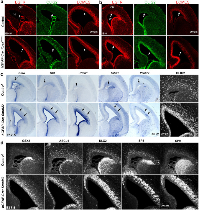
Human's robust cognitive abilities, including creativity and language, are made possible, at least in large part, by evolutionary changes made to the cerebral cortex. This paper reviews the biology and evolution of mammalian cortical radial glial cells (primary neural stem cells) and introduces the concept that a genetically step wise process, based on a core molecular pathway already in use, is the evolutionary process that has molded cortical neurogenesis. The core mechanism, which has been identified in our recent studies, is the extracellular signal-regulated kinase (ERK)-bone morphogenic protein 7 (BMP7)-GLI3 repressor form (GLI3R)-sonic hedgehog (SHH) positive feedback loop. Additionally, I propose that the molecular basis for cortical evolutionary dwarfism, exemplified by the lissencephalic mouse which originated from a larger gyrencephalic ancestor, is an increase in SHH signaling in radial glia, that antagonizes ERK-BMP7 signaling. Finally, I propose that: (1) SHH signaling is not a key regulator of primate cortical expansion and folding; (2) human cortical radial glial cells do not generate neocortical interneurons; (3) human-specific genes may not be essential for most cortical expansion. I hope this review assists colleagues in the field, guiding research to address gaps in our understanding of cortical development and evolution.

Keywords: BMP7; Cortical evolution; Cortical expansion; Cortical gliogenesis; Cortical neurogenesis; FGF-ERK signaling; Human-specific gene; Interneuron; Radial glia; SHH signaling.


Conflict of interest statement

Conflict of interest: The author claims that there are no conflicts of interest.

Figures

Fig. 1
Fig. 1
Mammalian cortical expansion and evolution. Mammalian cortical neuron numbers range from 0.014 billion in mice to 16.3 billion in humans. Brain volumes range from 0.4 cc in mice to 1250 cc in humans. I suggest that there is a fundamental conserved genetic/biochemical engine that drives the increase in the number of cortical neurons, which further drives cortical size and brain volume expansion during evolution. Note that the mouse is lissencephalic but originated from a larger and gyrencephalic ancestor, as gyrencephalic ferrets separated before mice from the human phylogenetic tree.
Fig. 2
Fig. 2
Mouse and human cortical RG cell lineage progression. a, b Mouse cortical neurogenesis and gliogenesis. Upon entry into the neurogenic phase, a single mouse cortical fRG cell may divide 5-6 times and generate PyN-IPCs, which produce 8–9 PyNs distributed in both deep and upper layers. Cortical gliogenesis occurs after neurogenesis. At the gliogenic stage, mouse fRG cells first generate bMIPCs, which then give rise to cortical APCs, OPCs, and OBIN-IPCs. c Mammalian cortical PyNs are first generated in the ventral and lateral pallium, followed by the dorsal and medial pallium (cortex). Tbr1 and Neurod6 mRNA in situ hybridization on mouse brain sections at E12.5. Images are reprinted with permission from Moreau et al., 2021 [92]. d Human cortical neurogenesis and gliogenesis. I propose that a single human cortical neurogenic fRG cell may divide ~30 times and generate PyN-IPCs, and these PyN-IPCs may produce 30-45 PyNs distributed in deep layers. Around GW16, fRG cells give rise to tRG and oRG cells. A single human oRG cell in the lateral OSVZ may divide ~30 times and generate PyN-IPCs, and these PyN-IPCs may produce 30-45 PyNs distributed in upper layers. Human tRG cells undergo a neurogenesis-to-gliogenesis switch and generate bMIPCs, which divide several times and give rise to cortical APCs, OPCs, and OBIN-IPCs. e A single human cortical fRG cell may generate 60-90 PyNs. f, g Signaling pathways and transcription factors regulate mouse and human cortical RG cell neurogenesis, quiescence, proliferation, and self-renewal. Those signals that are stronger in human cortical RG cells, than in mice, are highlighted in magenta.
Fig. 3
Fig. 3
Human cortical oRG cells and tRG cells exhibit distinct transcriptional signatures. a Schematic summarizing the process of mouse and human cortical RG neurogenesis and gliogenesis. b Immunohistochemical analysis shows that human oRG cells have a larger soma (more than 10 µm in diameter, arrowheads) and do not express EGFR. Note that HOPX+ EGFR+ cells are cortical APCs, as they have smaller soma size. c PAX6+ oRG cells produce TBR2 (EOMES)+ PyN IPCs that maintain PAX6 expression (arrows). d Very strong HOPX-expressing oRG cells generate TBR2 (EOMES)+ PyN IPCs (arrowheads) that never express HOPX, suggesting that oRG cells have longer cell cycles. e scRNA-Seq analysis of molecular profiles of human cortical RG cells at GW22, GW23, and GW26. Heat map of selected differentially expressed genes for oRG cells versus tRG cells. f Selected GO terms supporting that oRG cells are neurogenic while tRG cells are gliogenic. Images (a, e, f) are reprinted with permission from Li et al., (2024) [55]. Images (bd) are adapted from Yang et al., 2022 [60].
Fig. 4
Fig. 4
ERK signaling drives the evolutionary expansion of the mammalian cerebral cortex. a, b The ANR expressed FGFs in hemichordates (500 million years ago) and mice. c During mouse early forebrain development, FGFs are expressed in the rostral patterning center, whereas SHH is expressed in POA stem/progenitor cells and MGE-derived neurons. d, e At the beginning of cortical neurogenesis in the most recent ancestor to all mammals, it is assumed that there was already a subset of cortical fRG cells that expressed relatively higher levels of pERK. Elevated ERK signaling in these cortical fRG cells promotes BMP7 expression, which increases GLI3R generation and represses SHH signaling. A decrease in SHH signaling in cortical fRG cells further enhances ERK signaling. Therefore, the ERK-BMP7-GLI3R-SHH signaling pathway in cortical fRG cells participates in a positive feedback loop, which expands the cortical fRG cell pool, increases the length of the cortical neurogenic period, and thus can drive increases in cortical surface area and neurogenesis; this may underlie these features characteristically observed in evolutionarily more advanced mammals. f SHH signaling underlies cortical evolutionary dwarfism in mice. During mouse cortical development and evolution, smaller brains with smaller lateral ventricle volumes may result in higher SHH concentration in the cerebral spinal fluid and thus can increase SHH signaling in the cortex, which antagonizes ERK signaling. Relatively weak ERK signaling fails to induce Bmp7 expression in the dorsal cortical RG cells, resulting in a shortened period of cortical neurogenesis (for example, from more than 130 days in humans to about 7 days in mice). g ERK signaling is elevated in human cortical fRG and oRG cells during development and evolution and induces BMP7 expression that antagonizes SHH signaling. Images are reprinted with permission from Sun et al., 2024 [54].
Fig. 5
Fig. 5
Constitutive ERK signaling induces Bmp7 expression in cortical RG cells and can drive cortical expansion. af Expression of pERK (phosphorylated ERK), BMP7, HOPX, ETV5, EGFR, and OLIG2 is increased in the cortex of hGFAP-Cre; RosaMEK1DD mice at E14.5 (arrows); reprinted with permission from Sun et al., 2024 [54]. g Expression patterns of Fgfr1, Fgfr2, and Fgfr3 in E12-E13 mouse cerebral cortex; reprinted with permission from Iwata and Hevner (2009) [190]. Note the low rostral-high caudal Fgfr3 expression gradient. h The mutational spectrum of human FGFR3. Gain of function FGFR3 mutations result in dwarfism syndromes, ranging from achondroplasia and hypochondroplasia to the more severe neonatal lethal thanatophoric dysplasias TDI and TDII; reprinted with permission from Ornitz and Legeai-Mallet (2015) [204]. ik Temporal lobe enlargement and abnormal sulci in GW18, GW22, and GW40 TD fetuses; reprinted with permission from Hevner (2005) [205]. i, m Brain morphology of TD mouse at P0 and P28. Note that TD mice have an enlarged cerebral cortex; reprinted with permission from Lin et al., (2003) [211].
Fig. 6
Fig. 6
Constitutive SHH-SMO signaling promotes cortical OBIN genesis. ad Higher levels of SHH-SMO signaling promote cortical RG cell and IPC proliferation, induce EGFR and OLIG2 expression, expand the cortical VZ/SVZ, and strongly induce GSX2 and DLX2 expression, favoring the generation of OBINs. Images are reprinted with permission from Sun et al., (2024) [54], Li et al., (2024) [55], and Zhang et al., (2020) [85].
Fig. 7
Fig. 7
Constitutive SHH-SMO signaling promotes cortical abnormal folding. a Higher levels of SHH-SMO signaling reduce the number of TBR1+ PyNs in the deep cortical layers in hGFAP-Cre; RosaSmoM2 mice at P0. Note that the thickness of the SVZ is expanded and the cortex is enlarged. b Higher levels of SHH-SMO signaling induce abnormal medial cortical folding and cortical expansion, reduce the number of TBR1+ PyNs in the deep cortical layers, and increase SATB2+ PyNs in the upper cortical layers at P10; reprinted with permission from Wang et al., (2016) [218].
Fig. 8
Fig. 8
Mouse cortical interneurons (CINs) are derived from subcortical ganglionic eminences (GEs). a tSNE plot from scRNA-seq data (a total of 98,047 mouse cortical cells) was obtained using mouse E10.5 to the P4 cortex [306]. Individual cell types are labeled, adapted from Di Bella DJ et al., 2021 [306]. MGE-, POA-, and/or CGE-derived Dlx1+, and/or Dlx2+, Gad2+, Dlx5+, Sp8+, Sp9+, Nr2f2+, Lhx6+ CINs are clearly identified. Note that only a small number of IPCs for interneurons that express Egfr, Gsx2, Dlx1, Dlx2, and Gad2 are identified (arrows) adjacent to the bMIPC cluster that express Ascl1, Egfr, Olig1, and Olig2. Genetic labeling of the bMIPC experiments demonstrates that bMIPCs produce OBINs but not CINs. b It is well accepted that mouse CINs are derived from the MGE, POA, and CGE. c Mouse OBINs are derived from fRG cells in the developing MGE, LGE, cortex, and septum (SP). After birth, mouse OBINs are derived from B1 NSCs in the SVZ of the lateral ventricle. d Mouse cortical fRG cells first give rise to bMIPC, which then give rise to cortical APCs, OPCs, and OBIN-IPCs.
Fig. 9
Fig. 9
Virtually all human CINs are derived from subcortical ganglionic eminences (GEs), but not from the cortex. a tSNE plot from scRNA-seq data (a total of 57,868 human cortical cells) obtained using human GW18 (19,373 cells), GW22 (12,567 cells), GW23 (12,557 cells), and GW26 (13,371 cells) cortical tissue [88]. Individual cell types are labeled, adapted from Trevino et al., 2021 [88]. MGE-, POA-, and/or CGE-derived DLX1+, and/or DLX2+, GAD2+, DLX5+, SP8+, SP9+, NR2F2+, LHX6+ CINs are clearly identified. A small number of IPCs for interneurons express EGFR, GSX2, DLX1, DLX2, and GAD2 (arrows) are in the bMIPC cluster (ASCL1, EGFR, OLIG1, and OLIG2). This provides evidence that EGFR+, DLX1+, DLX2+, and GAD2+ IPCs are OBIN-IPCs, but not CIN-IPCs. Note that DLX5, SP8, and SP9 expression is not observed in the OBIN-IPC cluster, suggesting that very few immature OBINs are generated by GW23-26. b I propose that virtually all human CINs are derived from the MGE, POA, and CGE, similar to mice. c I propose that human OBINs are derived from fRG cells in the developing MGE, LGE, cortex, and septum (SP). After birth, human OBIN-IPCs are derived from B1 NSCs in the SVZ of the lateral ventricle. d Human cortical tRG cells first give rise to bMIPC, which then give rise to cortical APCs, OPCs, and OBIN-IPCs.

References

    1. Rakic P. A century of progress in corticoneurogenesis: From silver impregnation to genetic engineering. Cereb Cortex 2006, 16: i3–i17. - PubMed
    1. Hoch RV, Rubenstein JLR, Pleasure S. Genes and signaling events that establish regional patterning of the mammalian forebrain. Semin Cell Dev Biol 2009, 20: 378–386. - PubMed
    1. Fernandes M, Hébert JM. The ups and Downs of holoprosencephaly: Dorsal versus ventral patterning forces. Clin Genet 2008, 73: 413–423. - PubMed
    1. Hébert JM, Fishell G. The genetics of early telencephalon patterning: Some assembly required. Nat Rev Neurosci 2008, 9: 678–685. - PMC - PubMed
    1. Grove EA, Fukuchi-Shimogori T. Generating the cerebral cortical area map. Annu Rev Neurosci 2003, 26: 355–380. - PubMed

Substances

LinkOut - more resources