. 2024 Sep 1;327(3):F532-F542.
doi: 10.1152/ajprenal.00316.2023. Epub 2024 Jul 18.

Renin-angiotensin system-mediated nitric oxide signaling in podocytes

Affiliations

Renin-angiotensin system-mediated nitric oxide signaling in podocytes

Marharyta Semenikhina et al. Am J Physiol Renal Physiol. .

Abstract

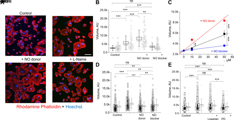
Nitric oxide (NO) is widely recognized for its role in regulating renal function and blood pressure. However, the precise mechanisms by which NO affects renal epithelial cells remain understudied. Our previous research has shown that NO signaling in glomerular podocytes can be initiated by Angiotensin II (ANG II) but not by ATP. This study aims to elucidate the crucial interplay between the renin-angiotensin system (RAS) and NO production in podocytes. To conduct our research, we used cultured human podocytes and freshly isolated rat glomeruli. A variety of RAS peptides were used, alongside confocal microscopy, to detect NO production and NO/Ca2+ cross talk. Dynamic changes in the podocyte cytoskeleton, mediated by RAS-NO intracellular signaling, were observed using fluorescent labeling for F-actin and scanning probe microscopy. The experiments demonstrated that ANG II and ANG III generated high levels of NO by activating the angiotensin II type 2 receptor (AT2R). We did not detect functional MAS receptor presence in podocytes, and the moderate NO response to ANG 1-7 was also mediated through AT2R. Furthermore, NO production impacted intracellular Ca2+ signaling and correlated with an increase in podocyte volume and growth. Scanning probe experiments revealed that AT2R activation and the corresponding NO generation are responsible for the protrusion of podocyte lamellipodia. Taken together, our data indicate that AT2R activation enhances NO production in podocytes and subsequently mediates changes in Ca2+ signaling and podocyte volume dynamics. These mechanisms may play a significant role in both physiological and pathophysiological interactions between the RAS and podocytes.NEW & NOTEWORTHY The renin-angiotensin system plays a crucial role in the production of intracellular nitric oxide within podocytes. This mechanism operates through the activation of the angiotensin II type 2 receptor, leading to dynamic modifications in intracellular calcium levels and the actin filament network. This intricate process is vital for linking the activity of angiotensin receptors to podocyte function.

Keywords: NO bioavailability; angiotensin; cytoskeleton; redox signaling.


Conflict of interest statement

Christine A. Klemens and Alexander Staruschenko are editors of the American Journal of Physiology-Renal Physiology and were not involved and did not have access to information regarding the peer-review process or final disposition of this article. An alternate editor oversaw the peer review and decision-making process for this article. None of the other authors has any conflicts of interest, financial or otherwise, to disclose.

Figures

None
Graphical abstract
Figure 1.
Figure 1.
Renin-angiotensin system (RAS) peptides mediate nitric oxide (NO) production in human podocytes. A: schematic demonstrating the enzymatic conversion of angiotensin (ANG) I to RAS peptides and predicted receptor pathways for intracellular signaling. B: representative confocal images of NO production in response to acute application of ANG II or ANG III in human podocyte culture cells loaded with DAF-FM fluorescent dye. Scale bar = 50 µm for both experiments. C: representative NO generation comparison in response to different RAS peptides (ANG II, ANG III, ANG 1–7, and ANG 1–9 at a concentration of 100 µM).
Figure 2.
Figure 2.
Nitric oxide (NO) production in response to renin-angiotensin system (RAS) peptides in human podocytes. A: changes in DAF-FM fluorescence maximum amplitude from baseline after acute application of the corresponding RAS peptide. Note the absence of difference for angiotensin (ANG) IV peptide. n ≥20 cells for each group. ANOVA, **P < 0.01 and ***P < 0.001. B and C: statistical summary representing changes in NO maximum amplitude (B) and total NO production (AUC) (C) for the 7 min after stimulation of the RAS pathway. n ≥ 20 cells for each group. One-way ANOVA with Dunnett’s post hoc test, **P < 0.01 and ***P < 0.001 vs. ANG II. AUC, area under the curve.
Figure 3.
Figure 3.
Classical renin-angiotensin system (RAS) hormones [angiotensin (ANG) II and ANG III] mediate nitric oxide (NO) production through angiotensin II type 2 receptor (AT2R) in human podocytes. A: the dose-response curve for ANG II, the most potent RAS peptide for NO production in human podocytes (EC50 = 13 μM, ligand-receptor binding fit modeled with Levenberg–Marquardt algorithm, adj. R2 = 0.93). B: representative NO responses to the acute application of ANG II in the absence (black) or presence (red) of the selective non-peptide antagonist PD123319 (PD, 10 µM). C: statistical summary of ANG II (left) and ANG III (right) mediated NO responses in the absence or presence of vehicle or AT1R or AT2R inhibitors (losartan or PD, respectively). Note that the presence of losartan resulted in nonsignificant changes (ns) in the ANG II or ANG III response. n ≥ 20 cells for each group. One-way ANOVA with Tuckey post hoc test, **P < 0.01, ***P < 0.001, and ****P < 0.0001.
Figure 4.
Figure 4.
Angiotensin (ANG) 1–7 mediates nitric oxide (NO) production through the angiotensin II type 2 receptor (AT2R), but not the Mas receptor, in human podocytes. A: representative confocal images showing NO generation in response to the acute application of ANG 1–7 in podocyte culture cells loaded with DAF-FM fluorescent dye. Scale bar = 50 µm. B: summary graphs for the ANG 1–7 mediated NO response in the presence of AT2R or Mas receptor antagonists [PD (10 µM) or A-779 (10 µM), respectively]. n ≥ 13 cells for each group. One-way ANOVA with post hoc Tukey, ***P < 0.001. Note that the presence of the Mas receptor antagonist resulted in nonsignificant changes (ns) in the ANG 1–7 response.
Figure 5.
Figure 5.
Renin-angiotensin system (RAS) peptides activate nitric oxide (NO) signaling in podocytes of freshly isolated glomeruli from Wistar rats. A: representative NO responses to angiotensin (ANG) II and ANG III (50 µM) in podocytes of rat glomeruli. B: representative confocal images of NO production in response to the acute application of ANG II and ANG III in podocytes loaded with DAF-FM dye. Scale bar = 50 µm. C: ANG II-mediated NO production in glomerular podocytes was blocked by pretreatment (1 h) with the selective nonpeptide antagonist PD123319 (PD, 10 µM). n ≥ 11 cells for each group. ANOVA, **P < 0.01.
Figure 6.
Figure 6.
The relationship between intracellular Ca2+ and nitric oxide (NO) production mediated by angiotensin (ANG) III. A: representative confocal imaging experiments for the simultaneous detection of Ca2+ (red) and NO (black) changes (using CalBryte 590 and DAF-FM, respectively) in response to acute ANG III application (100 µM) in the presence of vehicle or selective nonpeptide antagonist of angiotensin II type 2 receptor (AT2R) [PD123319 (PD, 10 µM)]. B: statistical summary for the experiments shown in A. n ≥ 24 cells for each group. One-way ANOVA, **P < 0.01.
Figure 7.
Figure 7.
Renin-angiotensin system (RAS)-mediated nitric oxide (NO) production regulates podocyte volume. A: representative rhodamine-phalloidin staining of the actin cytoskeleton demonstrated that angiotensin (ANG) II-mediated (50 µM) NO production promoted an increase in volume in cultured human podocytes. B: statistical summary of the experiment shown in A, indicating that preincubation with the NO donor DETA-NONOate (50 µM) resulted in the highest change in podocyte volume. This effect could be eliminated by the presence of the nonselective inhibitor of NO synthases l-NAME (10 µM). n ≥ 39 cells for each group. KS ANOVA with post hoc Tukey, **P < 0.01 and ***P < 0.001. C: dose-dependent production of NO correlated with the volume increase in podocytes. n ≥ 3 experiments for each group. One-way ANOVA with post hoc Tukey, ***P < 0.001. D: statistical summary indicating the changes in podocyte volume during the preincubation with the NO donor or l-NAME in the absence of ANG II stimulation. n ≥ 250 cells for each group. One-way ANOVA with post hoc Tukey, **P < 0.01 and ***P < 0.001. E: statistical summary of ANG II-mediated podocyte volume changes in the presence of AT1R and AT2R antagonists [losartan (30 µM) and PD123319 (PD; 10 µM)] showing that AT2R signaling is responsible for the increase in podocyte volume. n ≥ 250 cells for each group. One-way ANOVA with Tukey post hoc test, ***P < 0.001.
Figure 8.
Figure 8.
Activation of angiotensin II type 2 receptor (AT2R) promotes lamellipodium growth in cultured human podocytes. A: scanning ion conductance microscopy revealed a 3-D topographical image of podocyte lamellipodia before and after angiotensin (ANG) II application (0 and 30 min, respectively). B: the dynamics of podocyte lamellipodium growth significantly increased in the presence of ANG II (50 µM). Preincubation with AT1R or AT2R antagonists [losartan (30 µM) and PD123319 (PD; 10 µM), respectively] did not affect lamellipodium growth (top). ANG II application significantly increased lamellipodium growth, and this effect could be blocked by AT2R but not AT1R inhibition (bottom). C: statistical summary for the experiment shown in B, indicating that the dynamics of lamellipodium growth are mediated through the AT2R pathway. n ≥ 3 for each group. One-way ANOVA with Tukey post hoc test, *P < 0.05.

References

    1. Pérez-Torres I, Manzano-Pech L, Rubio-Ruíz ME, Soto ME, Guarner-Lans V. Nitrosative stress and its association with cardiometabolic disorders. Molecules 25: 2555, 2020. doi: 10.3390/molecules25112555. - DOI - PMC - PubMed
    1. Palygin O, Ilatovskaya DV, Levchenko V, Endres BT, Geurts AM, Staruschenko A. Nitric oxide production by glomerular podocytes. Nitric Oxide 72: 24–31, 2018. doi: 10.1016/j.niox.2017.11.005. - DOI - PMC - PubMed
    1. Thomas SR, Chen K, Keaney JF Jr. Hydrogen peroxide activates endothelial nitric-oxide synthase through coordinated phosphorylation and dephosphorylation via a phosphoinositide 3-kinase-dependent signaling pathway. J Biol Chem 277: 6017–6024, 2002. doi: 10.1074/jbc.M109107200. - DOI - PubMed
    1. Searles CD, Ide L, Davis ME, Cai H, Weber M. Actin cytoskeleton organization and posttranscriptional regulation of endothelial nitric oxide synthase during cell growth. Circ Res 95: 488–495, 2004. doi: 10.1161/01.RES.0000138953.21377.80. - DOI - PubMed
    1. Hsu HH, Hoffmann S, Endlich N, Velic A, Schwab A, Weide T, Schlatter E, Pavenstädt H. Mechanisms of angiotensin II signaling on cytoskeleton of podocytes. J Mol Med (Berl) 86: 1379–1394, 2008. doi: 10.1007/s00109-008-0399-y. - DOI - PubMed

LinkOut - more resources