. 2024 Jul 19;385(6706):282-288.
doi: 10.1126/science.adn3796. Epub 2024 Jul 18.

Sculpting conducting nanopore size and shape through de novo protein design

Affiliations

Sculpting conducting nanopore size and shape through de novo protein design

Samuel Berhanu et al. Science. .

Abstract

Transmembrane β-barrels have considerable potential for a broad range of sensing applications. Current engineering approaches for nanopore sensors are limited to naturally occurring channels, which provide suboptimal starting points. By contrast, de novo protein design can in principle create an unlimited number of new nanopores with any desired properties. Here we describe a general approach to designing transmembrane β-barrel pores with different diameters and pore geometries. Nuclear magnetic resonance and crystallographic characterization show that the designs are stably folded with structures resembling those of the design models. The designs have distinct conductances that correlate with their pore diameter, ranging from 110 picosiemens (~0.5 nanometer pore diameter) to 430 picosiemens (~1.1 nanometer pore diameter). Our approach opens the door to the custom design of transmembrane nanopores for sensing and sequencing applications.


Conflict of interest statement

Competing interests: A.A.V. and C.B. are inventors on an E.U. provisional patent application submitted by the Flanders Institute of Biotechnology that covers the sequences of the square-shaped TMB12 designs.

Figures

Fig. 1.
Fig. 1.. Sculpting β-barrel geometry.
(A) Barrel diameter can be controlled through the number of β-strands in the β-barrel blueprint. (B) β-barrel 2D interaction map. Strong bends in the β-strands (< 90° bend, right) are achieved by stacking several glycine kink residues (yellow spheres) along the β-barrel axis, as opposed to placing one kink (>90° bend, left). (C and D) Cross sections of explicitly assembled β-barrel backbones without [cylinder in (C)] and with (D) glycine kinks. The Cβ atoms of the residues facing the pore are shown as spheres and colored according to their respective repulsion energy. Glycine kink positions are shown with arrows; placement at the corners of the embedded rectangular, oval, and triangular shapes [dashed lines in (D)] generates the desired backbone geometries. (E) Polar threonine residues are tolerated on the membrane-exposed surface of TMBs (right) as they can form a hydrogen bond to the backbone, mimicking the interactions with water molecules observed in similarly curved areas of water-exposed β-strands (left).
Fig. 2.
Fig. 2.. Biophysical characterization of designed nanopores.
Top row: 10-stranded design (TMB10_163). Bottom row: 12-stranded design with a square cross section (TMB12_3). Both designs elute as one major species with retention time consistent with a monomeric protein in complex with DPC detergent (A) and show distinct negative maxima in far UV CD spectra at 215 nm (B) that remain stable up to >70°C (C), and cooperative and reversible folding/unfolding transitions in DUPC LUVs [where <λ> is the average tryptophan fluorescence emission wavelength in nanometers (see methods)] (D).
Fig. 3.
Fig. 3.. Experimentally determined nanopore structures closely align with the computational design models.
(A to C) Crystal structure of TMB10_163. (A) Backbone superposition. The seven surface residues mutated in TMB10_165 are shown as sticks with the substitution label. (B) Superposition of side chains involved in key folding motifs in the lumen, including 2Fo to Fc, omit electron density contoured at 1.0 s. A water molecule crystallized in the pore is shown as a red sphere. (C) Cross section superposition with residues shown as spheres to highlight the water-accessible pore. (D and E) TMB2_13 structure in LDAO micelles. (D) Long-range NMR NOE contacts mapped to the expected TMB12_3 hydrogen bonds (dashed black lines). Residues with amide assignment are shown in white and green, unassigned residues are shown in ash gray. Residues with β-sheet secondary structure are shown as squares, all others as circles. Bold outlines indicate available methyl assignments. NOE contacts are shown as red lines (long-range amide-amide, dashes indicate diagonal overlap) and blue lines (contacts involving side chain methyl groups). (E) Ensemble of the 20 lowest-energy solution NMR structures (β-sheets shown in brown).
Fig. 4.
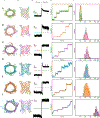
Fig. 4.. Conductance of designed nanopores.
Designs: (A) TMB10_165, (B) TMB12_3, (C) TMB12_oval_4, (D) TMB12_rect_8, (E) TMB12_tri_12, (F) TMB14_8. (i) Top view representation. (ii) Vertical cross sections of the pore. (iii) single channel conductance (smallest observed conductance jump). (iv) sequential insertions of designed pore in planar lipid bilayer membrane from detergent solubilized sample at low concentrations. (v) histogram of smallest measured current jumps for each design, up to 50 pA. The applied voltage across the bilayer was 100 mV and experiments were performed in a buffer containing 500 mM NaCl. A Gaussian fit was carried out for the single channel current histograms for each design. For TMB10_165, 38 independent single channel jumps were identified from three recordings to plot the histogram shown. Similarly, 44 single channel insertions were identified for TMB12_3 (four recordings), 29 insertions for TMB12_oval_4 (three recordings), 30 insertions for TMB12_rect_8 (three recordings), 45 insertions for TMB12_tri_12 (five recordings), and 32 insertions for TMB14_8 (three recordings) to plot the above depicted histograms.

Update of

References

    1. Dal Peraro M, van der Goot FG, Nat. Rev. Microbiol 14, 77–92 (2016). - PubMed
    1. Ranava D, Caumont-Sarcos A, Albenne C, Ieva R, FEMS Microbiol. Lett 365, fny087 (2018). - PubMed
    1. Diederichs KA, Buchanan SK, Botos I, J. Mol. Biol 433, 166894 (2021). - PMC - PubMed
    1. Day PM, Inoue K, Theg SM, Plant Cell 31, 1845–1855 (2019). - PMC - PubMed
    1. Harrington L, Alexander LT, Knapp S, Bayley H, ACS Nano 13, 633–641 (2019). - PubMed