. 2024 Jul 18;15(1):6059.
doi: 10.1038/s41467-024-50178-3.

WRN inhibition leads to its chromatin-associated degradation via the PIAS4-RNF4-p97/VCP axis

Affiliations

WRN inhibition leads to its chromatin-associated degradation via the PIAS4-RNF4-p97/VCP axis

Fernando Rodríguez Pérez et al. Nat Commun. .

Abstract

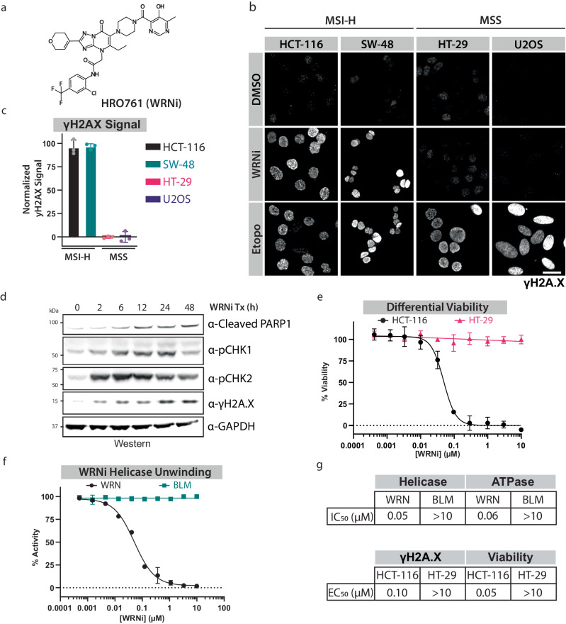
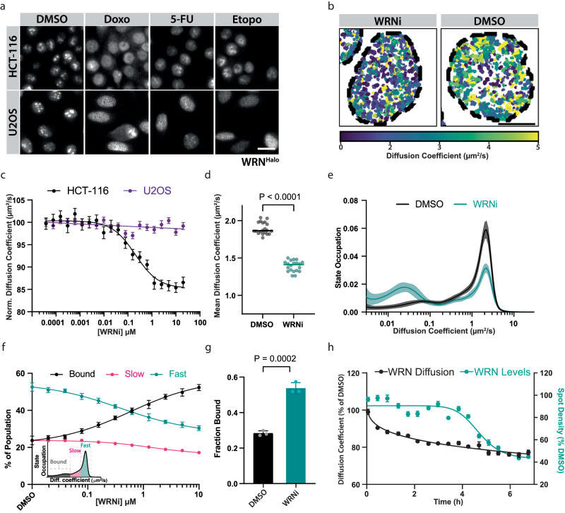
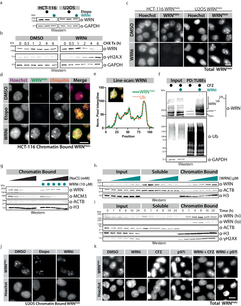
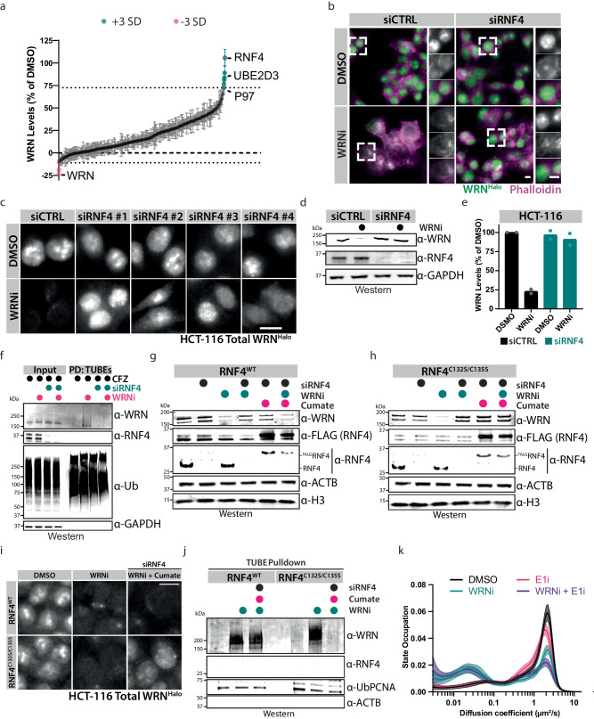
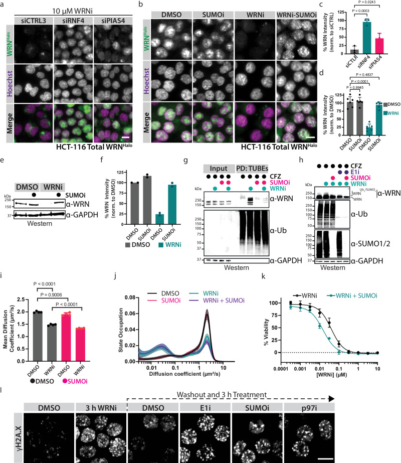
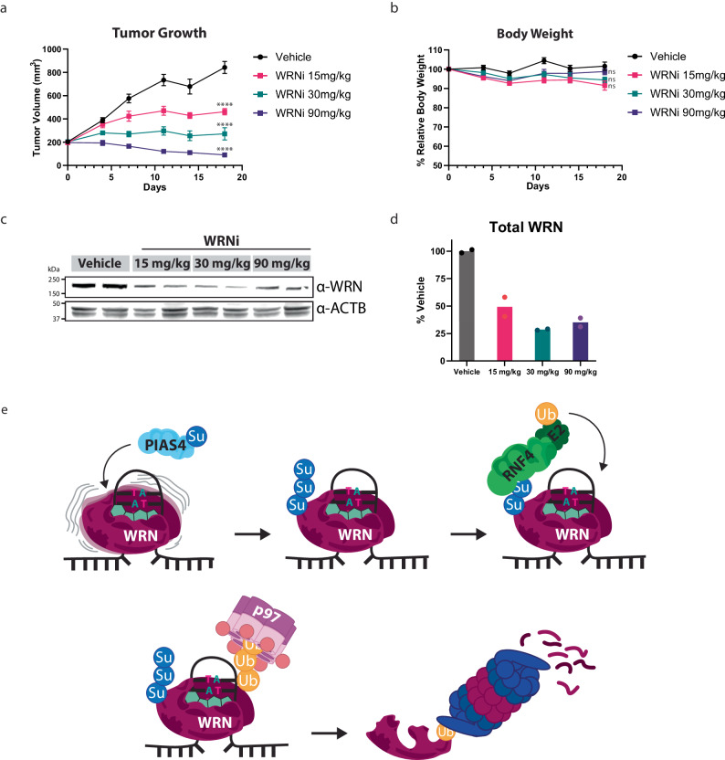
Synthetic lethality provides an attractive strategy for developing targeted cancer therapies. For example, cancer cells with high levels of microsatellite instability (MSI-H) are dependent on the Werner (WRN) helicase for survival. However, the mechanisms that regulate WRN spatiotemporal dynamics remain poorly understood. Here, we used single-molecule tracking (SMT) in combination with a WRN inhibitor to examine WRN dynamics within the nuclei of living cancer cells. WRN inhibition traps the helicase on chromatin, requiring p97/VCP for extraction and proteasomal degradation in a MSI-H dependent manner. Using a phenotypic screen, we identify the PIAS4-RNF4 axis as the pathway responsible for WRN degradation. Finally, we show that co-inhibition of WRN and SUMOylation has an additive toxic effect in MSI-H cells and confirm the in vivo activity of WRN inhibition using an MSI-H mouse xenograft model. This work elucidates a regulatory mechanism for WRN that may facilitate identification of new therapeutic modalities, and highlights the use of SMT as a tool for drug discovery and mechanism-of-action studies.


Conflict of interest statement

The authors are employees and/or shareholders of Eikon Therapeutics, Inc. Furthermore, a patent application related to the subject matter described in the manuscript has been filed by Eikon Therapeutics, Inc. F.R.P. and S.B. are listed as co-inventors on US provisional patent application 63/599,976. The authors declare no other competing interests.

Figures

Fig. 1
Fig. 1. HRO761 is a specific and potent WRN inhibitor.
a Chemical structure of HRO761 (WRNi). b WRN inhibition leads to induction of the DNA damage response in MSI-H cells. Phospho-histone H2A.X (Ser139) (γH2A.X) staining was used to visualize DNA damage in MSI-H (HCT-116, SW-48) and MSS (HT-29, U2OS) cells after 24 h treatment with indicated compounds. Scale bar = 20 μm. c Quantification of γH2A.X signal levels in cells in (b), normalized to DMSO and Etoposide. Graphs represent averages of n = 3 plate replicates. d WRN inhibition by WRNi leads to an induction of the DNA damage response resulting in apoptosis. HCT-116 cells were treated with 10 μM WRNi and analyzed by Western Blot (WB) to assess DNA damage response markers. GAPDH was used as a loading control. e Dose-response curves are measuring the viability of HCT-116 cells or HT-29 cells after treatment with WRNi for 4 days. Graphs represent averages from n = 6 plates. f Dose-response curves measuring in vitro WRN or BLM unwinding activity after treatment with WRNi. Graphs represent averages from n = 4 replicates. WRN and BLM unwinding activity is normalized to DMSO. g Summary of IC50 or EC50 values of WRNi in the indicated biochemical and cellular assays. All curve fits were done by fitting a 4-parameter logarithmic regression curve. All error bars represent standard deviation (s.d.). DMSO is dimethyl sulfoxide; BLMi is BLM inhibitor Compound 2; Etopo is etoposide. MW is molecular weight. Source data are provided as a Source Data file.
Fig. 2
Fig. 2. Single-molecule tracking shows a change in WRN cellular dynamics in an MSI-H-dependent manner.
a Validation of WRNHalo cell lines, HCT-116 and U2OS. Subcellular localization of WRN in the presence or absence of DNA-damaging compounds. WRNHalo successfully translocates across compartments, suggesting a functional protein. Scale bar = 20 μm. b Representative SMT tracks overlaid with Hoechst nuclear stain outlines, in the presence or absence of 10 μM WRNi. Each track is colored according to the diffusion coefficient of the molecule it represents. c Inhibition of WRN only affects its mean diffusion coefficient in MSI-H cells. Dose-response curves with WRNi measuring the diffusion coefficient of WRNHalo after 4 h treatments in HCT-116WRN-Halo or U2OSWRN-Halo. The graph represents the average from n = 4 plates per condition. Error bars represent the standard error of the mean (s.e.m.). d Dot plot quantification of WRN diffusion coefficient after treatment with 10 μM WRNi. Each point represents the average WRN diffusion coefficient across all the nuclei in each FOV. n = 20 plates. Lines represent sample medians. P values were calculated using a two-tailed, unpaired t-test e WRNi shifts a large fraction of molecules from the free-diffusing state (“fast”) to the chromatin-bound (“bound”) state. Distribution of diffusive states in HCT-116WRN-Halo cells showing the relative proportion of WRN molecules as a function of diffusion coefficient occupation, in the presence and absence of 10 μM WRNi. Plot line represents sample means, shaded area represents s.d. f Dose-response curves with WRNi measuring the different diffusive states of WRN protein. Inset is a representation of how diffusive states are classified. Error bars represent s.d., n = 8 plates. g Quantification from (f) of the chromatin-bound fraction of WRNHalo protein in the presence or absence of 10 μM WRNi. Error bars represent s.d., n = 3 plates. h Overlay of WRNHalo diffusion coefficient and molecule spot densities after treatment with 10 μM WRNi over the indicated timepoints. Values are normalized to DMSO. n = 2 plates. Error bars represent s.e.m. All P values were calculated using a two-tailed, unpaired Student’s t-test. DMSO is dimethyl sulfoxide; WRNi is HRO761; 5-FU is 5-fluorouracil, Doxo is doxorubicin. Source data are provided as a Source Data file.
Fig. 3
Fig. 3. WRN Inhibition leads to its chromatin-associated degradation.
a WB analysis of HCT-116 or U2OS cells treated with 10 μM WRNi or etoposide for 16 h. b Treatment of HCT-116 cells with 100 μg/mL of cycloheximide (CHX) in the presence or absence of 10 μM WRNi, followed by WB analysis. Quantifications of CHX chase are in Supplementary Fig. 5b. c Loss of WRN protein after treatment with WRNi can be visualized by microscopy. HCT-116WRN-Halo or U2OSWRN-Halo were treated with 10 μM WRNi for 16 h and imaged. Quantification is in Supplementary Fig. 5c. Scale bar = 10 μm. d WRN inhibition leads to WRN trapping on chromatin and its ubiquitylation. HCT-116WRN-Halo cells were treated with 10 μM WRNi or etoposide for 6 h, followed by imaging. Trapped WRN quantification is in Supplementary Fig. 5b. Scale bar = 10 μm. e Line-scan quantification of WRNi treated cells from (d). Line-scan quantifications of etoposide treatments are in Supplementary Fig. 5f. f WRN inhibition leads to its ubiquitylation. Tandem ubiquitin-binding entities (TUBEs) pulldown (PD) of HCT-116 cells after treatment with 10 μM WRNi for 6 h in the presence of 1 μM carfilzomib (CFZ), and subsequent blotting with indicated antibodies. g WB analysis of chromatin fractionations of HCT-116 cells in the presence or absence of WRNi treated for 2 h, performed under various NaCl concentrations (25, 75, 150, 300, or 500 mM NaCl). Inputs and soluble fractions in Supplementary Fig. 5i. h Subcellular fractionations of HCT-116 cells as in (g), performed under 300 mM CSK buffer with increasing concentrations of WRNi (0, 0.03, 0.1, 0.3, 1.0, 3.0 μM). i WB analysis of HCT-116 cells subjected to a 10 μM WRNi time course, followed by subcellular fractionation as in (h). “hi” indicates a high WB exposure, and “lo” a low exposure. j WRN chromatin trapping upon its inhibition is MSI-H dependent. Cells were treated with WRNi as in (d) but using the MSS cell line U2OSWRN-Halo. Trapped WRN Quantification is in Supplementary Fig. 5e. k Degradation of WRN is dependent on the p97/VCP-proteasome axis. HCT-116WRN-Halo were treated with 10 μM of WRNi and 1 μM of either CB-5083 (p97i) or CFZ for 6 h, then imaged. Quantifications are in Supplementary Fig. 5c. DMSO is dimethyl sulfoxide; WRNi is HRO761; CHX is cycloheximide; CFZ is carfilzomib; Etopo is etoposide. All P values were calculated using a two-tailed, unpaired Student’s t-test. ns not significant. MW is molecular weight. Source data are provided as a Source Data file. For WBs in a, b, f GAPDH was used as a loading control; for (g–i), ACTB and H3 were used as processing controls.
Fig. 4
Fig. 4. Phenotypic siRNA screen identified RNF4 as the ubiquitin E3 ligase targeting WRN for degradation.
a A phenotypic screen identified genes involved in WRN degradation. Colored circles indicate hits that are 3 standard deviations (SD) from the mean; error bars represent s.d., n = 2 biological replicates. Quantification of WRN protein levels after treatment with siRNAs in the presence or absence of 10 μM WRNi for 24 h. b Representative images of HCT-116WRN-Halo cells from the siRNA screen performed in (a). c siRNA SMARTpool decomplexification and validation. Representative images of HCT-116WRN-Halo treated with 10 uM WRNi. Quantifications are in Supplementary Fig. 7c. d WB analysis of HCT-116 cells treated with siRNF4 oligos for 24 h, then treated with or without 10 μM WRNi for an additional 24 h. e Quantifications of (d). Graphs represent the mean value of n = 2 biological replicates. f WB analysis of TUBEs pulldowns (PD) after depletion of RNF4. HCT-116 cells were treated with siRNF4 oligos for 24 h, then treated with or without 10 μM WRNi for 6 h. All samples were treated with CFZ. g WB analysis of HCT-116WRN-Halo;FLAG-RNF4 cells treated with 10 μM WRNi after depletion of RNF4, following induction of stably integrated RNF4. h WB analysis as in (g) but using an RNF4C132S/C135S catalytic mutant. i Samples were treated as in (g) with the addition of JF549, and imaged. Images are representative of n = 3 biological replicates j WB analysis of HCT-116WRN-Halo under the indicated drug treatments and induction of indicated RNF4 constructs. All samples are treated with CFZ. Inputs and total ubiquitin loading controls are in Supplementary Fig. 7g. Ub-PCNA was used as a processing control. k Distribution of diffusive states in HCT-116WRN-Halo cells showing the relative proportion of WRN molecules as a function of diffusion coefficient occupation after treatment with 1 μM E1i in the presence or absence of 10 μM WRNi. Plot line represents sample means, shaded area represents s.d. DMSO is dimethyl sulfoxide; WRNi is HRO761; E1i is TAK-243; CFZ is carfilzomib. MW is molecular weight. All scale bars = 10 μm. WB data shown in this figure are representative images of biological duplicates (n = 2). Source data are provided as a Source Data file. For WBs in d, f GAPDH was used as a loading control; for g, h, ACTB and H3 were used as loading controls. In f, UB was used as a processing control.
Fig. 5
Fig. 5. The PIAS4-RNF4 axis is responsible for the chromatin-associated degradation of WRN.
a HCT-116WRN-Halo cells were treated with the indicated siRNA oligos for 24 h, and subsequently treated with 10 μM of WRNi for 24 h before imaging. b Treatment of HCT-116WRN-Halo with 1 μM of SUMOi in the presence or absence of 10 μM WRNi for 6 h before imaging. c, d Quantifications of a, b, respectively. Graphs represent the average of n = 3 plates for siRNA experiments and n = 6 plates for the small molecule experiments. Each dot represents the average of six wells. For both c and d, error bars represent s.d. e. WB analysis of HCT-116 cells treated with 1 μM SUMOi in the presence or absence of 10 μM WRNi for 16 h, at which point cells were lysed and analyzed. f Quantification of (e). Graphs represent the mean value of n = 2 WB runs. g WB analysis of TUBE pulldowns of HCT-116WRN-Halo cells after treatment with 1 μM SUMOi in the presence or absence of 10 μM WRNi (6 h). All samples were treated with CFZ. h WB analysis of HCT-116 cells co-treated with WRNi in the presence or absence of E1i and/or SUMOi. i Dot plots of WRN diffusion coefficient via SMT after co-treatment with SUMOi and either DMSO or WRNi. Each point represents the average diffusion coefficient across all the nuclei in an FOV. n = 4 plates. Lines represent sample medians. j Distribution of diffusive states for WRNHalo in HCT-116WRN-Halo after treatment with 1 μM of SUMOi in the presence or absence of 10 μM WRNi. The plot line represents sample means, shaded area represents s.d. k Dose-response of WRNi in HCT-116 cells treated with or without 10 nM SUMOi. Error bars represent s.d., n = 3 plates. l Failure to extract trapped WRN leads to persistent DNA damage. HCT-116 cells were treated with WRNi for 3 h, washed, and replenished with fresh growth medium containing 1 μM of the indicated inhibitors, then imaged. Scale bar = 10 μm. DMSO is dimethyl sulfoxide; WRNi is HRO761; E1i is TAK-243; SUMOi is ML-792; CFZ is carfilzomib. All P values were calculated using a two-tailed, unpaired Student’s t-test. MW is molecular weight. Source data are provided as a Source Data file. For all WBs, GAPDH was used as a loading control. In g, UB was used as a processing control; in h, SUMO1/2 and UB were used as processing controls.
Fig. 6
Fig. 6. WRN Inhibition has potent antitumor effects in an MSI-H in vivo mouse model.
a HCT-116 xenograft tumor model in nude mice shows that WRN inhibition has robust antitumor effects. Mice were dosed daily with the indicated doses for 18 days total. Error bars represent s.e.m., treatment cohorts of n = 8 mice. Significance was assessed via a two-way ANOVA with Dunnett’s multiple comparison test; dosing groups were compared against the vehicle control at the terminal day 18 timepoint. b In vivo WRN inhibition is well tolerated. Body weights of mice across different treatment groups were taken throughout the study duration, showing no signs of malnourishment. Error bars represent s.e.m., treatment cohorts of n = 8 mice Significance was assessed via a two-way ANOVA with Dunnett’s multiple comparison test; dosing groups were compared against the vehicle control at the terminal day 18 timepoint. c Pharmacodynamic analysis by WB of HCT-116 mice xenografts tumors after treatment with WRNi, showing the degradation of WRN. ACTB was used as a loading control. d Quantifications of (c). Lines indicate the sample median. e Proposed model for targeted proteasomal degradation of trapped WRN upon treatment with WRNi. Chromatin-bound WRN that is actively surveying DNA damage in MSI-H cells becomes trapped upon inhibition by WRNi. This stalled WRN is SUMOylated by the SUMO ligase PIAS4. SUMOylated WRN recruits the STUbL RNF4, leading to its ubiquitylation. Ubiquitylated WRN is extracted from chromatin by p97/VCP, leading to its degradation by the proteasome. Significance was defined as follows: ns P > 0.01; ****P < 0.0001. Source data are provided as a Source Data file.

References

    1. Opresko PL, Cheng WH, von Kobbe C, Harrigan JA, Bohr VA. Werner syndrome and the function of the Werner protein; what they can teach us about the molecular aging process. Carcinogenesis. 2003;24:791–802. doi: 10.1093/carcin/bgg034. - DOI - PubMed
    1. Croteau DL, Popuri V, Opresko PL, Bohr VA. Human RecQ helicases in DNA repair, recombination, and replication. Annu. Rev. Biochem. 2014;83:519–552. doi: 10.1146/annurev-biochem-060713-035428. - DOI - PMC - PubMed
    1. Constantinou A, et al. Werner’s syndrome protein (WRN) migrates Holliday junctions and co-localizes with RPA upon replication arrest. EMBO Rep. 2000;1:80–84. doi: 10.1093/embo-reports/kvd004. - DOI - PMC - PubMed
    1. Bendtsen KM, et al. Dynamics of the DNA repair proteins WRN and BLM in the nucleoplasm and nucleoli. Eur. Biophys. J. 2014;43:509–516. doi: 10.1007/s00249-014-0981-x. - DOI - PMC - PubMed
    1. von Kobbe C, Bohr VA. A nucleolar targeting sequence in the Werner syndrome protein resides within residues 949-1092. J. Cell Sci. 2002;115:3901–3907. doi: 10.1242/jcs.00076. - DOI - PubMed

MeSH terms

LinkOut - more resources