. 2024 Jul 18;148(1):8.
doi: 10.1007/s00401-024-02756-4.

Amyloid-β peptide signature associated with cerebral amyloid angiopathy in familial Alzheimer's disease with APPdup and Down syndrome

Affiliations

Amyloid-β peptide signature associated with cerebral amyloid angiopathy in familial Alzheimer's disease with APPdup and Down syndrome

Amal Kasri et al. Acta Neuropathol. .

Abstract

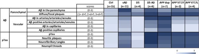
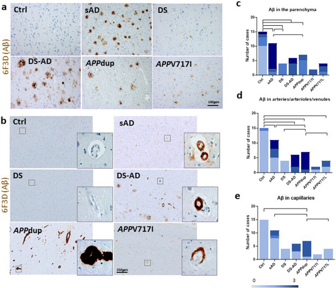
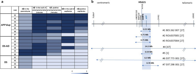
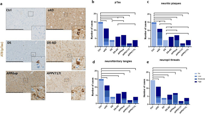
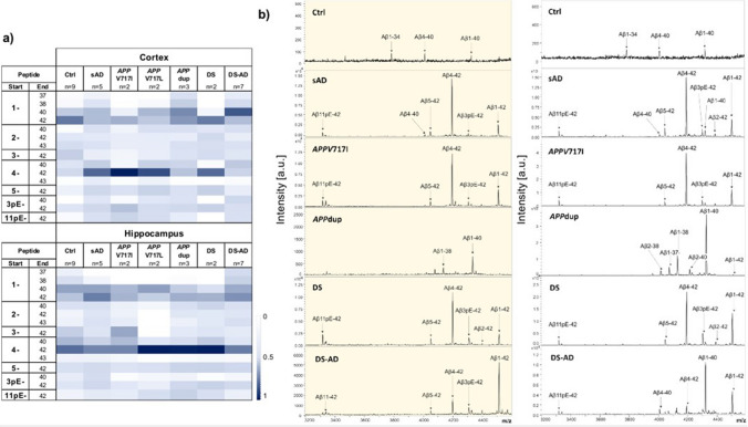
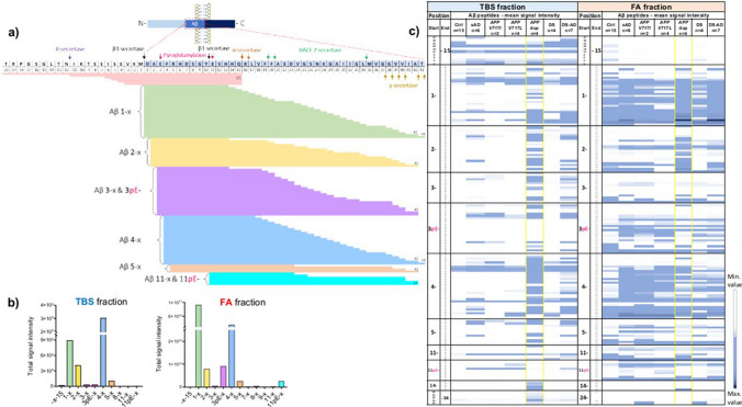
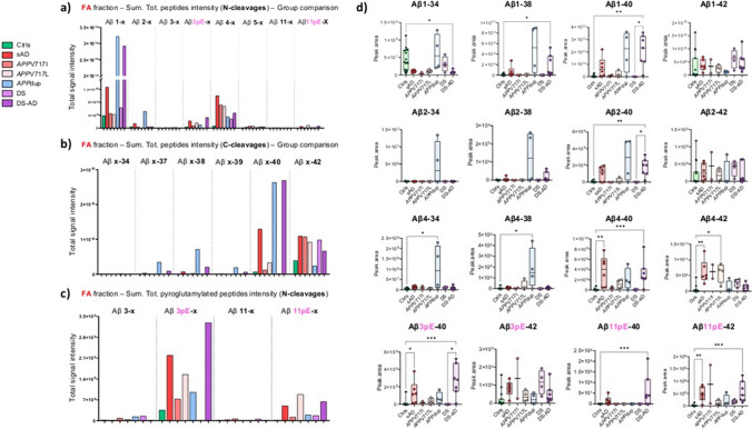
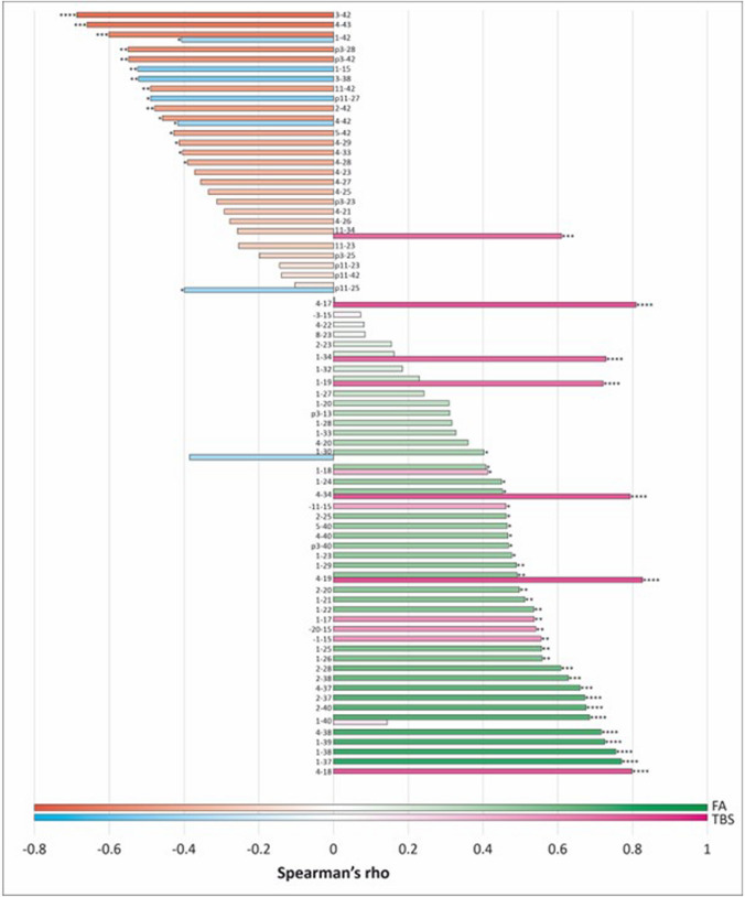
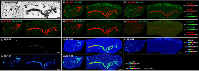
Alzheimer's disease (AD) is characterized by extracellular amyloid plaques containing amyloid-β (Aβ) peptides, intraneuronal neurofibrillary tangles, extracellular neuropil threads, and dystrophic neurites surrounding plaques composed of hyperphosphorylated tau protein (pTau). Aβ can also deposit in blood vessel walls leading to cerebral amyloid angiopathy (CAA). While amyloid plaques in AD brains are constant, CAA varies among cases. The study focuses on differences observed between rare and poorly studied patient groups with APP duplications (APPdup) and Down syndrome (DS) reported to have higher frequencies of elevated CAA levels in comparison to sporadic AD (sAD), most of APP mutations, and controls. We compared Aβ and tau pathologies in postmortem brain tissues across cases and Aβ peptides using mass spectrometry (MS). We further characterized the spatial distribution of Aβ peptides with MS-brain imaging. While intraparenchymal Aβ deposits were numerous in sAD, DS with AD (DS-AD) and AD with APP mutations, these were less abundant in APPdup. On the contrary, Aβ deposits in the blood vessels were abundant in APPdup and DS-AD while only APPdup cases displayed high Aβ deposits in capillaries. Investigation of Aβ peptide profiles showed a specific increase in Aβx-37, Aβx-38 and Aβx-40 but not Aβx-42 in APPdup cases and to a lower extent in DS-AD cases. Interestingly, N-truncated Aβ2-x peptides were particularly increased in APPdup compared to all other groups. This result was confirmed by MS-imaging of leptomeningeal and parenchymal vessels from an APPdup case, suggesting that CAA is associated with accumulation of shorter Aβ peptides truncated both at N- and C-termini in blood vessels. Altogether, this study identified striking differences in the localization and composition of Aβ deposits between AD cases, particularly APPdup and DS-AD, both carrying three genomic copies of the APP gene. Detection of specific Aβ peptides in CSF or plasma of these patients could improve the diagnosis of CAA and their inclusion in anti-amyloid immunotherapy treatments.

Keywords: Alzheimer’s disease; Aβ peptides; Cerebral amyloid angiopathy; Down syndrome; Mass spectrometry; Neuropathology.


Conflict of interest statement

KB has served as a consultant and at advisory boards for Acumen, ALZPath, AriBio, BioArctic, Biogen, Eisai, Lilly, Moleac Pte. Ltd, Novartis, Ono Pharma, Prothena, Roche Diagnostics, and Siemens Healthineers; has served at data monitoring committees for Julius Clinical and Novartis; has given lectures, produced educational materials and participated in educational programs for AC Immune, Biogen, Celdara Medical, Eisai and Roche Diagnostics; and is a co-founder of Brain Biomarker Solutions in Gothenburg AB (BBS), which is a part of the GU Ventures Incubator Program, outside the work presented in this paper. HZ has served at scientific advisory boards and/or as a consultant for Abbvie, Acumen, Alector, Alzinova, ALZPath, Amylyx, Annexon, Apellis, Artery Therapeutics, AZTherapies, Cognito Therapeutics, CogRx, Denali, Eisai, Merry Life, Nervgen, Novo Nordisk, Optoceutics, Passage Bio, Pinteon Therapeutics, Prothena, Red Abbey Labs, reMYND, Roche, Samumed, Siemens Healthineers, Triplet Therapeutics, and Wave, has given lectures in symposia sponsored by Alzecure, Biogen, Cellectricon, Fujirebio, Lilly, and Roche, and is a co-founder of Brain Biomarker Solutions in Gothenburg AB (BBS), which is a part of the GU Ventures Incubator Program (outside submitted work). AS has served on scientific advisory boards for AC-Immune, and ProMIS Neurosciences.

Figures

Fig. 1
Fig. 1
Quantification of Aβ and Tau pathologies in postmortem human cortex from controls, sAD, DS, DS-AD, APPdup and APP mutations. Heatmap showing mean scores for 10 AD neuropathological features across groups. sAD = sporadic AD, APPV717I and APPV717L = AD with APP mutations at codon 717, APPdup = APP microduplication, DS = Down syndrome, DS-AD = Down syndrome with AD. Lightest blue corresponds to score 0, and darkest blue to score 3
Fig. 2
Fig. 2
Immunohistochemical quantification of parenchymal and vascular Aβ deposits in postmortem human cortex from controls, sAD, DS, DS-AD, APPdup and APP mutations. Representative images of anti-Aβ 6F3D immunohistochemical staining in the parenchyma (a) and blood vessels (b) (scale bar is 100 µm in A and 250 µm in B). Distribution of the number of cases in each group with Absence = score 0, Low = score 1, Moderate = score 2, High = score 3 levels of Aβ deposits in brain parenchyma (c), in the wall of arteries/arterioles/venules (d) and capillaries (e). Chi2-Square tests, α = 5%, comparisons with p-values p < 0.05 considered as significant are indicated as bars. Ctrl = control, sAD = poradic AD, APPV717I and APPV717L = AD with APP mutations at codon 717, APPdup = APP microduplication, DS = Down syndrome, DS-AD = Down syndrome with AD
Fig. 3
Fig. 3
Parenchymal and vascular Aβ deposit differences between cases with APPdup, DS and DS-AD. (a). Heatmap of scores across cases revealed striking differences between APPdup and DS-AD. Lightest blue corresponds to score 0, and darkest blue to score 3. (b) Overlap of the duplicated segment from HSA21 in various cases with APPdup includes two genes: APP and GABPA. APPdup = APP microduplication, DS = Down syndrome, DS-AD = Down syndrome with AD
Fig. 4
Fig. 4
Immunohistochemical quantification of pTau pathology in postmortem human cortex from controls, sAD, DS, DS-AD, APPdup and APP mutations. (a) Representative images of anti-pTau AT8 immunohistochemistry (scale bar 800 µm). (b). Distribution of the number of cases in each group with No = score 0, Low = score 1, Moderate = score 2, High = score 3 levels of pTau deposits (c), neurofibrillary tangles (d) and neuritic plaques (e). Chi2 tests, α = 5%, comparisons with p-values p < 0.05 considered as significant are indicated. Ctrl = control, sAD = sporadic AD, APPV717I and APPV717L = AD with APP mutations at codon 717, APPdup = APP microduplication, DS = Down syndrome, DS-AD = Down syndrome with AD
Fig. 5
Fig. 5
MALDI analysis showing relative abundance of Aβ peptides in the hippocampus and cortex of controls, sAD, ADAD, APPdup, DS and DS-AD cases. Tables in panel (a) show normalized mean areas of the most abundant Aβ peptides in the cortex and hippocampus. Panel (b) shows one representative mass spectra per group side by side, respectively in hippocampus (yellow panel on the left) and in the cortex (on the right). The identity of the most intense peaks are indicated with the label. Ctrl = control, sAD = sporadic AD, APPV717I and APPV717L = AD with APP mutations at codon 717, APPdup = APP microduplication, DS = Down syndrome, DS-AD = Down syndrome with AD
Fig. 6
Fig. 6
Overview of the Aβ peptides detected by LC/MS–MS analysis in the soluble and insoluble fractions. (a) Schematics of the Aβ peptide and possible enzymatic cleavages, followed by a graphic representation of the main peptides detected by LC/MS–MS, and (b) different abundance of Aβ peptide in the two fractions. Bar graphs represent the sum of the detected peptides in all groups. (c) Comparison of the mean abundance of the different peptides in the two fractions analyzed. Tables are color-coded based on lowest, 50 percentile and highest value. TBS fraction = soluble fraction, FA fraction = insoluble fraction, Ctrl = control, sAD = sporadic AD, APPV717I and APPV717L = AD with APP mutations at codon 717, APPdup = APP microduplication, DS = Down syndrome, DS-AD = Down syndrome with AD
Fig. 7
Fig. 7
Detailed analysis of C- and N-cleavages of Aβ peptides in the insoluble FA fraction. A, b) From top to bottom, three bar-graphs representing the sum of the total intensity of different peptide-groups with the same N-terminal or C-terminal end and different cleavages. Panel (c) focus on pyroglutamylated Aβ forms at position 3- and 11-. (d) Scatter plots showing data distribution of the most abundant peptides in the different groups. P-values determined using Kruskal–Wallis followed by Dunn’s test to adjust for multiple comparison (ap < 0.033, bp < 0.002, cp < 0.001). Ctrl = controls, sAD = sporadic AD, APPV717I and APPV717L = AD with APP mutations at codon 717, APPdup = APP microduplication, DS = Down syndrome, DS-AD = Down syndrome with AD. Aβ3pE, 11pE = pyroglutamylated peptides
Fig. 8
Fig. 8
Correlation between the most abundant Aβ peptides and the CAA score in TBS and FA fractions. Bar lengths and color intensity represent intensity of correlations of the single Aβ peptide and the CAA score. The TBS fraction is colour-coded in pink for positive correlations and light blue for negative correlation. The FA fraction is represented in green for positive correlations and orange for negative correlations. Correlations were assessed using Spearman rho non-parametric test. Statistically significant correlations are indicated (ap < 0.05, bp < 0.01, cp < 0.001, dp < 0.0001)
Fig. 9
Fig. 9
MALDI-TOF–MS representative ion images of Aβ peptides in postmortem brain of an APPdup case. MALDI ion images generated by visualizing the intensity distribution of individual ion signals (m/z) over the tissue array. (a) An optical image of brain sections. (b–f) MALDI overlay images of Aβ1-38, Aβx-40 and Aβx-42 peptides and (g–k) single ion images of different Aβ peptides revealing pathology specific chemical localization patterns

Similar articles

Cited by

References

    1. Antonell A, Gelpi E, Sanchez-Valle R, Martinez R, Molinuevo JL, Llado A. Breakpoint sequence analysis of an AbetaPP locus duplication associated with autosomal dominant Alzheimer's disease and severe cerebral amyloid angiopathy. J Alzheimers Dis. 2012;28:303–308. doi: 10.3233/JAD-2011-110911. - DOI - PubMed
    1. Attems J. Sporadic cerebral amyloid angiopathy: pathology, clinical implications, and possible pathomechanisms. Acta Neuropathol. 2005;110:345–359. doi: 10.1007/s00401-005-1074-9. - DOI - PubMed
    1. Baksh RA, Pape SE, Chan LF, Aslam AA, Gulliford MC, Strydom A, Consortium G-D Multiple morbidity across the lifespan in people with Down syndrome or intellectual disabilities: a population-based cohort study using electronic health records. Lancet Public Health. 2023;8:e453–e462. doi: 10.1016/S2468-2667(23)00057-9. - DOI - PubMed
    1. Bayer TA. Pyroglutamate Abeta cascade as drug target in Alzheimer's disease. Mol Psychiatry. 2022;27:1880–1885. doi: 10.1038/s41380-021-01409-2. - DOI - PMC - PubMed
    1. Blennow K, Mattsson N, Scholl M, Hansson O, Zetterberg H. Amyloid biomarkers in Alzheimer's disease. Trends Pharmacol Sci. 2015;36:297–309. doi: 10.1016/j.tips.2015.03.002. - DOI - PubMed

Publication types

MeSH terms

LinkOut - more resources