Review
. 2024 Jul 4:15:1402139.
doi: 10.3389/fimmu.2024.1402139. eCollection 2024.

Hem1 inborn errors of immunity: waving goodbye to coordinated immunity in mice and humans

Affiliations
Review

Hem1 inborn errors of immunity: waving goodbye to coordinated immunity in mice and humans

Alexandra Christodoulou et al. Front Immunol. .

Abstract

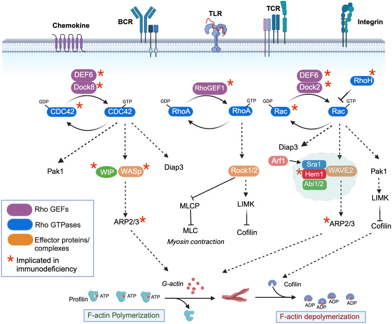
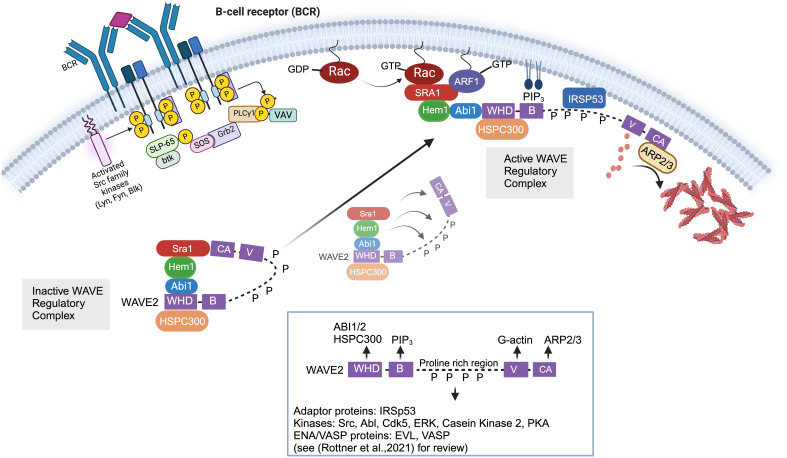
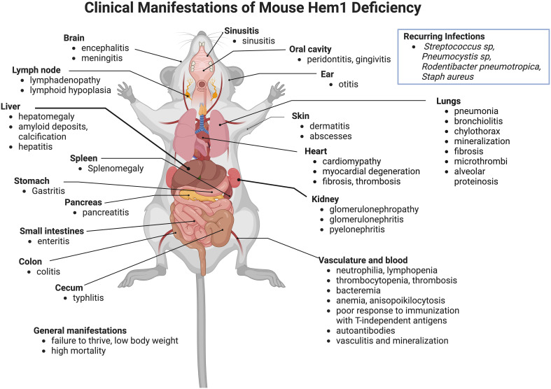
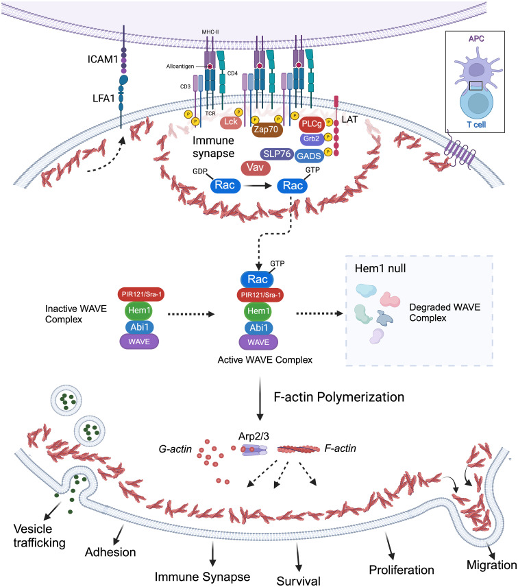
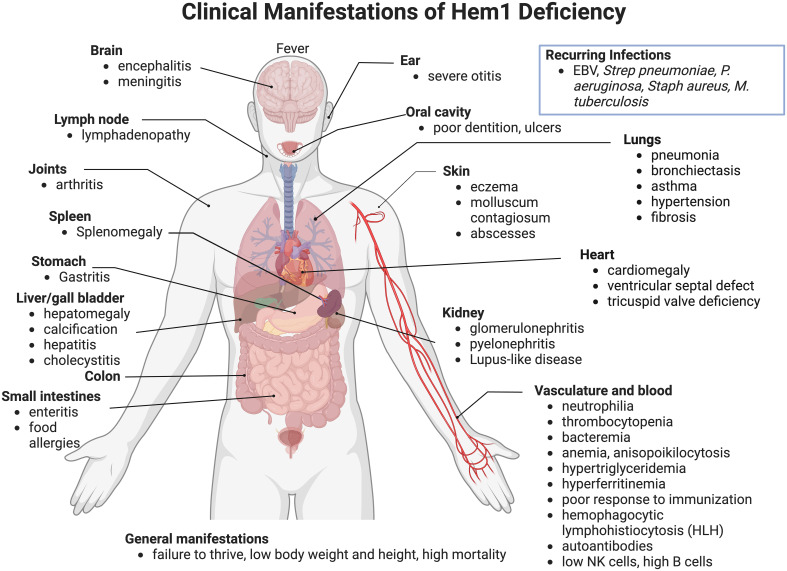
Inborn errors of immunity (IEI) are a group of diseases in humans that typically present as increased susceptibility to infections, autoimmunity, hyperinflammation, allergy, and in some cases malignancy. Among newly identified genes linked to IEIs include 3 independent reports of 9 individuals from 7 independent kindreds with severe primary immunodeficiency disease (PID) and autoimmunity due to loss-of-function mutations in the NCKAP1L gene encoding Hematopoietic protein 1 (HEM1). HEM1 is a hematopoietic cell specific component of the WASp family verprolin homologous (WAVE) regulatory complex (WRC), which acts downstream of multiple immune receptors to stimulate actin nucleation and polymerization of filamentous actin (F-actin). The polymerization and branching of F-actin is critical for creating force-generating cytoskeletal structures which drive most active cellular processes including migration, adhesion, immune synapse formation, and phagocytosis. Branched actin networks at the cell cortex have also been implicated in acting as a barrier to regulate inappropriate vesicle (e.g. cytokine) secretion and spontaneous antigen receptor crosslinking. Given the importance of the actin cytoskeleton in most or all hematopoietic cells, it is not surprising that HEM1 deficient children present with a complex clinical picture that involves overlapping features of immunodeficiency and autoimmunity. In this review, we will provide an overview of what is known about the molecular and cellular functions of HEM1 and the WRC in immune and other cells. We will describe the common clinicopathological features and immunophenotypes of HEM1 deficiency in humans and provide detailed comparative descriptions of what has been learned about Hem1 disruption using constitutive and immune cell-specific mouse knockout models. Finally, we discuss future perspectives and important areas for investigation regarding HEM1 and the WRC.

Keywords: Hem-1; Hem1; NCKAP1L; actin cytoskeleton; hematopoietic protein-1; immunodeficiency disease; inborn errors of immunity; wave.


Conflict of interest statement

The authors declare that the research was conducted in the absence of any commercial or financial relationships that could be construed as a potential conflict of interest.

Figures

Figure 1
Figure 1
Regulation of filamentous actin polymerization by the rho-family GTPases, Hem1, and the WAVE regulatory complex. Activation of immune receptors upon ligand binding results in activation of Rho guanine nucleotide exchange factors (Rho-GEFs), which catalyze the release of GDP for GTP on Rho-GTPases (CDC42, RhoA, Rac1/2/3), resulting in GTPase activation. Conversely, Rho-GTPase activating proteins (Rho-GAPs) cause GTP to be hydrolyzed back to the inactive GDP-bound form. CDC42-GTP specifically binds and activates the WIP-WASp complex (left), which facilitates ARP2/3-mediated nucleation of ATP-bound G-actin, resulting in its release from Profilin and polymerization into filamentous actin (F-actin). GTP-bound Rac (right) predominantly interacts with and activates the WAVE regulatory complex (WRC), which also facilitates ARP2/3 nucleation of ATP-G-actin into F-actin. The Arf1 GTPase interaction with Sra1 enhances WRC activation. Both CDC42-GTP and Rac-GTP activate Pak-dependent Lim kinase (Pak1), which stabilizes F-actin by preventing Cofilin from severing ADP-bound actin and promoting actin depolymerization, and Diap3, which is required for nucleation of branched actin filaments. RhoA-GTP (middle) controls dynamic aspects of actin regulation by activating Rho-associated coiled-coil-containing kinase (ROCK), which inhibits myosin light chain phosphatase (MCLP), leading to increased phosphorylation of myosin light chains (MLCs), which bind actin and stimulate contraction at the “tail” during migration. RhoA-GTP also activates LIMK. An additional atypical RhoGTPase RhoH that is not regulated by GTP exchange is also present in hematopoietic cells and it believed to act in part by inhibiting the membrane association and activation of Rac-GTP. *Denotes genes where variants have been associated with Inborn Errors of Immunity (IEI).
Figure 2
Figure 2
Cellular and molecular activation of the WAVE regulatory complex. In immune cells, the Wave Regulatory Complex (WRC) consists of Sra1, Hem1, Abi1/2, WAVE2, and HSPC300. The WAVE2 protein has multiple domains including an amino-terminal WAVE homology domain (WHD, which is important for interactions with other WRC members), a basic region (B) (which bonds phospholipids), proline rich region (PRR, which binds multiple SH3 containing proteins), a c-terminal Verprolin-homology domains (V), Cofilin domain (C), and acidic domain (A). The c-terminal V, C, and A domains (collectively known as the VCA domain) are central in binding G-actin monomers and ARP2/3 complex. The WRC exists basally in an autoinhibited “closed” state due to interaction of the “meander” and VCA regions of WAVE with Sra1. Stable activation of the WRC involves relocation of the WRC from the cytosol to the plasma membrane where it interacts with PIP3 via the WAVE basic region (B), IRSp53 via the WAVE PRR, and Rac-GTP and ARF1 via Sra1. These synergistic interactions collectively promote the allosteric release of the VCA domain from the “autoinhibiting” interaction with Sra1, allowing the VCA domain to interact with actin monomers and the ARP2/3 complex resulting in actin nucleation and formation of branched actin networks. In non-immune cells, it is known that multiple phosphorylation events additionally regulate the stability of WRC activation. For example, SH3 domain containing kinases such as Abl, Src, and Cdk5 interact with the proline rich region of WAVE via SH3 domains. Phosphorylation of WAVE2 by Abl kinase has been shown to be important for WRC-actin assembly and lamellipodia formation, and phosphorylation by Src or Cdk5 alters actin dynamics. However, we have not yet obtained a full understanding of the kinases that interact with and phosphorylate WAVE in immune cells. Of note, various transmembrane receptors may also contribute to WRC recruitment to (or its regulation at) the plasma membrane using a specific peptide motif called the WIRS (WRC Interacting Receptor Sequence) which binds a pocket on the surface of the WRC via Abi2 and Sra1 [not shown in Figure but reviewed in (45)].
Figure 3
Figure 3
Clinicopathological features of Hem1 deficiency in mice. Shown are clinical and pathological findings noted in mice containing either a non-coding point mutation in Hem1 (Hem1pt/pt) (36, 77, 78), or Cre-mediated deletion of Hem1 (Hem1-/-) (65, 66, 74). As noted in Figure 8 , the lesions and clinical findings noted in mice are remarkably similar to HEM1 deficient human patients. For example, despite being housed under Specific Pathogen Free (SPF) conditions, Hem1 deficient mice fail to thrive, exhibit low body weight, are more susceptible to opportunistic infections, and exhibit higher mortality. Inflammation is noted in multiple tissues including the gastrointestinal tract, kidney, pancreas, heart, lung, liver, skin, oral cavity, inner ear, sinuses etc. Signs of autoimmunity including increased autoantibodies were also noted.
Figure 4
Figure 4
Importance of Hem1 and the WAVE regulatory complex in murine T Lymphocytes. The T cell receptor is (TCR) is activated following recognition of antigen displayed by an antigen presenting cell (APC), resulting in downstream activation of Rac from the Rho family of GTPases. GTP-bound Rac interacts with the WAVE regulatory complex (WRC) which facilitates F-actin polymerization via the ARP2/3 complex. Hem1 is a crucial component of the WRC and Hem1 deficiency results in destabilization and degradation of WAVE complex proteins, resulting in defective F-actin polymerization and branching in T cells. Branched actin has many important roles in T lymphocytes including formation of lamellipodia required for migration; integrin activation (e.g. LFA1 etc.) and focal adhesion formation for cell-cell adhesion; immune synapse formation required for APC interactions and concentrating T cell signaling molecules for optimal T cell activation; creating a cortical actin barrier to prevent inappropriate release of vesicles containing cytokines and other effector molecules; proliferation and survival.
Figure 5
Figure 5
Hem1 regulates murine B lymphocyte development. B cells can be broadly divided into conventional B-2 B cells, which constitute the majority of B cells that interact with T follicular cells to produce specific antibodies, and fetal liver derived self-renewing innate-like B1a B cells, which are predominantly found in peritoneal and pleural cavities and produce natural antibodies. B2 B cells develop in the BM and fetal liver, where HSCs become committed pro-B cells, which have their Ig genes in germline configuration. Following Ig heavy chain gene rearrangement, the IgH protein is expressed at the surface of pre-B cells in conjunction with Igα and Igβ as the pre-BCR, which stimulates IgL chain rearrangement and formation of the BCR on immature B cells. Immature B cells which are deemed non-autoreactive migrate to the spleen where development continues through the transitional (T1,T2) stages, followed by differentiation into innate-like marginal zone (MZ) B cells or mature recirculating Follicular (FO) B cells, which migrate to LN and BM and produce specific antibodies. B1a B cells develop predominantly in the fetal liver and follow a similar developmental progression. B cell specific disruption of Hem1 in mice results in significant reduction of transitional B, MZ, FO, and B1 B cells in bone marrow, spleen, and lymph node tissues.
Figure 6
Figure 6
Hem1 deficiency results in B cell hyperactivation in mice. (Top left). In the absence of cognate Ag, BCRs are maintained at the cell surface in lipid-bound nanoclusters. (bottom left) Ag binding results in cortical actin depolymerization, resulting in increased BCR diffusion into microclusters and formation of the immune synapse, which then recruits and activates key signaling molecules resulting in B cell activation. (top right) One model is that in following disruption of Hem1, diminished cortical actin allows increased BCR diffusion and interactions in the absence of cognate antigen, resulting in basal or increased B cell activation. (bottom right) An alternative model is that disruption of Hem1 results in diminished immune synapse and microcluster formation, resulting in selection for stronger signaling autoreactive B cells during positive selection.
Figure 7
Figure 7
Hem1 regulates the development and/or functions of macrophages and neutrophils. (A) Engagement of phagocytic receptors upon binding to targets leads to activation of signaling pathways that stimulate formation of phagocytic cups, which contain adhesion molecules and are rich in branched actin foci, colocalized ARP2/3 and WRC. In the absence of Hem1, phagocytic cups fail to form properly and adhesion to targets are weak, leading to deficient phagocytosis. (B) Actin branching is important for forming lamellipodia, actin-filled protrusive structures which are critical for cell migration. The WRC accumulates and becomes anchored at the cell membrane, where it stimulates actin branching and forward driving force. Hem1 deficient macrophages and neutrophils are deficient in their abilities to migrate through complex matrices. (C) Alveolar macrophages (AMs) are a specialized subset that reside in lung airways. They develop via a unique process during fetal life, where fetal liver monocytes migrate to the fetal lung around embryonic day 15-18 (E15-18). In response to GM-CSF produced by bronchial epithelial cells around embryonic day 19 through post-natal day 3 (PND3), fetal monocytes develop into pre-alveolar macrophages. Thereafter, pre-AMs in response to autocrine TGF-β upregulate PPARγ which stimulates differentiation into mature AMs. In the absence of Hem1, AMs fail to develop efficiently, leading to an accumulation of debris and protein in airways (“alveolar proteinosis”).
Figure 8
Figure 8
Clinical and pathological manifestations of Hem1 deficiency in humans. Shown are clinical and pathological findings in nine patients with loss of function mutations in Hem1 (62, 64, 65, 82). As noted in Figure 3 , the lesions and clinical findings noted in Hem1 deficient patients are remarkably similar to murine models. Hem1 deficiency results in a syndrome characterized by immunodeficiency as well as hyperinflammation and autoimmunity. Although clinical presentations were varied among patients, all patients presented with recurrent infections including otitis media, skin abscesses and pneumonia. Systemic inflammation is also common resulting in hepatosplenomegaly and lymphadenopathy in several patients. Few patients also have autoimmune disorders resulting in autoantibodies, similar to what was seen in Hem1-/- mice models (36, 65, 66).

Similar articles

Cited by

References

    1. Dupre L, Boztug K, Pfajfer L. Actin dynamics at the T cell synapse as revealed by immune-related actinopathies. Front Cell Dev Biol. (2021) 9:665519. doi: 10.3389/fcell.2021.665519 - DOI - PMC - PubMed
    1. Papa R, Penco F, Volpi S, Gattorno M. Actin remodeling defects leading to autoinflammation and immune dysregulation. Front Immunol. (2020) 11:604206. doi: 10.3389/fimmu.2020.604206 - DOI - PMC - PubMed
    1. Dupre L, Prunier G. Deciphering actin remodelling in immune cells through the prism of actin-related inborn errors of immunity. Eur J Cell Biol. (2023) 102:151283. doi: 10.1016/j.ejcb.2022.151283 - DOI - PubMed
    1. Burns SO, Zarafov A, Thrasher AJ. Primary immunodeficiencies due to abnormalities of the actin cytoskeleton. Curr Opin Hematol. (2017) 24:16–22. doi: 10.1097/MOH.0000000000000296 - DOI - PubMed
    1. Rottner K, Faix J, Bogdan S, Linder S, Kerkhoff E. Actin assembly mechanisms at a glance. J Cell Sci. (2017) 130:3427–35. doi: 10.1242/jcs.206433 - DOI - PubMed

LinkOut - more resources