[Preprint]. 2024 Jul 13:2024.07.12.603173.
doi: 10.1101/2024.07.12.603173.

PRC1.6 localizes on chromatin with the human silencing hub (HUSH) complex for promoter-specific silencing

Affiliations

PRC1.6 localizes on chromatin with the human silencing hub (HUSH) complex for promoter-specific silencing

Tomás C Rodríguez et al. bioRxiv. .

Abstract

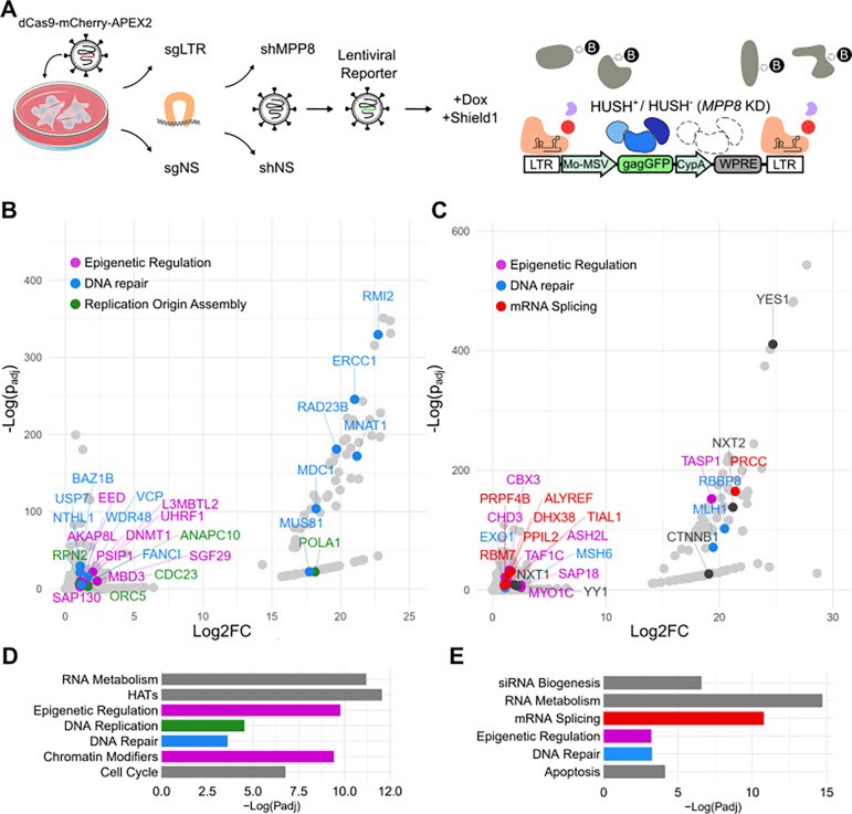
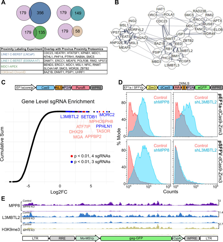
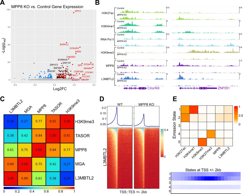
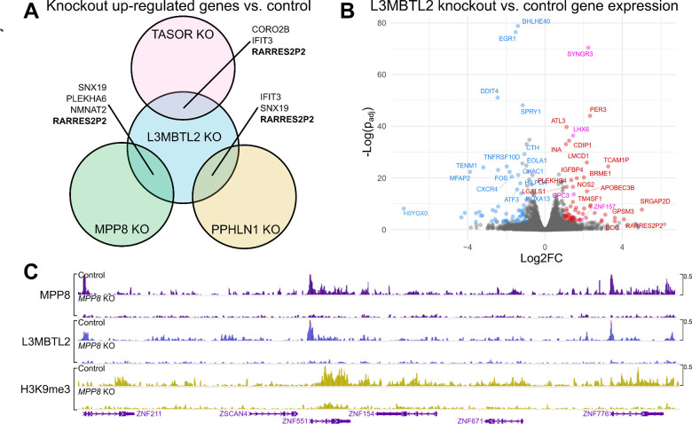
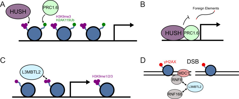
An obligate step in the life cycle of HIV-1 and other retroviruses is the establishment of the provirus in target cell chromosomes. Transcriptional regulation of proviruses is complex, and understanding the mechanisms underlying this regulation has ramifications for fundamental biology, human health, and gene therapy implementation. The three core components of the Human Silencing Hub (HUSH) complex, TASOR, MPHOSPH8 (MPP8), and PPHLN1 (Periphilin 1), were identified in forward genetic screens for host genes that repress provirus expression. Subsequent loss-of-function screens revealed accessory proteins that collaborate with the HUSH complex to silence proviruses in particular contexts. To identify proteins associated with a HUSH complex-repressed provirus in human cells, we developed a technique, Provirus Proximal Proteomics, based on proximity labeling with C-BERST (dCas9-APEX2 biotinylation at genomic elements by restricted spatial tagging). Our screen exploited a lentiviral reporter that is silenced by the HUSH complex in a manner that is independent of the integration site in chromatin. Our data reveal that proviruses silenced by the HUSH complex are associated with DNA repair, mRNA processing, and transcriptional silencing proteins, including L3MBTL2, a member of the non-canonical polycomb repressive complex 1.6 (PRC1.6). A forward genetic screen confirmed that PRC1.6 components L3MBTL2 and MGA contribute to HUSH complex-mediated silencing. PRC1.6 was then shown to silence HUSH-sensitive proviruses in a promoter-specific manner. Genome wide profiling showed striking colocalization of the PRC1.6 and HUSH complexes on chromatin, primarily at sites of active promoters. Finally, PRC1.6 binding at a subset of genes that are silenced by the HUSH complex was dependent on the core HUSH complex component MPP8. These studies offer new tools with great potential for studying the transcriptional regulation of proviruses and reveal crosstalk between the HUSH complex and PRC1.6.


Conflict of interest statement

COMPETING INTERESTS The authors declare no competing interests.

Figures

Figure 1:
Figure 1:. Provirus Proximity Proteomics in human cells.
(A) Schematic describing the Provirus Proximity Proteomics approach targeting the provirus of a lentivector after transduction of HEK293T cells. An sgRNA targeting the provirus LTRs (sgLTR) recruited dCas9-mCherry-APEX2, then a pulse of biotin-phenol was conjugated to proteins in the immediate vicinity. HUSH indicates cells transduced with an shRNA targeting MPP8 (MPP8 KD). HUSH+ indicates cells transduced with a non-targeting control shRNA. (B,C) Proteins enriched at the provirus (sgLTR) in HUSH+ and HUSH cells, respectively. Named proteins are significantly enriched over non-specific nuclear background (padj < 0.05; Log2FC > 1) and colored according to the function annotated in D and E. (D,E) Enrichment of Reactome [24] pathways in HUSH+ and HUSH backgrounds, respectively. Colors correspond to member proteins in panels B and C. For information associated with this figure please see Extended Data Tables 1, 2, 3, 4, and 5.
Figure 2:
Figure 2:. Validation of HUSH+ proteins enriched by Provirus Proximity Proteomics using orthogonal assays.
(A) Comparison of the 179 proteins enriched at the provirus under HUSH+ conditions with those proteins enriched in published proximity labeling datasets [19,34,36]. (B) STRING network analysis of proteins enriched at the provirus in HUSH+ cells. Line widths are in proportion to the evidence supporting interaction between two nodes, as described in [35]. (C) CRISPR knockout screen conducted in Jurkat cells using single vector Toronto KnockOut (TKO) CRISPR library Version 3 with a puromycin selection cassette. Cumulative sum plot depicts the comparison of sgRNA relative quantity in cell populations transduced with library and treated with 10 μg puromycin versus a population transduced with library and treated with 1 μg puromycin. Individual Log2FC values represent aggregate counts of multiple sgRNAs corresponding to a single gene calculated by MAGeCK [37]. Red and blue points meet significance criteria (p < 0.01) for 3 (blue) or 4 (red) sgRNAs. (D) Flow cytometry histogram depicting GFP-positive population of Jurkat cells transduced with pEF1a-Zim3-dCas9-P2A-EGFP (top panels) or pSFFV-Zim3-dCas9-P2A-EGFP (bottom panels). Each population was treated with shRNA vectors targeting MPP8 (left panels) or L3MBTL2 (right panels) in blue, vs the non-targeting shRNA control in red. (E) Normalized CUT&Tag signal (counts-per-million) for MPP8, L3MBTL2, and H3K9me3 at the pscALP-PLXIN-gagGFP-WPRE provirus in HEK293T cells. For data associated with this figure please see Extended Data Table 6 and SRA Bioproject PRJNA869850.
Figure 3:
Figure 3:. Genome-wide characterization of loci bound by PRC1.6 and HUSH in Jurkat cells.
(A) Total RNA sequencing of Jurkat cells harboring L3MBTL2 knockout versus control Jurkat cells. Colored/labeled points meet significance thresholds (FDR 0.1). (B) Normalized (counts-per-million) CUT&Tag signal targeting named factors at CXorf49 and ZNF551 loci in unmodified Jurkat cells (Control) and MPP8 knockout Jurkat cells (MPP8 KO). (C) Pearson correlation of counts-per-million-normalized CUT&Tag signal of named factors at annotated human genes (hg38 RefSeq transcription start to end site). (D) L3MBTL2 CUT&Tag coverage map at human genes; transcription start site (TSS) to transcription end site (TES) regions are scaled by length. 2 kilobases upstream and downstream of scaled regions are also depicted. (E) ChromHMM [40] emission states using CUT&Tag from named factors as input (top panel). Coverage of each emission state with respect to transcription start sites (TSSs). For data associated with this figure please see Extended Data Tables 7 and 8 and SRA Bioproject PRJNA869850.
Figure 4:
Figure 4:. Transcriptome-wide and genome-level effects of HUSH and L3MBTL2 depletion in HEK293T cells.
(A) Venn diagram depicting genes that are upregulated vs control (padj<0.05, log2FC>1) in TASOR, MPP8, PPHLN1, and L3MBTL2 knock out HEK293T cells. (B) Volcano plot showing differential gene expression (padj<0.05, log2FC>1) in L3MBTL2 knockout cells compared to control knockouts. Colored points depict genes meeting the significance threshold. Magenta points indicate genes that are also upregulated in Jurkat L3MBTL2 knockouts. (C) MPP8, L3MBTL2, and H3K9me3 CUT&Tag in control knockouts and MPP8 knockouts. Genomic region depicted is the chromosome 19 ZNF gene cluster. For data associated with this figure please see Extended Data Table 9 and SRA Bioproject PRJNA869850.
Figure 5:
Figure 5:. Possible mechanisms underlying co-localization of HUSH and PRC1.6 complexes.
(A) The HUSH and PRC1.6 complexes transcriptionally repress developmental genes and overlap at select loci. (B) Both complexes reside at endogenous promoters to safeguard against perturbation by retroviruses and retrotransposons. (C) L3MBTL2 may be recruited to HUSH loci through recognition of H3K9me3 marks previously established by the HUSH complex. (D) DNA damage may independently recruit the HUSH and PRC1.6 complexes to overlapping loci (adapted from [54]).

References

    1. Seczynska M, Lehner PJ. The sound of silence: mechanisms and implications of HUSH complex function. Trends Genet. 2023;39: 251–267. - PubMed
    1. Müller I, Helin K. Keep quiet: the HUSH complex in transcriptional silencing and disease. Nat Struct Mol Biol. 2024;31: 11–22. - PubMed
    1. Tchasovnikarova IA, Timms RT, Matheson NJ, Wals K, Antrobus R, Göttgens B, et al. Epigenetic silencing by the HUSH complex mediates position-effect variegation in human cells. Science. 2015;348: 1481–1485. - PMC - PubMed
    1. Douse CH, Tchasovnikarova IA, Timms RT, Protasio AV, Seczynska M, Prigozhin DM, et al. TASOR is a pseudo-PARP that directs HUSH complex assembly and epigenetic transposon control. Nat Commun. 2020;11: 4940. - PMC - PubMed
    1. Schultz DC, Ayyanathan K, Negorev D, Maul GG, Rauscher FJ 3rd. SETDB1: a novel KAP-1-associated histone H3, lysine 9-specific methyltransferase that contributes to HP1-mediated silencing of euchromatic genes by KRAB zinc-finger proteins. Genes Dev. 2002;16: 919–932. - PMC - PubMed

Publication types

LinkOut - more resources