. 2021 Oct 13;59(Suppl 1):e17-e21.
doi: 10.1055/s-0041-1735140. eCollection 2024 Jul.

High-grade Pleomorphic Sarcoma Associated with Metallosis in a Patient with Total Hip Arthroplasty

Affiliations

High-grade Pleomorphic Sarcoma Associated with Metallosis in a Patient with Total Hip Arthroplasty

Roni Serra Campos et al. Rev Bras Ortop (Sao Paulo). .

Abstract

Although the relationship between hip arthroplasty and the development of sarcoma was first described in the literature about forty years ago, this association is extremely rare. In the present case report, we describe the association between orthopedic implants and soft tissue sarcoma in a 79-year-old man who underwent primary total hip arthroplasty (THA) for coxarthrosis 24 years ago. In the present case report, we describe the clinical evolution and the radiographic and histopathological findings of the lesion. In the intraoperative period of the second revision surgery, loosening of the acetabular and femoral components in association with extensive areas of necrosis and metallosis was evidenced. We performed debridement of the hip and right thigh region and removed the implants. Due to the extent of the lesion and to necrosis, it was not possible to perform a new joint reconstruction. The histopathological diagnosis of high-grade undifferentiated pleomorphic sarcoma associated with extensive areas of metallosis was confirmed in tissue adjacent to the implant. The patient developed pulmonary metastases and died 6 months after the diagnosis. Despite the rarity of this association, sarcomas should be considered in the differential diagnosis of aseptic loosening, especially in the presence of metallosis in the peri-implant tissue. To our knowledge, the 24-year latency period between primary THA and the establishment of a sarcoma diagnosis is one of the longest reported to date.

Keywords: arthroplasty, replacement, hip; hip prosthesis; metals; sarcoma.


Conflict of interest statement

Conflito de Interesses Os autores declaram não haver conflito de interesses.

Figures

Fig. 1
Fig. 1
Right hip revision surgery in May 2016. ( A ) Anteroposterior panoramic radiograph of the pelvis showing bilateral total hip arthroplasty. On the right, there are signs of resorption of the proximal femur and migration of the acetabular component into the iliac bone, which presents diffuse osteolysis. There is also an important increase in soft tissue volume in the periprosthetic region. The left uncemented prosthesis presents loosening of the acetabular component, which is medialized and verticalized. The femoral component is fixed. ( B ) Intraoperative images showing metallosis (blackened areas). ( C ) Extensive areas of tissue necrosis adjacent to the implant (arrows) that were debrided to remove the acetabular and femoral components, making it impossible to perform a new joint reconstruction.
Fig. 2
Fig. 2
Histopathological aspect and immunohistochemical profile of the lesion associated with the implant in the right hip joint. ( A ) High-grade undifferentiated pleomorphic sarcoma characterized by a marked degree of cell anaplasia (arrows). ( B ) Wear debris of prosthetic components identified by blackened pigments (arrows) in association with tumor cells (*). ( C ) Absence of immunostaining for desmin. ( D ) Weak immunopositivity for S-100 protein. ( E ) Diffuse positive expression of anti-CD68 antibodies and ( F ) Anti-Vimentin. ( A and B ) Hematoxylin and Eosin (H&E) staining. Scale bar: 50 µm ( A, B ), 70 µm ( C-E ) and 100 µm ( F ).
Fig. 3
Fig. 3
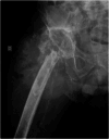
Right hip radiograph performed in July 2016 after revision arthroplasty procedure through posterolateral access with the removal of prosthetic components. Presence of a large tumoral mass in soft tissues in association with extensive destruction of the proximal third of the femur.
Fig. 4
Fig. 4
Chest computed tomography scan. In July 2016, the patient had pleural effusion and multiple metastatic nodules in both lungs and died in August 2016.
Fig. 1
Fig. 1
Cirurgia de revisão do quadril direito em maio de 2016. (A) Radiografia panorâmica da bacia em incidência anteroposterior evidenciando artroplastia total bilateral de quadril. À direita, observa-se sinais de reabsorção proximal do fêmur e migração do componente acetabular para dentro do ilíaco, que apresenta osteólise difusa. Nota-se ainda importante aumento de volume de partes moles na região periprotética. A protése não cimentada à esquerda apresenta afrouxamento do componente acetabular, que se encontra medializado e verticalizado. O componente femoral está fixo. (B) Imagens do intraoperatório mostrando metalose (áreas enegrecidas). (C) Áreas extensas de necrose do tecido adjacente ao implante (setas) que foi desbridado para a retirada dos componentes acetabular e femoral, não sendo possível realizar nova reconstrução articular.
Fig. 2
Fig. 2
Aspecto histopatológico e perfil imuno-histoquímico da lesão associada ao implante da articulação do quadril direito. (A) Sarcoma pleomórfico indiferenciado de alto grau caracterizado por grau acentuado de anaplasia celular (setas). (B) Debris de desgaste dos componentes da prótese identificado pelos pigmentos enegrecidos (setas) em associação com as células tumorais (*). (C) Ausência de imunomarcação para desmina. (D) Imunopositividade fraca para proteína S-100. (E) Expressão positiva difusa para os anticorpos anti-CD 68 e (F) Anti-Vimentina. (A e B) Coloração hematoxilina e eosina (H&E). Barra de escala: 50µm (A,B), 70µm (C-E) e 100µm (F).
Fig. 3
Fig. 3
Radiografia do quadril direito realizada em julho de 2016 após procedimento de artroplastia de revisão via acesso posterolateral com remoção dos componentes protéticos. Presença de volumosa massa tumoral em partes moles em associação com extensa destruição do terço proximal do fêmur.
Fig. 4
Fig. 4
Tomografia computadorizada do tórax. Em julho de 2016, o paciente apresentava derrame pleural e múltiplos nódulos metastáticos em ambos os pulmões e evoluiu para o óbito em agosto de 2016.

References

    1. Purdue P E, Koulouvaris P, Potter H G, Nestor B J, Sculco T P. The cellular and molecular biology of periprosthetic osteolysis. Clin Orthop Relat Res. 2007;454(454):251–261. - PubMed
    1. Bagó-Granell J, Aguirre-Canyadell M, Nardi J, Tallada N. Malignant fibrous histiocytoma of bone at the site of a total hip arthroplasty. A case report. J Bone Joint Surg Br. 1984;66(01):38–40. - PubMed
    1. Visuri T, Pulkkinen P, Paavolainen P. Malignant tumors at the site of total hip prosthesis. Analytic review of 46 cases. J Arthroplasty. 2006;21(03):311–323. - PubMed
    1. EUROCARE-5 Working Group . Stiller C A, Botta L, Brewster D H et al.Survival of adults with cancers of bone or soft tissue in Europe-Report from the EUROCARE-5 study. Cancer Epidemiol. 2018;56:146–153. - PubMed
    1. Lamovec J, Zidar A, Cucek-Plenicar M. Synovial sarcoma associated with total hip replacement. A case report. J Bone Joint Surg Am. 1988;70(10):1558–1560. - PubMed

LinkOut - more resources