. 2024 Jun 24:40:445-459.
doi: 10.1016/j.bioactmat.2024.06.026. eCollection 2024 Oct.

Concerting magnesium implant degradation facilitates local chemotherapy in tumor-associated bone defect

Affiliations

Concerting magnesium implant degradation facilitates local chemotherapy in tumor-associated bone defect

Qingqing Guan et al. Bioact Mater. .

Abstract

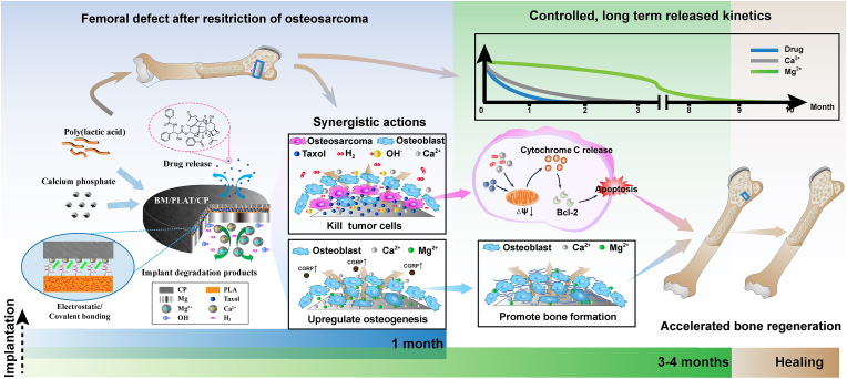
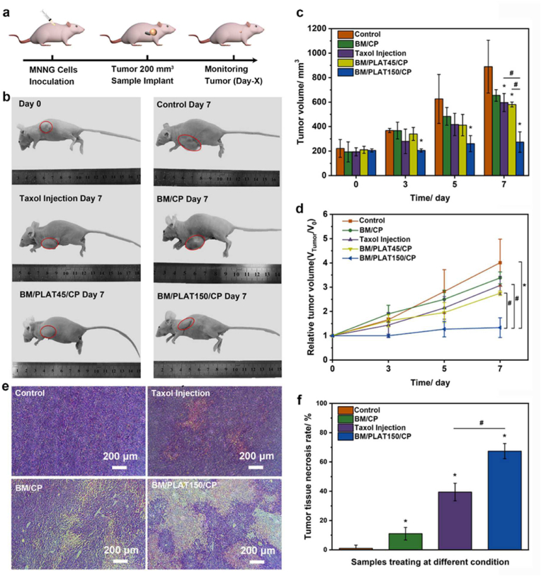
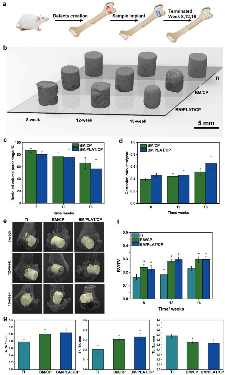
Effective management of malignant tumor-induced bone defects remains challenging due to severe systemic side effects, substantial tumor recurrence, and long-lasting bone reconstruction post tumor resection. Magnesium and its alloys have recently emerged in clinics as orthopedics implantable metals but mostly restricted to mechanical devices. Here, by deposition of calcium-based bilayer coating on the surface, a Mg-based composite implant platform is developed with tailored degradation characteristics, simultaneously integrated with chemotherapeutic (Taxol) loading capacity. The delicate modulation of Mg degradation occurring in aqueous environment is observed to play dual roles, not only in eliciting desirable osteoinductivity, but allows for modification of tumor microenvironment (TME) owing to the continuous release of degradation products. Specifically, the sustainable H2 evolution and Ca2+ from the implant is distinguished to cooperate with local Taxol delivery to achieve superior antineoplastic activity through activating Cyt-c pathway to induce mitochondrial dysfunction, which in turn leads to significant tumor-growth inhibition in vivo. In addition, the local chemotherapeutic delivery of the implant minimizes toxicity and side effects, but markedly fosters osteogenesis and bone repair with appropriate structure degradation in rat femoral defect model. Taken together, a promising intraosseous administration strategy with biodegradable Mg-based implants to facilitate tumor-associated bone defect is proposed.

Keywords: Biodegradable magnesium-based implant; Bone regeneration; Local tumor therapy; Multifunctional composite coating; Tumor-associated bone defect.


Conflict of interest statement

The authors declare that they have no known competing financial interests or personal relationships that could have appeared to influence the work reported in this paper.

Figures

Image 1
Graphical abstract
Scheme 1
Scheme 1
Schematic diagram of BM/PLAT/CP implant design and bio-application in treating tumor-induced bone defects.
Fig. 1
Fig. 1
Preparation and characterization of BM/PLAT/CP. a) The process of the bilayer coatings on Mg-implant. b) SEM image of the top view of BM/PLAT implants. c, d) SEM images of the top view and cross-section view of BM/PLAT/CP implants. e) pH and osmolality and f) cumulative Mg2+ amount profiles of BM/PLAT/CP during 60 days of immersion in DMEM. g) In vitro H2 release profile of BM -based implants in phosphate-buffered saline with 0.1 % Tween 20 (PBST) for 21 days. h) Calculated in vitro corrosion rate of BM/MgF2, BM/PLA and BM/PLAT/CP. i) In vitro Taxol release profile of BM/PLAT/CP implants in PBST for 2 weeks. *p < 0.05.
Fig. 2
Fig. 2
In vitro effect of different implant extract on MC3T3-E1 cell response. a) Fluorescent microscopy images and b) adherent cell density of MC3T3-E1 osteoblastic cells incubated with different implants. Scale bar = 200 and 100 μm, respectively. c) Cell viability and d) ALP activities of MC3T3-E1 cells cultured in samples extracts. e) RT-PCR results of expression of osteogenic genes Alp, Opn, Col I and Runx 2 after 14 days of culture. nsp>0.05 vs. Control group, *p < 0.05 vs. Control group; #p < 0.05.
Fig. 3
Fig. 3
In vitro killing effect on MNNG osteosarcoma cells. a) Fluorescent microscopy images and b) adherent cell density of MNNG osteosarcoma cells on control, BM/MgF2, BM/CP and BM/PLAT/CP samples, respectively. Inserted images in a) with higher magnification showed the cell spreading morphology, with scale bar represents 100 μm. c) Cell viability results of MNNG cells cultured with extract of BM/MgF2, BM/CP and BM/PLAT/CP, respectively. *p < 0.05, **p < 0.01.
Fig. 4
Fig. 4
Viability results of a) MNNG cells vs. b) MC3T3-E1 cells cultured in the BM extracts with dilutions and BM/PLAT/CP extract. c, d) pH value, osmolality and Mg2+ concentration of BM extracts with dilutions (100 % BM, 80 % BM, 50 % BM and 20 % BM) and BM/PLAT/CP extract. e) Schematic illustration on the effect of degradation extracts of BM/PLAT/CP on differentiating viability between bone tissue cell and tumor cell.
Fig. 5
Fig. 5
The mechanism evaluations of BM/PLAT/CP implant induced osteosarcoma cytotoxicity. a, b) MMP of MNNG cells were treated with different samples. Scale bar: 50 μm. c, d) Fluorescence microscope images and the corresponding ratio analysis of Ca2+ ions released from different samples in MNNG cells. Scale bar: 100 μm. e) ROS level of MNNG cells treating with BM/CP extract. Scale bar:100 μm. f) Intracellular ATP content analysis with different treatments (n = 3). g) Western blotting analysis of Cyt-c, and Bcl-2 proteins. h) Schematic diagram of proposed mechanism of BM/PLAT/CP. *p < 0.05, **p < 0.01, ***p < 0.001 vs. Control group. #p < 0.05, ##p < 0.01, ###p < 0.001.
Fig. 6
Fig. 6
The anti-tumor effect of BM-based implant on osteosarcoma tumor-bearing mice. a) Schematic illustration of MNNG-bearing mice and therapeutic process of implants. b) Tumor photographs of nude mice for the control, Taxol, BM/CP, Mg/PLAT45/CP, and Mg/PLAT150/CP groups at Day 0 and Day 7. c) Tumor volume and d) relative tumor volume change of five groups at each time point. e, f) H&E stained images of tumor tissue and tumor tissue necrosis rate for the control, Taxol, BM/CP, and Mg/PLAT150/CP groups. *p < 0.05 vs. Control group. #p < 0.05.
Fig. 7
Fig. 7
Micro-CT images and histomorphometry analyses. a) Schematic illustration of femoral defect model establishment and therapy process of BM/PLAT/CP. b) 3D-reconstructed micro-CT images of Ti, BM/CP, and BM/PLAT/CP implants at different weeks post-surgery, respectively. c) The volume change and d) calculated corrosion rate of implants at different weeks post-surgery. e) Micro-CT images of different groups at week 8, 12, and 16 after implantation f) Bone volume fraction (BV/TV) of implants groups at different weeks post-surgery. g) Trabecular thickness (Tb. Th), trabecular spacing (Tb. Sp), and trabecular number (Tb. N) of different implant group at 16 weeks post-surgery. *p < 0.05.
Fig. 8
Fig. 8
In vivo analysis of the new bone formation of implants. a) Histologic overview of Ti, BM/CP, and BM/PLAT/CP implanted in femoral defects and b) quantification of the new bone area and BIC at different weeks post-surgery. Van Gieson's picrofuchsin staining (scale bar = 200 μm). The asterisk represents the implant. c, d) Sequential fluorescent labeling observation of Ti, BM/CP, and BM/PLAT/CP groups. The percentage of stained bone area is presented correspondingly. Scale bar = 100 μm *p < 0.05.

References

    1. Coleman R.E., Croucher P.I., Padhani A.R., Clézardin P., Chow E., Fallon M., Guise T., Colangeli S., Capanna R., Costa L. Nat. Rev. Dis. Prim. 2020;6:83. doi: 10.1038/s41572-020-00216-3. - DOI - PubMed
    1. Gill J., Gorlick R. Nat. Rev. Clin. Oncol. 2021;18:609–624. doi: 10.1038/s41571-021-00519-8. - DOI - PubMed
    1. Bian Y.X., Cai X.J., Lv Z.H., Xu Y.M., Wang H., Tan C.L., Liang R.Z., Weng X.S. Adv. Sci. 2023;10 doi: 10.1002/advs.202301806. - DOI - PMC - PubMed
    1. Jing Z.H., Yuan W.Q., Wang J.D., Ni R.H., Qin Y., Mao Z.N., Wei F., Song C.L., Zheng Y.F., Cai H., Liu Z.J. Bioact. Mater. 2024;33:223–241. doi: 10.1016/j.bioactmat.2023.11.001. - DOI - PMC - PubMed
    1. Sun P.F., Qu F., Zhang C., Cheng P.F., Li X.Y., Shen Q.M., Li D.F., Fan Q.L. Adv. Sci. 2022;9 doi: 10.1002/advs.202204718. - DOI - PMC - PubMed

LinkOut - more resources