Review
. 2024 Jul 4:15:1394725.
doi: 10.3389/fphys.2024.1394725. eCollection 2024.

Unraveling neurovascular mysteries: the role of endothelial glycocalyx dysfunction in Alzheimer's disease pathogenesis

Affiliations
Review

Unraveling neurovascular mysteries: the role of endothelial glycocalyx dysfunction in Alzheimer's disease pathogenesis

Nicholas O'Hare et al. Front Physiol. .

Abstract

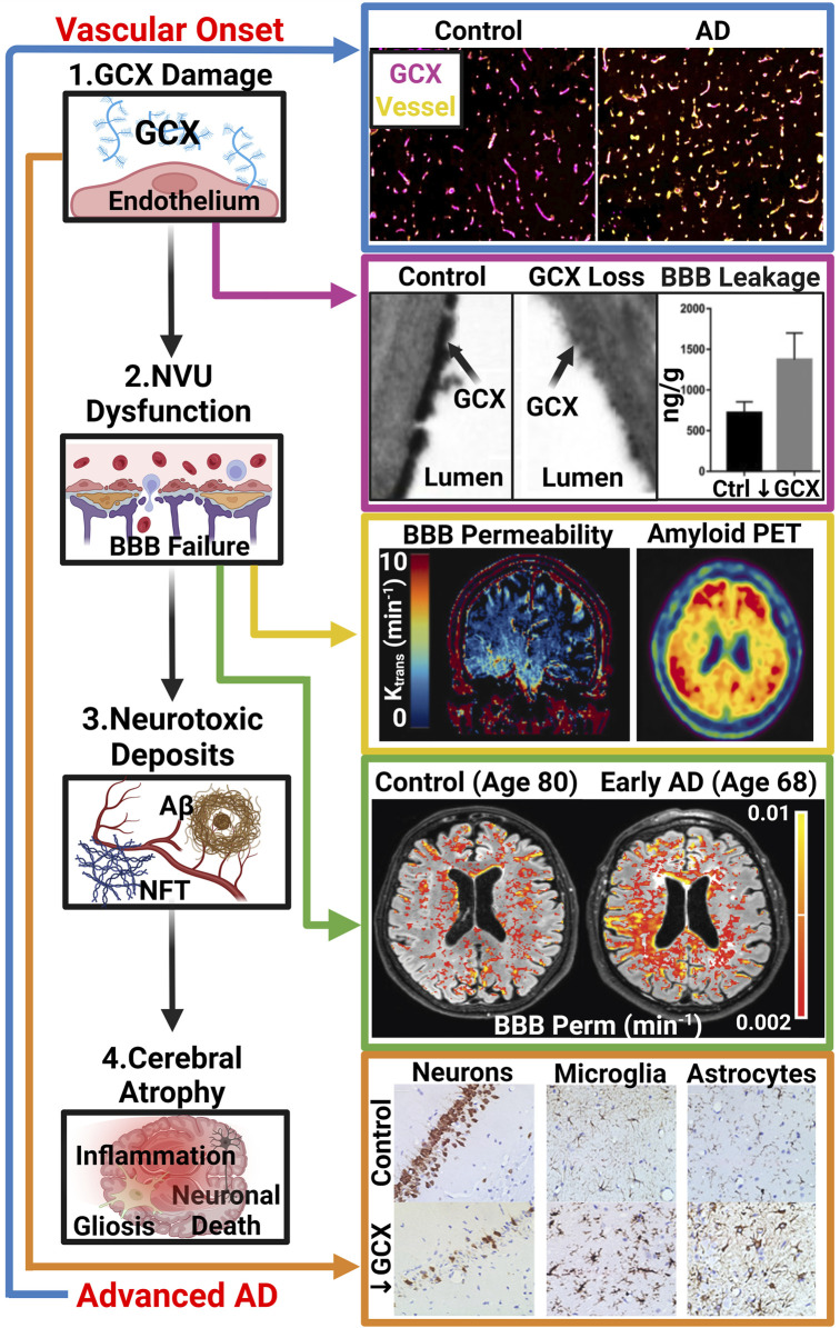
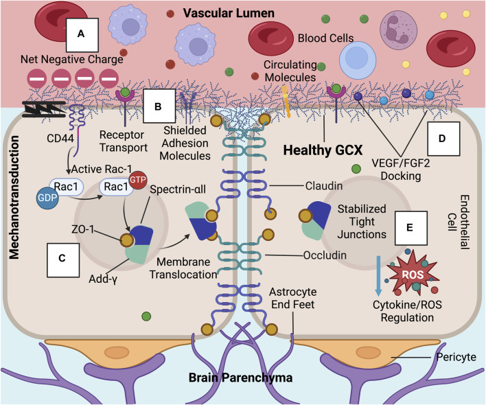
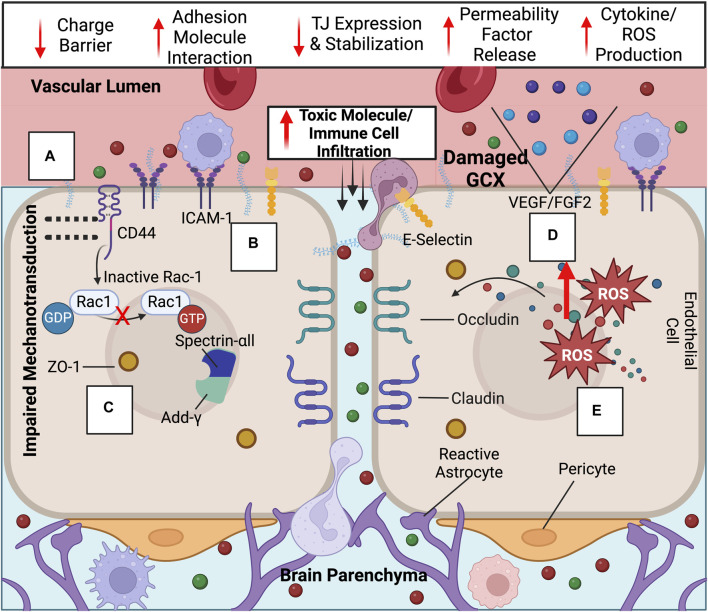
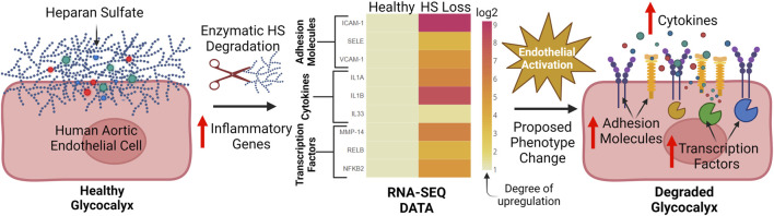
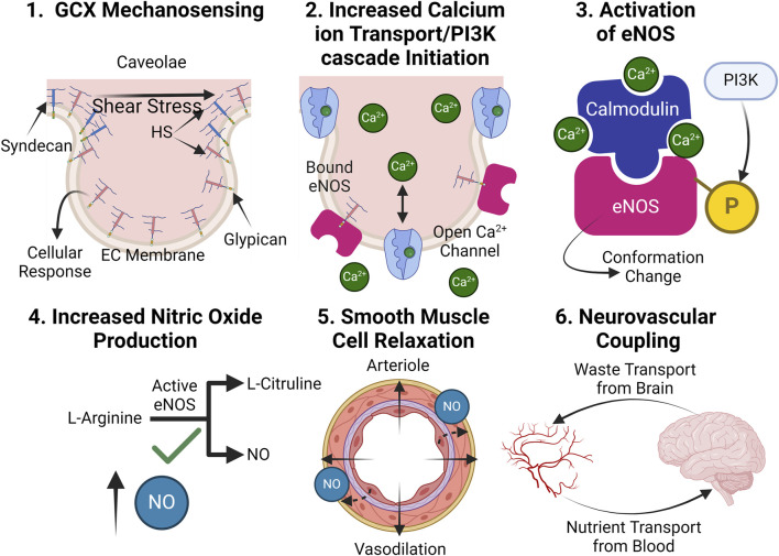
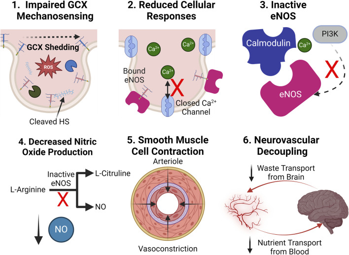
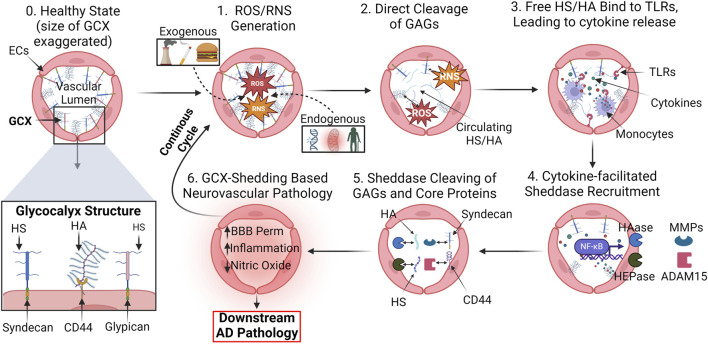
While cardiovascular disease, cancer, and human immunodeficiency virus (HIV) mortality rates have decreased over the past 20 years, Alzheimer's Disease (AD) deaths have risen by 145% since 2010. Despite significant research efforts, effective AD treatments remain elusive due to a poorly defined etiology and difficulty in targeting events that occur too downstream of disease onset. In hopes of elucidating alternative treatment pathways, now, AD is commonly being more broadly defined not only as a neurological disorder but also as a progression of a variety of cerebrovascular pathologies highlighted by the breakdown of the blood-brain barrier. The endothelial glycocalyx (GCX), which is an essential regulator of vascular physiology, plays a crucial role in the function of the neurovascular system, acting as an essential vascular mechanotransducer to facilitate ultimate blood-brain homeostasis. Shedding of the cerebrovascular GCX could be an early indication of neurovascular dysfunction and may subsequently progress neurodegenerative diseases like AD. Recent advances in in vitro modeling, gene/protein silencing, and imaging techniques offer new avenues of scrutinizing the GCX's effects on AD-related neurovascular pathology. Initial studies indicate GCX degradation in AD and other neurodegenerative diseases and have begun to demonstrate a possible link to GCX loss and cerebrovascular dysfunction. This review will scrutinize the GCX's contribution to known vascular etiologies of AD and propose future work aimed at continuing to uncover the relationship between GCX dysfunction and eventual AD-associated neurological deterioration.

Keywords: Alzheimer’s disease; blood-brain barrier; endothelial glycocalyx; neurovascular dysfunction; vascular etiology; vascular mechanobiology.


Conflict of interest statement

The authors declare that the research was conducted in the absence of any commercial or financial relationships that could be construed as a potential conflict of interest.

Figures

FIGURE 1
FIGURE 1
The GCX-Vascular Hypothesis of AD: Hypothesized sequence of events linking GCX degradation to AD pathology: (1.) Various vascular insults induce damage to the GCX. (2.) GCX damage initiates NVU dysfunction, notably affecting the BBB. (3.) NVU dysregulation leads to abnormal accumulation of neurotoxic deposits including Aβ and NFTs within the brain, characteristic of AD. (4.) Subsequently, these protein aggregates induce brain inflammation, gliosis, and neuronal death, facilitating cognitive decline. Several recent studies have supported this GCX-rooted hypothesis. (Blue Arrow): Smyth et al. observed a significant reduction in GCX integrity in post-mortem AD tissue compared to healthy controls, suggesting a correlation between late-stage AD and GCX integrity via immunostaining of UEA1 lectin for GCX and Collagen type IV for cerebral vasculature. Adapted from Figure 5 in Smyth LCD, et al. Acta Neuropathologica Communications. 2022; 10(1), Copyright (2022) by Acta Neuropathologica Communications (Smyth et al., 2022). This figure is sourced from an open-access publication. (Magenta Arrow): Zhu et al. demonstrated increased leakage of Evan’s blue dye into the brain parenchyma upon GCX degradation using hyaluronidase treatment in mice, indicating GCX loss triggers NVU dysfunction. Adapted from Figure 2 in Zhu J., et al., J Cereb Blood Flow Metab. 2018; 38(11):1979-92, Copyright (2018) by the Journal of Cerebral Blood Flow (32). Used with permission. (Yellow Arrow): Moon et al. showed a regional correlation between increased BBB permeability and AD protein aggregation in early AD patients using DCE MRI and amyloid positron emission tomography. Adapted from Figure 1 in Moon Y., et al. J Cereb Blood Flow Metab. 2023; 43(11):1813-25, Copyright (2023) by the Journal of Cerebral Blood Flow ((Moon et al., 2023)). Used with permission. (Green Arrow): Furthermore, van de Haar et al. observed global increases of BBB permeability in patients suffering from early cognitive decline associated with AD compared to controls independent of age. Adapted from van de Haar HJ, et al. Radiology. 2016; 281(2):527-35, Copyright (2016) by the Journal of Radiology ((van de Haar et al., 2016)). Used with Permission. (Orange Arrow) Finally, Zhu et al. investigated the impact of GCX degradation on neuronal loss and glial cell activation through immunostaining of neurons (NeuN), microglia (Iba1), and astrocytes (GFAP), revealing significant reductions in neuron count and activated glial cells resembling cerebral pathologies in AD. Adapted from Figure 5 in Zhu J., et al. J Cereb Blood Flow Metab. 2018; 38(11):1979-92, Copyright (2018) by the Journal of Cerebral Blood Flow ((Zhu et al., 2018)). Used with permission. While these studies support the proposed cascade, further research is needed to elucidate whether GCX damage is a causative factor in AD manifestation or a consequence of an already pathological state (De La Torre, 2002; van de Haar et al., 2016; Zhu et al., 2018; Govindpani et al., 2019; Zhao et al., 2021; Smyth et al., 2022; Moon et al., 2023). Created with BioRender.com.
FIGURE 2
FIGURE 2
In healthy conditions, the GCX acts as a regulator of permeability and inflammation at the BBB: These GCX specific functions include (A) acting as a physical and charge barrier to circulating molecules, (B) adhesion molecule shielding, (C) stabilizing tight junction complexes and localizing ZO-1 to the cell membrane through mechanotransduction, (D) docking permeability inducing factors such as VEGF and FGF2, and (E) regulating cytokine and ROS production (Jin et al., 2021; Zhao et al., 2021; Deore et al., 2022). Created with BioRender.com.
FIGURE 3
FIGURE 3
Upon GCX degradation, a variety of transport mechanisms are compromised at the BBB, leading to ultimate increased neurotoxic molecule and immune cell infiltration into the brain: Compromised endothelial functions include the loss of the (A) GCX’s negative charge barrier, (B) adhesion molecule upregulation and exposure, (C) decreased tight junction expression and ZO-1 membrane localization impairment due to failed mechanotransduction, (D) permeability factor release, and (E) cytokine/ROS production. Created with BioRender.com.
FIGURE 4
FIGURE 4
Our lab has found that key inflammatory genes which contribute to endothelial activation are broadly upregulated after HS enzymatic degradation: After enzymatic cleavage of the HS chain on human aortic endothelial cells, 40 out of 60 relevant pro-inflammatory genes involved in endothelial activation were found to be upregulated via RNA-SEQ analysis. These genes include adhesion molecules (ICAM-1, SELE. VCAM-1), cytokines (IL1A, IL1B, IL33), and inflammation promoting transcription factors (MMP-14, RELB, NFKB-2). Data was normalized to the healthy condition (no HS degradation) and displayed through a heat map. This pronounced upregulation of inflammatory markers suggests a shift towards an activated endothelium after HS degradation and possible downstream vascular dysfunction. To the best of our knowledge, no previous studies have holistically interrogated the role of HS in inflammation through RNA-SEQ (Harding et al., 2019a; O Hare et al., 2022). Created with BioRender.com.
FIGURE 5
FIGURE 5
In healthy conditions, the GCX plays an essential role in facilitating proper NO production: (1.) The GCX acts a mechanosensor upon shear stress exposure, facilitating a variety of essential cellular responses. (2.) Such responses include the opening of calcium ion channels, which increases calcium ion availability within the cell and PI3K pathway initiation, leading to eNOS phosphorylation. (3.) Calcium ions form a complex with calmodulin and phosphorylated eNOS, converting inactive eNOS into its active form. (4.) Active eNOS catalytically converts L-arginine into NO and L-citruline. (5.) NO release relaxes smooth muscle cells, leading to blood vessel dilation. (6.) Proper dilation allows for proper blood supply of nutrients to the brain and waste clearance out of it, effectively coupling the two systems (Tarbell and Pahakis, 2006; Dudzinski and Michel, 2007; Kumagai et al., 2009). Created with BioRender.com.
FIGURE 6
FIGURE 6
Upon GCX degradation, NO production is impaired, leading to neurovascular decoupling: (1.) Upon degradation, improper mechanotransduction of fluid shear stress stimulus occurs. (2.) Impaired cellular function leaves calcium ion channels closed, and the PI3K pathway inactive. (3.) eNOS fails to conform into its active state. (4.) NO is not generated without an active eNOS catalysis. (5.) With a lack of NO bioavailability, smooth muscle cells contract, restricting blood flow. (6.) A reduction in blood flow causes the nutrient transport to the brain to not meet its demand while waste transport out of the brain slows, leading to toxic accumulations, decoupling the two systems. Created with BioRender.com.
FIGURE 7
FIGURE 7
Proposed mechanism of GCX degradation resulting in neurovascular pathology: (0.) In a healthy state, the GCX robustly lines the surface of endothelial cells to regulate physiology. A zoomed in view of key GCX components is also shown: Syndecans and glypicans bind heparan sulfate (HS) and CD44 binds hyaluronic acid (HA). The proposed mechanism of degradation is as follows: (1.) ROS/RNS upregulation in cerebral vasculature due to a variety of endogenous and exogenous factors. (2.) ROS/RNS directly cleave key GCX GAGs HS and HA from the endothelium. (3.) Circulating HS and HA bind to toll-like-receptors (TLRs) on endothelial and immune cells, facilitating the upregulation of cytokines. (4.) GCX sheddases heparinase (HEPase) and hyaluronidase (HAase), as well as cleavers of core proteins like matrix metalloproteinases (MMPs) and ADAM15, are recruited. (5.) Sheddases continue to deteriorate the GCX. (6.) With a compromised GCX, vascular permeability, inflammation, and impairment of mechanotransduction-dependent responses like NO production ensue, leading to eventual AD pathology and a continuous, ferocious cycle of endothelial dysfunction (van Golen et al., 2014; Qu et al., 2021; Zhao et al., 2021). Created with BioRender.com.

Similar articles

Cited by

References

    1. Abrahimi P., Chang W. G., Kluger M. S., Qyang Y., Tellides G., Saltzman W. M., et al. (2015). Efficient gene disruption in cultured primary human endothelial cells by CRISPR/Cas9. Circ. Res. 117 (2), 121–128. 10.1161/CIRCRESAHA.117.306290 - DOI - PMC - PubMed
    1. Ahmad W., Ijaz B., Shabbiri K., Ahmed F., Rehman S. (2017). Oxidative toxicity in diabetes and Alzheimer's disease: mechanisms behind ROS/RNS generation. J. Biomed. Sci. 24 (1), 76. 10.1186/s12929-017-0379-z - DOI - PMC - PubMed
    1. Al-Ahmad A. J., Patel R., Palecek S. P., Shusta E. V. (2019). Hyaluronan impairs the barrier integrity of brain microvascular endothelial cells through a CD44-dependent pathway. J. Cereb. Blood Flow. Metab. 39 (9), 1759–1775. 10.1177/0271678X18767748 - DOI - PMC - PubMed
    1. Alderton W. K., Cooper C. E., Knowles R. G. (2001). Nitric oxide synthases: structure, function and inhibition. Biochem. J. 357 (Pt 3), 593–615. 10.1042/0264-6021:3570593 - DOI - PMC - PubMed
    1. Alphonsus C. S., Rodseth R. N. (2014). The endothelial glycocalyx: a review of the vascular barrier. Anaesthesia 69 (7), 777–784. 10.1111/anae.12661 - DOI - PubMed

LinkOut - more resources