. 2024 Oct:69:101397.
doi: 10.1016/j.dcn.2024.101397. Epub 2024 May 31.

UNITY: A low-field magnetic resonance neuroimaging initiative to characterize neurodevelopment in low and middle-income settings

F Abate  1 A Adu-Amankwah  2 K A Ae-Ngibise  3 F Agbokey  3 V A Agyemang  3 C T Agyemang  3 C Akgun  4 J Ametepe  5 T Arichi  6 K P Asante  3 S Balaji  7 L Baljer  8 P J Basser  9 J Beauchemin  10 C Bennallick  8 Y Berhane  1 Y Boateng-Mensah  2 N J Bourke  8 L Bradford  11 Mmk Bruchhage  12 R Cano Lorente  10 P Cawley  6 M Cercignani  5 V D Sa  10 A de Canha  13 N de Navarro  14 D C Dean 3rd  15 J Delarosa  16 K A Donald  11 A Dvorak  7 A D Edwards  6 D Field  14 H Frail  17 B Freeman  18 T George  19 J Gholam  5 J Guerrero-Gonzalez  15 J V Hajnal  6 R Haque  20 W Hollander  21 Z Hoodbhoy  22 M Huentelman  23 S K Jafri  22 D K Jones  5 F Joubert  24 T Karaulanov  21 M P Kasaro  25 S Knackstedt  16 S Kolind  7 B Koshy  26 R Kravitz  27 S Lecurieux Lafayette  6 A C Lee  28 B Lena  29 N Lepore  30 M Linguraru  31 E Ljungberg  32 Z Lockart  33 E Loth  34 P Mannam  26 K M Masemola  35 R Moran  8 D Murphy  34 F L Nakwa  36 V Nankabirwa  37 C A Nelson  38 K North  28 S Nyame  3 R O Halloran  17 J O'Muircheartaigh  6 B F Oakley  34 H Odendaal  39 C M Ongeti  40 D Onyango  40 S A Oppong  2 F Padormo  17 D Parvez  14 T Paus  41 M S Pepper  13 K S Phiri  42 M Poorman  17 J E Ringshaw  11 J Rogers  17 M Rutherford  6 H Sabir  43 L Sacolick  17 M Seal  44 M L Sekoli  13 T Shama  20 K Siddiqui  17 N Sindano  25 M B Spelke  18 P E Springer  45 F E Suleman  46 P C Sundgren  47 R Teixeira  17 W Terekegn  1 M Traughber  17 M G Tuuli  40 J van Rensburg  13 F Váša  8 S Velaphi  36 P Velasco  4 I M Viljoen  19 M Vokhiwa  42 A Webb  29 C Weiant  21 N Wiley  7 P Wintermark  48 K Yibetal  1 Scl Deoni  49 Scr Williams  50
Affiliations

UNITY: A low-field magnetic resonance neuroimaging initiative to characterize neurodevelopment in low and middle-income settings

F Abate et al. Dev Cogn Neurosci. 2024 Oct.

Abstract

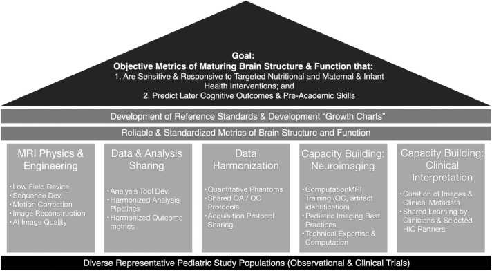
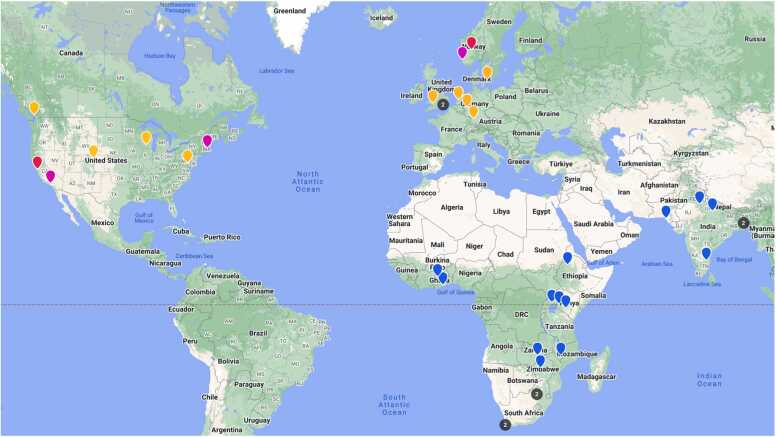
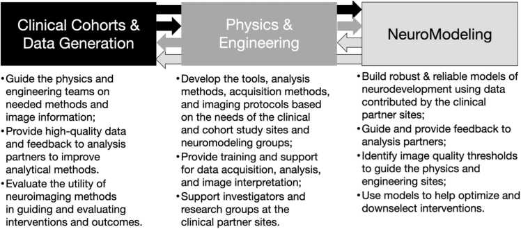
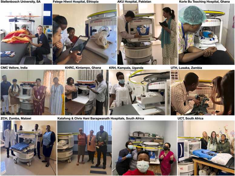
Measures of physical growth, such as weight and height have long been the predominant outcomes for monitoring child health and evaluating interventional outcomes in public health studies, including those that may impact neurodevelopment. While physical growth generally reflects overall health and nutritional status, it lacks sensitivity and specificity to brain growth and developing cognitive skills and abilities. Psychometric tools, e.g., the Bayley Scales of Infant and Toddler Development, may afford more direct assessment of cognitive development but they require language translation, cultural adaptation, and population norming. Further, they are not always reliable predictors of future outcomes when assessed within the first 12-18 months of a child's life. Neuroimaging may provide more objective, sensitive, and predictive measures of neurodevelopment but tools such as magnetic resonance (MR) imaging are not readily available in many low and middle-income countries (LMICs). MRI systems that operate at lower magnetic fields (< 100mT) may offer increased accessibility, but their use for global health studies remains nascent. The UNITY project is envisaged as a global partnership to advance neuroimaging in global health studies. Here we describe the UNITY project, its goals, methods, operating procedures, and expected outcomes in characterizing neurodevelopment in sub-Saharan Africa and South Asia.

Keywords: Child Health; Environmental Adversity; Global Health; Healthy Development; Low Field Magnetic Resonance Imaging; Neurodevelopment.


Conflict of interest statement

Declaration of Competing Interest The authors declare the following financial interests/personal relationships which may be considered as potential competing interests: H Frail, R O’Halloran, F Padormo, M Poorman, J Rogers, L Sacolick, K Siddiqui, R Teixeira, M Traughber are employees of Hyperfine.io W Hollander, T. Karaulanov, and C Weiant are employees of CaliberMRI. C Akgun, and P Velasco are employees of Flywheel.io

Figures

Fig. 1
Fig. 1
The UNITY project aims to identify sensitive, responsive, and predictive measures of maturing brain structure and function by characterizing patterns of neurodevelopment across a large and diverse meta-cohort of children. To achieve this aim, the GlobalMap project includes 5 areas or ‘pillars’ of focus: 1. MRI physics and engineering to develop and optimize novel acquisition methods tailored for low-field MRI; 2. Data and analysis method sharing; 3. Data harmonization through shared protocols, phantoms, and rigorous QA/QC protocols; 4. Capacity building in low-field pediatric neuroimaging through site-by-site training of research and clinical personnel on patient handling, data acquisition, and data analysis; and 5. Capacity building in MRI physics and low-field MR image interpretation.
Fig. 2
Fig. 2
Research sites that comprise the UNITY network include data collection and clinical partner sites (blue), physics and engineering development groups (orange), neuromodeling and analysis groups (pink), and education and clinical capacity groups (red). Black dots correspond to two sites in close proximity.
Fig. 3
Fig. 3
General flow of knowledge and interaction between the main UNITY network components.
Fig. 4
Fig. 4
A snapshot of images of the Hyperfine Swoop systems installed at many of the identified UNITY sites, including members of the study teams.
Fig. 5
Fig. 5
Pictures of Swoop arriving at a clinical cohort site. Following unloading of the scanner and accessory crate (a and b), the scanner crate is opened and protectors removed (c). The scanner can then be driven out of the grant and into the facility (d, e, and f) to its desired location. Here final set up and unpacking is performed (g) before being ready to image. From arrival to set up can take between 30 and 60 minutes depending on access and distance from delivery point to final scan room.
Fig. 6
Fig. 6
Pictures of In-person training beginning with a general safety orientation and introduction to the scanner (a), demonstrations and training on scanner driving and positioning (b), initial protocol setup and scanner interface (c), practice scanning on each other (d) and, finally, positioning and scanning of infants and toddlers (e and f).
Fig. 7
Fig. 7
Example (top) 3 T T1-weighted and (bottom) 64mT T2-weighted anatomical images of a 9-year old male child. Owing to differences in T1 and T2 relaxation parameters, T2-weighted imaging is preferred at low-field and is the primary image contrast of the UNITY common protocol.
Fig. 8
Fig. 8
Representative quantitative maps and images from the advanced acquisition methods, including single and multiple component relaxometry for quantitative T1, T2, and myelin water fraction imaging, magnetization transfer imaging, isotropic diffusion-weighted imaging, and diffusion tensor imaging and tractograhy of the cortico-spinal tracts and splenium of the corpus callosum. These methods are intended to provide increased sensitivity to microstructure change associated with early neurodevelopment and the impact of nutritional and other interventions.
Fig. 9
Fig. 9
Images of child participants in Hyperfine Swoop ranging in age from 1 to 3 months (a, b) using a baby tray positioning insert and foam head pads, 1 year (c) using a blue Med-Vac Immobilizer and head pads, and 5 years (d) using just ear and head pads to minimize motion.
Fig. 10
Fig. 10
The CaliberMRI UNITY phantom (a) outside and (b) inside the scanner. The phantom includes 3 sets of T1, T2, and ADC mimics arranged in parallel trays within the phantom (c) that provide differing contrast on T1 and T2-weighted and quantitative images (d). In addition, the phantom includes a spatial resolution grid, and internal and external temperature strips (a). A positioning cradle allows the phantom to be unambiguously positioned in the scanner (b).
Fig. 11
Fig. 11
Data Flow. MRI and health history information collected at each site may be spread across different devices, including Hyperfine Cloud for Hyperfine MRI data (if institutionally allowed), PACS (if available) or on a local laptop. From here, raw MRI and contextual data are uploaded to Flywheel for analysis and sharing. MRI data of questionable quality or with potential artifacts or incidental findings can also be shared with Collective Minds for community feedback. Cohort-specific and UNITY-wide analyses performed on Flywheel then form the basis of scientific reports and publications.
Fig. 12
Fig. 12
(Bottom to top) Creation of the age- and country-specific, and global templates from data generated at each of the clinical cohort sites. At least 15 male and female datasets at each age will be combined to generate age-specific templates within each country. These age-specific templates will then be used to generate a country-specific template. Finally, country-specific templates will be combined into the global ‘world’ template.
Fig. 13
Fig. 13
Examples of segmentations on low-field pediatric data with current segmentation tools. (A) Hippocampal segmentation using SynthSeg. (B) Segmentation of the cortex with iBeat. (C) Skull stripping performed using SynthStrip. On each image, white arrows point to examples of regions that were given the wrong tissue label.
Fig. 14
Fig. 14
Flowchart encompassing our overall strategy for the implementation of segmentation tools for the Hyperfine Swoop scanner.

References

    1. Anderson P.J., Burnett A. Assessing developmental delay in early childhood - concerns with the Bayley-III scales. Clin. Neuropsychol. 2017;31:371–381. - PubMed
    1. Anjos T., Altmae S., Emmett P., Tiemeier H., Closa-Monasterolo R., Luque V., Wiseman S., Perez-Garcia M., Lattka E., Demmelmair H., Egan B., Straub N., Szajewska H., Evans J., Horton C., Paus T., Isaacs E., van Klinken J.W., Koletzko B., Campoy C., Nutrimenthe Research Group Nutrition and neurodevelopment in children: focus on NUTRIMENTHE project. Eur. J. Nutr. 2013;52:1825–1842. - PubMed
    1. Ashburner J., Friston K.J. Voxel-based morphometry--the methods. Neuroimage. 2000;11:805–821. - PubMed
    1. Avants B.B., Tustison N.J., Wu J., Cook P.A., Gee J.C. An open source multivariate framework for n-tissue segmentation with evaluation on public data. Neuroinformatics. 2011;9:381–400. - PMC - PubMed
    1. Bick J., Nelson C.A. Early experience and brain development. Wiley Inter. Rev. Cogn. Sci. 2017;8 - PMC - PubMed

Publication types