. 2024 Sep:141:103715.
doi: 10.1016/j.dnarep.2024.103715. Epub 2024 Jul 18.

Human translesion DNA polymerases ι and κ mediate tolerance to temozolomide in MGMT-deficient glioblastoma cells

Affiliations

Human translesion DNA polymerases ι and κ mediate tolerance to temozolomide in MGMT-deficient glioblastoma cells

Marcela Teatin Latancia et al. DNA Repair (Amst). 2024 Sep.

Abstract

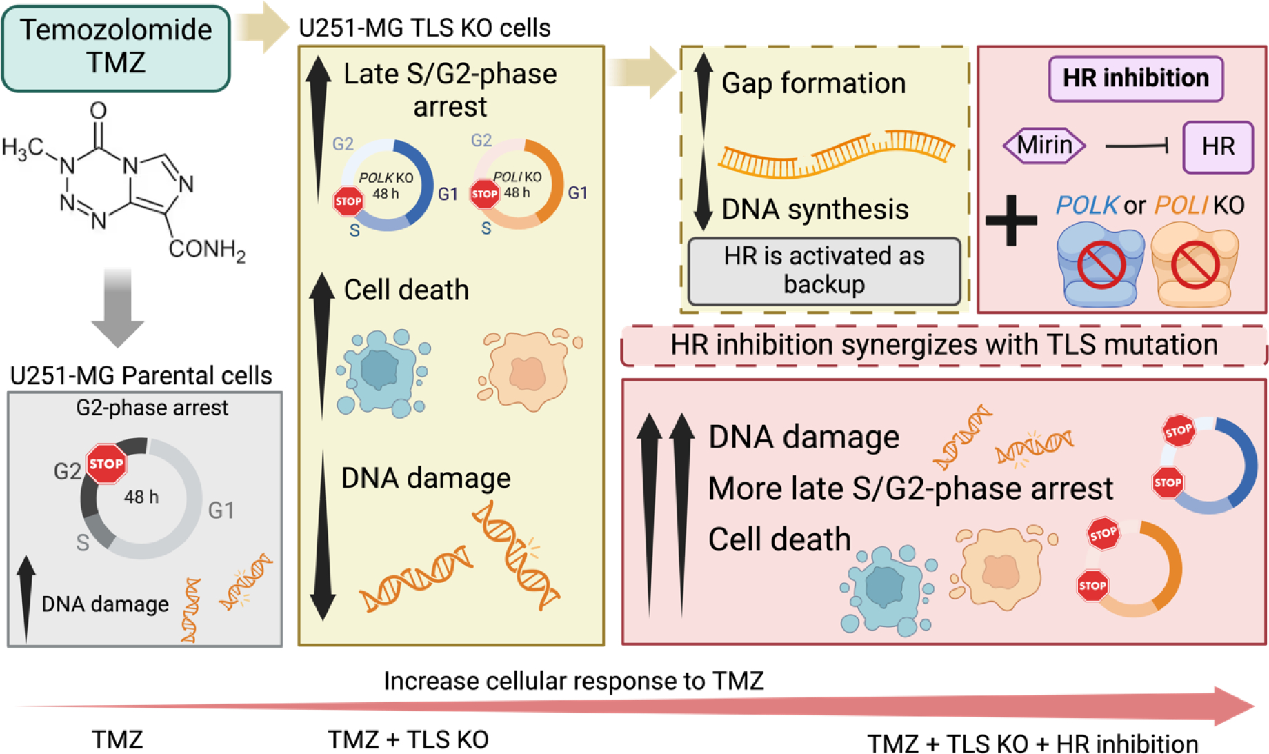
Glioblastoma (GBM) is a highly aggressive brain tumor associated with poor patient survival. The current standard treatment involves invasive surgery, radiotherapy, and chemotherapy employing temozolomide (TMZ). Resistance to TMZ is, however, a major challenge. Previous work from our group has identified candidate genes linked to TMZ resistance, including genes encoding translesion synthesis (TLS) DNA polymerases iota (Polɩ) and kappa (Polκ). These specialized enzymes are known for bypassing lesions and tolerating DNA damage. Here, we investigated the roles of Polɩ and Polκ in TMZ resistance, employing MGMT-deficient U251-MG glioblastoma cells, with knockout of either POLI or POLK genes encoding Polɩ and Polκ, respectively, and assess their viability and genotoxic stress responses upon subsequent TMZ treatment. Cells lacking either of these polymerases exhibited a significant decrease in viability following TMZ treatment compared to parental counterparts. The restoration of the missing polymerase led to a recovery of cell viability. Furthermore, knockout cells displayed increased cell cycle arrest, mainly in late S-phase, and lower levels of genotoxic stress after TMZ treatment, as assessed by a reduction of γH2AX foci and flow cytometry data. This implies that TMZ treatment does not trigger a significant H2AX phosphorylation response in the absence of these proteins. Interestingly, combining TMZ with Mirin (double-strand break repair pathway inhibitor) further reduced the cell viability and increased DNA damage and γH2AX positive cells in TLS KO cells, but not in parental cells. These findings underscore the crucial roles of Polɩ and Polκ in conferring TMZ resistance and the potential backup role of homologous recombination in the absence of these TLS polymerases. Targeting these TLS enzymes, along with double-strand break DNA repair inhibition, could, therefore, provide a promising strategy to enhance TMZ's effectiveness in treating GBM.

Keywords: Cancer; Chemotherapy resistance; DNA polymerase; Glioma; Temozolomide; Translesion synthesis.


Conflict of interest statement

Declaration of Competing Interest CFMM is an Editorial Board Member of DNA Repair. RW is an Associate Editor of DNA Repair. Neither were involved in the editorial review or the decision to publish this article.

Figures

Figure 1:
Figure 1:. Characterization of genetically modified U251-MG cells by CRISPR/Cas9.
Exon 3 of POLI and Exon 2 of POLK were targeted in U251-MG cell populations using the lentiCRISPR v2 Cas9 vector and the indicated gRNA sequences. (A). POLI KO clone B10 contains a homozygous combined indel – the blue residues on the parental sequence have been replaced by the red residues in the KO sequence and introduce a premature STOP codon. (B) POLK KO clone B11 contains different deletions of the two alleles, as indicated by dashes (−). The 2 bp deletion introduces a premature STOP codon, while the 12 bp deletion results in the deletion of residues R18, M19, G20 and L21. The inserts in the top left of each figure represent the western blot for the specific proteins (A- POLI, B- POLK).
Figure 2:
Figure 2:. Polκ and Polɩ TLS deficient cells are more sensitive to TMZ.
(A) After 72 h of TMZ treatment there is a significant decrease in cell viability, as visualized by the XTT colorimetric assay, of cells lacking POLK or POLI (KO), compared to parental cells. (n=5) (B) Apoptotic cells were determined by the presence of Annexin positive cells after 24 h or 48 h of TMZ treatment. (n=3) (C) Protein reconstitution in TLS KO cells shows that TMZ lethality is due to the absence of TLS polymerases. POLK WT-HA plasmids rescued the cell viability of POLK KO cells. POLI WT-HA plasmids also rescued the cell viability of POLI KO cells. The western blots presented in the right confirm that the transfections were successful. The pCMV6 vector is an empty plasmid used as a negative control plasmid in this over-expression assay. (n=3, * p < 0.05, ** p < 0.01, *** p < 0.001, **** p < 0.0001)
Figure 3:
Figure 3:. Wound Healing Assay showing that the absence of Polκ and Polɩ impacts cell migration and viability after exposure to TMZ.
The mutated cells have a much slower wound closure (thus, slower migration) following TMZ treatment, compared to parental cells. (n=3, * p < 0.05)
Figure 4:
Figure 4:. TLS KO U251-MG cells undergo cell cycle arrest in the late-S/G2 phase after TMZ treatment.
Cells in black color are parental cells, cells in blue color are POLK KO cells, and cells in orange color are POLI KO cells. The first column shows cells that were not treated, the second column shows cells treated with a low concentration of 50 μM TMZ and last, a higher concentration of TMZ was used: 400 μM TMZ. The first panel shows representative data after 24 h of TMZ treatment. The second panel shows representative data after 48 h of TMZ treatment. The quantification of n = 3 experimental replicates made with technical duplicates is presented in the bar graphic after 48 h of TMZ (50 μM) treatment, measured by flow cytometry. (* p < 0.05, ** p < 0.01, **** p < 0.0001)
Figure 5:
Figure 5:. POLK KO and POLI KO cells show impaired cell cycle progression.
High-content immunofluorescence assay was used to evaluate cell cycle progression. In black: U251-MG parental cells; in blue: U251-MG POLK KO cells; and in orange: U251-MG POLI KO cells. Non-treated result is indicated as NT and 50 μM TMZ treatment is indicated as TMZ. (A) Individual cells were analyzed by DNA content (DAPI fluorescent intensity) using DAPI stain microscopy and distributed in a histogram. TMZ treatment was performed for 24 h and 48 h, as indicated. (B) A click-it system to assess base incorporation with the thymidine analog EdU for 30 min by high-content microscopy to evaluate S phase among the cell lines in different times, 24 h and 48 h treated, or not, with 50 μM TMZ. Representative assay shows quantification of G1-phase cells (in red), G2-phase cells (in green) and S-phase (in dark blue) is divided between high incorporation of EdU or low incorporation of EdU.
Figure 6:
Figure 6:. TLS deficient cells present less γH2AX activation compared to parental cells.
In black: U251-MG parental cells; in blue: U251-MG POLK KO cells; and in orange: U251-MG POLI KO cells. (A) The γH2AX foci formation was assessed by immunofluorescence 24 h and 48 h after 50 μM TMZ treatment. The number of foci per cell increases with time but is lower in TLS KO cells compared to parental cells, particularly 48 h after treatment. Figure S2 shows the complete representative image without cropping. Three independent experiments, at least 100 cells counted/group. (B) Percentage of γH2AX positive cells determined by flow cytometry. Note that there is a reduction in the labelled γH2AX positive in the TLS KO cells compared to parental cells. (n=5, * p < 0.05, ** p < 0.01, **** p < 0.0001)
Figure 7:
Figure 7:. Combined treatment of TMZ and Mirin decreased cell viability in TLS KO cells.
(A) Mirin decreased cell viability in mutant cells after 3 days of treatment assessed by the XTT colorimetric assay, when combined with TMZ, but it did not change parental cells viability (n=3). (B) Colony formation assay (7 days after TMZ treatment) corroborates the results observed by the XTT colorimetric assay. Thus, the combined treatment of Mirin and TMZ is more effective in TLS KO cells compared to parental cells. (n=5, * p < 0.05, ** p < 0.01, *** p < 0.001, **** p < 0.0001, ns = non-significant)
Figure 8:
Figure 8:. TLS mutant cells upon combined treatment of Mirin plus TMZ show an increase in the DNA breaks.
(A) The alkaline comet assay shows that TMZ induces DNA breaks, but only when combined with exposure to Mirin, parental cells show a decrease in DNA breaks, while POLK KO cells show an important increased in DNA breaks. (n=4, **** p < 0.0001. Smaller **** showed on top of TLS mutant cells group are relative to parental cell on those same conditions); (B) Cells were harvested 48 h after cell treatment and marked for flow cytometry, and quantification was based on positive cells. (n=3, ** p < 0.01, *** p < 0.001, **** p < 0.0001)
Figure 9:
Figure 9:. Strategically inhibiting HR with Mirin, combined with TLS deficiency, enhances the vulnerability of glioblastoma cells to TMZ.
TMZ treated glioma cells, after the second round of replication, present increased DNA death. In the absence of Polκ or Polɩ, POLK or POLI KO cells, TMZ treatment leads to decreased cell viability and, after the second round of replication, an increase in late S/G2-phase arrest. However, there is a decrease in DNA damage. We propose that in these cells, the lack of TLS polymerases is compensated by other DNA repair mechanisms, possibly HR, which could be triggered by an increase in DNA gaps in the replicating DNA, as gaps are important substrates for HR. This backup by HR in TLS KO cells can explain the synergistic effect of using Mirin, an HR inhibitor. When Mirin is used in combination with TMZ, cells have decreased survival, increased in cell cycle arrest, and DNA damage.

References

    1. Menck CF, Munford V, DNA repair diseases: What do they tell us about cancer and aging?, Genet Mol Biol, 37 (2014) 220–233, 10.1590/s1415-47572014000200008. - DOI - PMC - PubMed
    1. Kannouche PL, Lehmann AR, Ubiquitination of PCNA and the polymerase switch in human cells, Cell Cycle, 3 (2004) 1011–1013. - PubMed
    1. Goodman MF, Woodgate R, Translesion DNA polymerases, Cold Spring Harb Perspect Biol, 5 (2013) a010363, 10.1101/cshperspect.a010363. - DOI - PMC - PubMed
    1. Aguilera A, Gomez-Gonzalez B, Genome instability: a mechanistic view of its causes and consequences, Nat Rev Genet, 9 (2008) 204–217, 10.1038/nrg2268. - DOI - PubMed
    1. Prakash S, Johnson RE, Prakash L, Eukaryotic translesion synthesis DNA polymerases: specificity of structure and function, Annu Rev Biochem, 74 (2005) 317–353, 10.1146/annurev.biochem.74.082803.133250. - DOI - PubMed

Publication types

MeSH terms

LinkOut - more resources