. 2024 Jul 18;12(7):e009070.
doi: 10.1136/jitc-2024-009070.

Generation of non-genetically modified, CAR-like, NK cells

Affiliations

Generation of non-genetically modified, CAR-like, NK cells

Loïs Coënon et al. J Immunother Cancer. .

Abstract

Background: Natural killer (NK) cell therapy is considered an attractive and safe strategy for anticancer therapy. Nevertheless, when autologous or allogenic NK cells are used alone, the clinical benefit has been disappointing. This is partially due to the lack of target specificity. Recently, CD19-specific chimeric antigen receptor (CAR)-NK cells have proven to be safe and potent in patients with B-cell tumors. However, the generation of CAR-NK cells is a complicated manufacturing process. We aim at developing a targeted NK cell therapy without the need for cellular genetic modifications. We took advantage of the natural expression of the IgG Fc receptor CD16a (FcγRIIIa) to induce strong antigen-specific effector functions through antibody-dependent cell-mediated cytotoxicity (ADCC). We have generated the new technology "Pin", which enables the arming of modified monoclonal antibodies (mAbs) onto the CD16a of ex vivo expanded NK (eNK) cells. Methods Ex vivo eNK were prepared from umbilical cord blood cells and expanded using interleukin (IL)-2/IL-15 and Epstein-Barr virus (EBV)-transformed B-lymphoblastoid feeder cells. mAbs were engineered with four substitutions called Pin mutations to increase their affinity to CD16a. eNK were incubated with anti-CD20 or anti-CD19 Pin-mAbs to generate "armed" eNK and were used to assess effector functions in vitro on cancer cell lines, lymphoma patient cells and in vivo.

Results: CD16a/Pin-mAb interaction is stable for several days and Pin-mAb eNK inherit the mAb specificity and exclusively induce ADCC against targets expressing the cognate antigen. Hence, Pin-mAbs confer long-term selectivity to eNK, which allows specific elimination of the target cells in several in vivo mouse models. Finally, we showed that it is possible to arm eNK with at least two Pin-mAbs simultaneously, to increase efficacy against heterogenous cancer cell populations.

Conclusions: The Pin technology provides an off-the-shelf NK cell therapy platform to generate CAR-like NK cells, without genetic modifications, that easily target multiple tumor antigens.

Keywords: Adoptive cell therapy - ACT; Immunotherapy; Monoclonal antibody; Natural killer - NK.


Conflict of interest statement

Competing interests: The patent of PINTM technology has been licensed to CYTEA BIO (WO2022023581A1 “Armed NK cells for universal cell therapy”). ER, HC, BF and JP are currently employees of CYTEA BIO. BR, PM and MV were initial creators of CYTEA BIO.

Figures
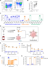
Figure 1
Figure 1. eNK cells armed with Pin-CD20 exhibit long-term binding in vitro and in vivo. (A) Representative dot plot showing UCB-derived NK cells purity before (left) and after 14 days of expansion (right). NK cell frequency is indicated within the dot plot. (B) eNK cytotoxicity against K562 cell lines at effector to target ratio 1:1 and 3:1 at 4 hours. n=7 individuals eNK donors represented, Wilcoxon matched-pairs test. (C) eNK phenotypic analysis of indicated markers. The bar graph shows individual eNK donors (n=6) and mean±SEM is shown. (D) Pin-mAbs eNK cells manufacturing scheme. Mutated Fc region (Pin-Fc): red; FcγR CD16a: blue. UCB-derived eNK cells are incubated with Pin-mAbs, allowing its arming onto CD16a expressing eNK cells. Created with BioRender.com. (E and F) Comparison of arming efficiency in rituximab-armed eNK (RTX eNK) and Pin-CD20-armed eNK (Pin-CD20 eNK) over a 72 hours kinetic, eNK cells represent the baseline. Representative histogram plot for one eNK donor (F) and bar graphs representing the frequency of armed cells (left) and the arming geometric mean of fluorescence intensity (gMFI, right) detected by an anti-rituximab idiotype antibody (E) are shown. n=9 eNK donors; two-way analysis of variance with Tukey’s test, difference between RTX eNK and Pin-CD20 eNK groups is shown; mean±SEM is shown. (G) Arming stability in vivo. Pin-CD20 eNK cells arming efficiency was assessed 24 hours after intraperitoneal injection in SCID mice. eNK, n=2; Pin-CD20 eNK, n=2. mean±SEM is shown. eNK, expanded NK; mAb, monoclonal antibodies; NK, natural killer; UCB, umbilical cord blood.
Figure 2
Figure 2. Pin-CD20 eNK cells display enhanced and specific antibody-dependent cellular cytotoxicity and activation on CD20 positive B-lymphoma cell lines recognition. (A) Schematic overview of in vitro functional assays performed to evaluate Pin-CD20 eNK cells. Flow cytometry: FC; perforin: Prf; granzyme B: GrzB. Created with BioRender.com. (B and C) Cytotoxicity assays comparing eNK versus Pin-CD20 eNK were performed on Daudi (n=6 eNK donors) and Toledo (n=3 eNK donors) B-lymphoma cell lines at different E:T ratios by flow cytometry. Two-way ANOVA with Sidak’s test and mean±SEM are shown (B). Four parameter logistic model was used to measure E:T ratios that lyse 20% (E:T20), 50% (E:T50) or 80% (E:T80) of the target cells (C). (D) eNK cells were armed with increasing concentration of Pin-CD20 mAbs and cytotoxicity was measured using E:T ratio ET80 measured for two eNK donors (from b, c). Mean±SEM is shown. (E) Cytotoxicity assay against Daudi Luciferase cell line using one eNK donor armed with Pin-CD20, Pin-EGFR, Pin-HER2 or unarmed. Mean±SEM is shown. (F) CD107a expression on eNK or Pin-CD20 eNK in the presence or absence of Daudi cells assessed by flow cytometry after 4 hours incubation at E:T ratio 3:1. n=5 eNK donors; Two-way ANOVA with Tukey’s test; mean±SEM is shown. (G) Cytotoxic mediators secretion from eNK or Pin-CD20 eNK in presence or absence of Daudi cells at E:T ratio 1:1. Data are shown as fold change normalized with eNK unstimulated (norm. eNK uns). n=8 eNK donors; Two-way ANOVA with Tukey’s test mean±SEM is shown. *p≤0.05; **p≤0.01; ***p≤0.001; ****p≤0.0001. ANOVA, analysis of variance; eNK, expanded natural killer; E:T, effector to target; IFN, interferon; TNF, tumor necrosis factor.
Figure 3
Figure 3. Pin-mAbs eNK are highly functional for several days, independently of the CD16a F158V polymorphism. (A and B) eNK cells were armed with Pin-CD20 or not and used in cytotoxicity assays against Daudi cells after 1 day (Day 1), 2 days (Day 2) incubation or no incubation time (Day 0) at E:T ratio 10:1. (A) Target cell death at E:T ratio 10:1 was measured by flow cytometry. N=10 eNK donors; paired t-test mean±SEM is shown; **p≤0.01. (B) Mean cytotoxicity of eNK and Pin-CD20 eNK over time. Mean±SEM is shown. (C) Comparison of Pin-CD20 arming efficiency in CD16a V/V, F/V or F/F donors over a 48 hours kinetic. Dot graphs represent the frequency of armed cells (left) and the arming geometric mean of fluorescence intensity (gMFI, right) detected by an anti-human Fab antibody. n=6 F/F, 6 F/V, 5 V/V eNK donors. Two-way analysis of variance with Tukey’s test mean±SEM is shown; ns=not significant. (D) Pin-ADCC effect of Pin-CD20 eNK at D0, D1 and D2 in CD16 V/V, F/V or F/F eNK donors at E:T ratio 10:1. Pin-ADCC was calculated by subtracting eNK natural cytotoxicity to Pin-CD20 eNK total cytotoxicity. n=4 F/F, 3 F/V and 3 V/V eNK donors. Mean±SEM is shown. (E, F and G) eNK and Pin-CD20 eNK were cultured or not with CFSE Daudi at E:T ratio 3:1 for 24 hours. CD16 expression (frequency and gMFI) was compared between t0h and t24h. (E) Frequency of armed eNK cells was measured at t1h and t24h (F). At 25 hours, eNK and Pin-CD20 eNK were challenged a second time with CellTrace Far Red Daudi at E:T ratio 3:1 for another 24 hours. Target cell count (CFSE, line; Far Red, dotted line) was measured at 24 hours and 48 hours (G). ADCC, antibody-dependent cellular cytotoxicity; CFSE, carboxyfluorescein succinimidyl ester; eNK, expanded NK; E:T, effector to target; NK, natural killer.
Figure 4
Figure 4. Pin-CD20 shows target specificity and enhanced cytotoxicity in vivo compared with unarmed eNK. (A) in vitro validation of CD20+ B cells targeting compared with other PBMC cell types (T cells, NK cells, monocytes) when eNK or Pin-CD20 eNK were co-incubated with PBMCs from healthy donors during 16 hours at effector to target ratio 1:1. PBMCs populations were then assessed by flow cytometry. n=6 samples (two experiments: one eNK donor tested on one PBMC donor and two eNK donors tested on two PBMC donors). Multiple paired t-tests and mean±SEM are shown. (B) 3×10⁶ eNK or Pin-CD20 eNK were injected intravenously into human CD34+reconstituted humanized NCG mice (Hu-NCG, 28 days after reconstitution). Relative percentage difference of peripheral blood CD20+B cells (left) and CD3+T cells (right) number at D7 compared with D1 was assessed by FC. eNK, n=6 mice; Pin-CD20 eNK, n=6 mice. Unpaired t-tests and mean±SEM are shown. (C) 1×10⁶ eNK or Pin-CD20 eNK were co-injected intraperitoneally in NSG mice with 1×10⁶ Raji and 1×10⁶ Nalm6 cells. Cells were harvested after 4 hours, and target cell number was measured by flow cytometry. Vehicle, n=8 mice; eNK, n=8 mice; Pin-CD20 eNK, n=7 mice. Two-way analysis of variance with Tukey’s test and mean±SEM are shown. *p≤0.05; **p≤0.01; ***p≤0.001; ****p≤0.0001. (D, E and F) In vivo leukemia mouse model. (D) In vivo model timeline. (E and F) At day 0, 2×10⁵ Raji cells were inoculated intravenous into SCID mice. At day 4 and 8, mice were treated intravenous with saline (vehicle), 30×106 eNK or Pin-CD20 eNK, followed by intravenous injection of IL-15 at day 11, 14 and 17. At day 28, bone marrow, lungs and liver were harvested for detection of CD20+ Raji cells by FC. (E) Bar graphs represent the frequency of Raji cells in each organ. (F) Table reporting the frequency of mice that present Raji cells in each organ (lymphoma incidence). Vehicle, n=4 mice; eNK, n=6 mice; Pin-CD20 eNK, n=7 mice. Kruskal-Wallis tests, p values and mean±SEM are shown. eNK, expanded NK; IL, interleukin; NK, natural killer; PBMC, peripheral blood mononuclear cells.
Figure 5
Figure 5. Pin-CD20 eNK and Pin-CD19 eNK efficiently kill patient with B-lymphoma cells. Patient with lymphoma cells from three B-lymphoma subtypes were used: mantle cell lymphoma (MCL), diffuse large B cell lymphoma (DLBCL) and follicular lymphoma (FL). Patient cells were co-cultured with eNK, Pin-CD20 eNK or Pin-CD19 eNK for 16 hours at effector to target ratio 3:1 and target cell death was assessed by FC. (A) Frequency of target cell death in the three lymphoma subtypes. MCL, n=6 patients; DLBCL, n=6 patients; FL, n=5 patients; for each patient sample, two to four eNK donors were tested. Two-way analysis of variance with Tukey’s test and mean±SEM are shown. *p≤0.05; ***p≤0.001; ****p≤0.0001. (B) Correlation between the average number of CD20 and CD19 molecules per cell and ADCC efficiency of Pin-CD20 eNK (left) and Pin-CD19 eNK (right). ADCC is the cytotoxicity measured above natural cytotoxicity from eNK cells. MCL, n=6 patients; DLBCL, n=4 patients; FL, n=4 patients. ADCC, antibody-dependent cellular cytotoxicity; eNK, expanded natural killer.
Figure 6
Figure 6. eNK cells can efficiently be armed with multiple Pin-monoclonal antibodies and remain actively cytotoxic. (A, B and C) eNK cells were armed with Pin-CD20, Pin-CD19 or both (double-armed eNK) using fluorescently labeled pin-CD20 (Pin-CD20-AF647) and pin-CD19 (Pin-CD19-AF488) and analyzed 1 hour, 8 hours, 24 hours, 48 hours and 72 hours after. (A) Representative dot plots of Pin-CD20-AF647 and Pin-CD19-AF488 arming for each condition. (B) Frequency of eNK cells armed with Pin-CD20 (left) or Pin-CD19 (right). (C) Frequency of eNK cells armed simultaneously with both Pin-CD20 and Pin-CD19 (double-armed). n=4 eNK donors. Data are shown as mean±SEM. (D) Patient with lymphoma cells were co-cultured with eNK or Pin-CD20 and/or Pin-CD19 eNK for 16 hours. Tumor cell death was assessed by flow cytometry. Mean±SEM is shown, MCL, n=3; DLBCL, n=4; FL, n=3. For each patient sample, two to three eNK donors were tested. (E) 1×10⁶ eNK, Pin-CD20 eNK, Pin-CD19 eNK or double-armed eNK were intraperitoneally co-injected with 1×10⁶ Daudi and 1×10⁶ Nalm6 cells into NSG mice. Peritoneal lavage were performed after 4 hours and the number of target cell was analyzed by flow cytometry. Vehicle, n=3 mice; eNK, n=5 mice; Pin-CD20 eNK, n=5 mice; Pin-CD19 eNK, n=5 mice; double-armed eNK, n=5 mice. Two-way analysis of variance with Sidak’s test and mean±SEM are shown, *p≤0.05; **p≤0.01; ***p≤0.001; ****p≤0.0001; ns, not significant. eNK, expanded natural killer; DLBCL, diffuse large B cell lymphoma; FC, follicular lymphoma; MCL, mantle cell lymphoma.

References

    1. Olson JA, Leveson-Gower DB, Gill S, et al. NK cells mediate reduction of GVHD by inhibiting activated, alloreactive T cells while retaining GVT effects. Blood. 2010;115:4293–301. doi: 10.1182/blood-2009-05-222190. - DOI - PMC - PubMed
    1. Bachanova V, Burns LJ, McKenna DH, et al. Allogeneic natural killer cells for refractory lymphoma. Cancer Immunol Immunother. 2010;59:1739–44. doi: 10.1007/s00262-010-0896-z. - DOI - PMC - PubMed
    1. Koehl U, Kalberer C, Spanholtz J, et al. Advances in clinical NK cell studies: donor selection, manufacturing and quality control. Oncoimmunology. 2016;5:e1115178. doi: 10.1080/2162402X.2015.1115178. - DOI - PMC - PubMed
    1. Villalba M, Alexia C, Bellin-Robert A, et al. Non-genetically improving the natural cytotoxicity of natural killer (NK) cells. Front Immunol. 2019;10:3026. doi: 10.3389/fimmu.2019.03026. - DOI - PMC - PubMed
    1. Allison M, Mathews J, Gilliland T, et al. Natural killer cell-mediated immunotherapy for leukemia. Cancers (Basel) 2022;14:843. doi: 10.3390/cancers14030843. - DOI - PMC - PubMed

MeSH terms

LinkOut - more resources