. 2024 Jul 19;15(1):6080.
doi: 10.1038/s41467-024-50437-3.

Pan-serotype dengue virus inhibitor JNJ-A07 targets NS4A-2K-NS4B interaction with NS2B/NS3 and blocks replication organelle formation

Affiliations

Pan-serotype dengue virus inhibitor JNJ-A07 targets NS4A-2K-NS4B interaction with NS2B/NS3 and blocks replication organelle formation

Dominik Kiemel et al. Nat Commun. .

Abstract

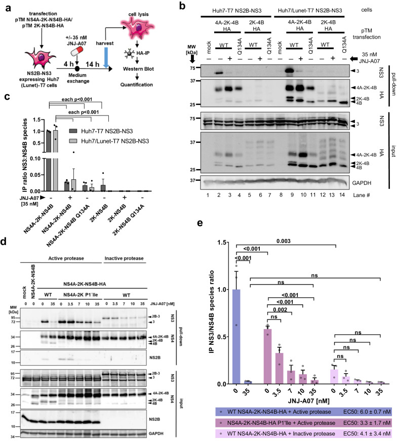
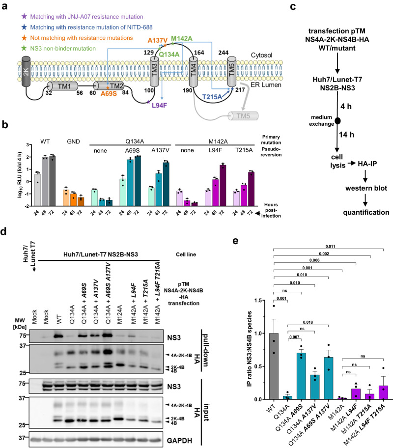
Dengue fever represents a significant medical and socio-economic burden in (sub)tropical regions, yet antivirals for treatment or prophylaxis are lacking. JNJ-A07 was described as highly active against the different genotypes within each serotype of the disease-causing dengue virus (DENV). Based on clustering of resistance mutations it has been assumed to target DENV non-structural protein 4B (NS4B). Using a photoaffinity labeling compound with high structural similarity to JNJ-A07, here we demonstrate binding to NS4B and its precursor NS4A-2K-NS4B. Consistently, we report recruitment of the compound to intracellular sites enriched for these proteins. We further specify the mechanism-of-action of JNJ-A07, which has virtually no effect on viral polyprotein cleavage, but targets the interaction between the NS2B/NS3 protease/helicase complex and the NS4A-2K-NS4B cleavage intermediate. This interaction is functionally linked to de novo formation of vesicle packets (VPs), the sites of DENV RNA replication. JNJ-A07 blocks VPs biogenesis with little effect on established ones. A similar mechanism-of-action was found for another NS4B inhibitor, NITD-688. In summary, we unravel the antiviral mechanism of these NS4B-targeting molecules and show how DENV employs a short-lived cleavage intermediate to carry out an early step of the viral life cycle.


Conflict of interest statement

J.-F.B., S.J.F.K., J.N., M.vL., and O.G. have been named inventor in a patent application claiming the discovery of this class of antiviral molecules as DENV replication inhibitors (WO 2017/167951), which was filed by applicants Janssen Pharmaceuticals, Inc. and Katholieke Universiteit Leuven, and has been granted in certain countries. O.G., J.-F.B., M.vL., S.J.F.K. and J.N. have been named inventors in a pending patent application relating to the use of substituted indole derivatives and substituted indoline derivatives in the manufacture of a medicament for the treatment or the prevention of dengue disease in an individual at risk of being infected by DENV and to a method for the treatment or the prevention of dengue in an individual at risk of being infected by DENV, which was filed by Applicants Janssen Pharmaceuticals, Inc. and Katholieke Universiteit Leuven (WO 2021/094563). O.G., B.G. and M.vL. are all full-time employees of Janssen and potential stockholders of Johnson and Johnson. The other authors declare no competing interests.

Figures

Fig. 1
Fig. 1. Compound A specifically localizes to intracellular sites enriched for DENV NS4B and NS3.
a Schematic representation of the workflow to perform click-labeling and immunofluorescence staining on fixed cells that were treated with one of the PAL enantiomers or DMSO control (vehicle). Note that a click reaction with Picolyl-azide-Cy5.5 was performed regardless of treatment condition. b Example confocal microscopic images (60x). Cells were transfected with the pIRO-D WT construct encoding the NS1-5 polyprotein or mock transfected and treated with either 1 µM Compound A, 1 µM Compound B or vehicle. Shown are the PAL fluorescence signal, the IF signals of NS3 and NS4B and a merge that includes nuclear DNA stain with DAPI. Scale bar = 10 µm. c For each sample transfected with pIRO-D WT, 21 cell profiles (7 from each of the 3 independent experiments) were examined for Pearson correlation coefficients between the PAL and the NS4B signal. Shown is the mean with standard deviation (SD). A one-way ANOVA followed by Tukey’s multiple comparisons test was used to determine statistical parameters. d Specificity of Compound A colocalization with NS4B was determined using cells transfected with a ZIKV polyprotein encoding construct (pIRO-Z). Example images obtained as described for b are shown. Scalebar = 10 µm. e Quantification of PAL and ZIKV NS4B signal overlap as described for c. Per sample, 21 cell profiles (7 from each of the 3 independent experiments) were analyzed and plotted with mean and SD. The indicated p-value resulted from an unpaired two-tailed t-test. f NS4B mutations conferring various degrees of resistance against JNJ-A07 were examined for their impact on colocalization between NS4B and PAL signals. The corresponding IF panel is shown in Supplementary Fig. 3b. Pearson correlation coefficients of a total of 21 cell profiles are plotted (7 each from three independent experiments). Statistical significance was calculated using a two-way ANOVA test and Šidák’s multiple comparisons. ns = non-significant.
Fig. 2
Fig. 2. Compound A binds to mature NS4B and the NS4A-2K-NS4B precursor.
a Schematic representation of the workflow. Note that although cells were treated with either Compound A or DMSO control (vehicle), the click reaction was performed on the beads with Picolyl-azide-Cy5.5 azide in both cases. Also note that different subclones of Huh7/Lunet-T7 cells were used: naïve ones in which 2K-NS4B-HA or NS4A-2K-NS4B-HA were transiently expressed and a subclone stably expressing Calnexin-HA. b Representative western blot showing the input controls. Due to its stable expression, the signal of Calnexin-HA is stronger than the ones of the transiently expressed viral proteins. Mock samples were derived from Huh7/Lunet-T7 NS2B-NS3 cells treated with transfection reagent only. c Representative images to analyze compound binding to individual proteins. Pull-down samples were loaded onto a gel, which was analyzed for Cy5.5 fluorescence signal using a LICOR Odyssey imaging system (middle plane). Thereafter, proteins were analyzed by HA-specific western blot (top panel). An overlay of both images is shown at the bottom. Mock transfected samples in lanes 1 and 2 reflect the unspecific background in the absence of HA-tagged proteins. For each construct, there is a Compound A-treated sample and a vehicle-treated sample to distinguish between non-specific labeling and specific binding to Compound A. Owing to high expression level, Calnexin-HA samples were diluted 1:500 prior to loading to reach western blot signal intensities in a linear range comparable with the other samples. d Quantification of compound binding efficiency; shown is the ratio of the fluorescence signals (reflecting Compound A bound proteins) to the corresponding HA western blot signal (reflecting the target protein). Non-specific compound labelling was subtracted by using the analogous ratio of the individual vehicle control. Bar graph shows individual data points, as well as mean and standard error of the mean (SEM; n = 4). Indicated p-values were calculated using one-way ANOVA with subsequent Tukey’s multiple comparisons test.
Fig. 3
Fig. 3. Cleavage kinetics of the NS4A-2K-NS4B precursor in the polyprotein context is not affected by JNJ-A07.
a Experimental workflow. Huh7/Lunet-T7 cells were transfected with a pIRO-D construct corresponding to the NS1-5 polyprotein and containing an HA-affinity tag in the N-terminal region of NS4B (HA*) after the 2 K peptide. After 60 minutes starvation in methionine and cysteine-free medium, cells were incubated in radioactive medium for 20 min (pulse) followed by incubation for various time spans in non-radioactive medium (chase). Proteins were enriched by HA-IP and after SDS-PAGE analyzed by autoradiography. b A representative autoradiogram from a total of 3 independent experiments is shown. Numbers above the lanes refer to chase time (minutes). c Signal intensities of NS4B-specific bands were quantified and adjusted to cysteine and methionine residues contained therein. The proportion of NS4A-2K-NS4B amongst all NS4B-containing species is plotted with mean and SEM (n = 3). No statistically significant differences were found between JNJ-A07 and vehicle control-treated samples for any time point as determined by a Sidak’s multiple comparisons test following a two-way ANOVA. d “One phase exponential decay”-curves were fitted to the result of each experiment (n = 3) to obtain three NS4A-2K-NS4B half-life values for both the JNJ-A07 and vehicle control-treated setup that are plotted here with mean and SEM. The difference was not significant as determined by a paired two-tailed two sample t-test.
Fig. 4
Fig. 4. NS2B/NS3 interacts with NS4A-2K-NS4B, but not with mature NS4B, and this interaction is prevented by JNJ-A07 in the trans-cleavage system.
a Experimental approach: Huh7-T7 or Huh7/Lunet-T7 cells, each stably expressing the T7 RNA polymerase and DENV NS2B/NS3 were transfected with T7-based expression plasmids encoding either NS4A-2K-NS4B-HA or 2K-NS4B-HA with NS4B corresponding to the wild-type (WT) or containing the NS3-nonbinder mutation Q134A. After 4 h, cells were treated with JNJ-A07 or vehicle, collected 14 h thereafter and lysates were used for HA-specific pull-down. b Side-by-side comparison of NS3 co-precipitation with either NS4A-2K-NS4B-HA or 2K-NS4B-HA. Eluates were analyzed by western blot along with 25% of the input. A representative experiment is shown. Arrowheads indicate named DENV non-structural proteins, whereby the usual “NS” nomenclature has been omitted here for reasons of space. c The ratios of NS3 to NS4B species (NS4A-2K-NS4B, 2K-NS4B and NS4B) were determined by quantifying 3 independent experiments and are plotted with mean and SEM. A two-way ANOVA preceded by Dunnet´s test was conducted to determine given p-values. d Interaction between NS2B/NS3 and stabilized NS4A-2K-NS4B precursors. Huh7-T7 cells expressing either the active DENV protease complex or an inactive version thereof (NS3 mutant S135A) were transfected with indicated expression constructs. Shown is a representative western blot result (n = 3). e IP ratios of HA-precipitated NS4A-2K-NS4B and co-captured NS3 were calculated from 3 independent experiments and normalized to vehicle-treated WT NS4A-2K-NS4B co-expressed with the active protease. Mean and SEM are shown; p-values were calculated by Šidák’s multiple comparisons test following one-way ANOVA (ns = non-significant). EC50 values given in the bottom lines were calculated by fitting dose-response curves or taken from Kaptein et al., 2021 (for “WT NS4A-2K-NS4B-HA + Active protease” shown here as reference).
Fig. 5
Fig. 5. JNJ-A07 neither affects the cleavage order of NS4A-2K-NS4B, nor its interaction with NS1.
a Constructs specified on the top were transfected into Huh7/Lunet-T7 cells stably expressing NS2B-NS3. 4 h post-transfection, cells were treated with either 35 nM JNJ-A07 or vehicle. After another 14 h, samples were harvested and used for HA-specific pull-down. Captured proteins were analyzed by western blot. The C-terminally HA-tagged NS4A-2K-NS4B precursor was included as reference. b Representative western blot showing HA-pull-down samples and total lysates (25% of input). GAPDH served as loading control for cell lysates. The section highlighted in panel d is indicated with a blue dashed line box. c Bar graph showing the ratio of NS3 to NS4 species (mean and SEM) as determined by quantification of independent western blots (n = 3). P-values were calculated using one-way ANOVA with subsequent Šidák’s multiple comparisons test. d Sections of the western blots highlighted in b adjusted for brightness and contrast to highlight NS4A species in pTM HA-NS4B-2K-NS4B transfected samples. e Huh7/Lunet-T7 cells stably expressing NS1-HA were transfected with constructs encoding NS4A-2K-NS4B. After 4 h, cells were treated with given concentrations of JNJ-A07 or vehicle, collected 14 h thereafter and lysates were used for HA-specific pull-down. Captured protein complexes were analyzed by western blot. A representative example of 3 independent experiments is shown. GAPDH served as loading control for cell lysates (input). f, g Ratios of NS1 to NS4A-2K-NS4B in input and pull-down samples were calculated with mean and SEM by analyzing signal intensities in the 3 independent experiments. For statistical analysis, one-way ANOVA plus post-hoc test was performed using Dunnett’s correction to account for multiple testing.
Fig. 6
Fig. 6. JNJ-A07 inhibits the formation of VPs in WT but not in compound-resistant NS4B mutants.
a Experimental workflow. Huh7/Lunet-T7 cells were transfected with pIRO-D plasmids encoding DENV NS1-NS5 WT or containing compound resistance mutations in NS4B. Cells were treated with JNJ-A07 or vehicle and 14 h later, fixed for immunofluorescence (IF) and transmission electron microscopy (TEM) analysis or lysed for western blot. b Transfection efficiencies as derived from IF analyses. Shown is a combined scatter plot of transfection efficiencies across all 14 specimens over the two independent experiments presented with mean and SD. c Western blot of DENV proteins; GAPDH served as loading control. d TEM analysis of cells transfected with the WT pIRO-D construct. Detected morphological alterations comprise convoluted membranes (CM) and vesicle packets (VP), which are string-like arrays of vesicles, here denoted as vesicle packet elements (VPEs). Right panel: enlargement of the red boxed area in the left panel. e Representative electron micrographs showing VPEs detected in cells after transfection with constructs specified on the top. Yellow scale bar = 100 nm. f Quantification of VPEs from 2 × 20 cell profiles per transfected cell sample (n = 2 independent experiments), after normalization to the transfection efficiency as determined by IF (see panel b). For each construct, VPE formation efficiency determined with vehicle-treated control cells was set to 100. Results are shown with mean and SEM. P-values were calculated by Dunn’s multiple comparison test following a Kruskal-Wallis test. g For each sample, the diameter of 50 randomly selected VPEs was determined. Shown are the individual values with mean and standard deviation. A two-way ANOVA and subsequent Šidák’s multiple comparisons test were employed for statistical analysis.
Fig. 7
Fig. 7. Pseudoreversions in the NS3 non-binder mutants NS4B Q134A and M142A rescue the NS3–NS4B interaction.
a Membrane topology of NS4B according to Miller et al. and position of NS3-non-binder mutations Q134A and M142A (in green), along with their corresponding pseudo-reversions. b Replication kinetics of sgDVs-R2A replicons harboring the NS4B mutations Q134A or M142A with and without second-site pseudo-reversions. Adapted from Chatel-Chaix et al. WT and GND mutant served as positive and negative control, respectively. Luciferase values were normalized to the 4 h time-point reflecting transfection efficiency. Data are n = 3 and are presented with mean and SD. c Experimental approach to study the capability of pseudo-reversions to restore the interaction between NS4A-2K-NS4B and NS2B/NS3. d Representative western blot (n = 3 independent experiments). HA-captured protein complexes were analyzed along with the corresponding input (20%). GAPDH served as loading control. MW, molecular weight. e IP ratio of NS3 to NS4B-containing species (mean and SEM) from 3 independent experiments. For statistical significance analysis, a repeated measures one-way ANOVA with subsequent Šidák’s multiple comparisons test was applied.
Fig. 8
Fig. 8. Interaction between NS2B/NS3 and NS4A-2K-NS4B is functionally linked to vesicle packets formation.
a Experimental approach to analyze VPs formation by NS3 non-binder mutants and pseudo-revertants. Huh7/Lunet-T7 cells were transfected with pIRO-D plasmids encoding DENV NS1-NS5 WT or NS3 nonbinder mutations in NS4B (with or without compensating pseudoreversions) and fixed 18 h later for immunofluorescence (IF) and transmission electron microscopy (TEM) analysis, or lysed for western blot. b Representative western blot (n = 2 independent experiments). c Representative electron micrographs. Scale bar = 100 nm d Quantification of VPEs (20 cell profiles for each experiment). Values were normalized for transfection efficiency and are plotted with mean and SEM. P-values were calculated using Dunn’s multiple comparison test after a Kruskal-Wallis test. e Example images from two independent cell profiles illustrating vesicle elongation in pIRO-D NS4B M142A L94F T215A transfected cells. Colored arrowheads are specified on the bottom. Scale bar = 300 nm. f Quantitative comparison of VPE morphotypes in WT and triple mutant transfected cells. 200 VPEs per sample were analyzed. Compensatory pseudo-reversions are highlighted by bold and italic letters.
Fig. 9
Fig. 9. JNJ-A07 blocks de novo formation of vesicle packet elements but does not disrupt existing ones.
a Experimental approach. Cells were harvested and analyzed by TEM, WB and IF to determine VPE formation, polyprotein expression and processing as well as transfection efficiency, respectively. b At the tested concentration of 35 nM, JNJ-A07 shows no apparent effect on polyprotein processing, regardless of the time of addition. Shown is one of two western blots. c Example electron micrographs. Scale bar = 100 nm. d VPE formation efficiency with mean and SEM for early and late timepoint of compound addition, relative to vehicle-treated control cells. VPEs from 20 cell profiles per condition and independent experiment (n = 2) were quantified and normalized to transfection efficiencies. P-values were calculated by Dunn’s multiple comparison test that was conducted after a Kruskal-Wallis test. e To discriminate between block of VPE de novo formation and active VPE disruption, cells were pre-treated with cycloheximide (CHX; 200 µg/ml) 30 min prior to JNJ-A07 addition. Cells treated only with CHX served as reference. f Representative western blot of a ribopuromycylation assay showing CHX-mediated inhibition of protein synthesis. Cells were treated with puromycin (1 g/ml) 5 minutes before harvest. Translation rate is reflected by the amount of puromycin incorporated into polypeptide chains, which is detected by puromycin-specific western blot. g Exemplary electron micrographs showing VPEs in transfected cells as outlined in e. Scale bar = 100 nm. h VPE formation efficiency (mean and SEM). Values were normalized to vehicle-treated cells (vehicle) that were set to 100%. Data acquisition and statistical analysis were performed as for d.
Fig. 10
Fig. 10. Graphical summary of the mechanism-of-action of JNJ-A07.
Upper panels (left to right): During DENV polyprotein processing, the cleavage intermediate NS4A-2K-NS4B is formed, which engages interaction with NS1 and the viral protease complex NS2B/NS3. This hypothetical multiprotein complex, together with other factors (e.g. certain NTR regions of the viral genome and putative host cell factors), induced de novo formation of VPs, the assumed sites of viral RNA replication. Lower panels (left to right): When applied early, JNJ-A07 might bind specifically to solitary NS4A-2K-NS4B and impairs its interaction with NS2B/NS3, whereas binding of NS1 on the ER luminal side is not affected. This prevents de novo formation of VPs, thus blocking viral RNA replication. When JNJ-A07 is added after VPs have already formed, they remain intact and decay with their natural half-life. This assumption is consistent with our previous observation that JNJ-A07 cannot break established NS4A-2K-NS4B - NS2B/NS3 complexes and with the known gradual loss of antiviral activity of JNJ-A07 with increasing time between the onset of viral replication and compound application.

References

    1. Ferreira-de-Lima, V. H. & Lima-Camara, T. N. Natural vertical transmission of dengue virus in Aedes aegypti and Aedes albopictus: a systematic review. Parasit. Vectors11, 77 (2018). 10.1186/s13071-018-2643-9 - DOI - PMC - PubMed
    1. Murray, N. E. A., Quam, M. B. & Wilder-Smith, A. Epidemiology of dengue: past, present and future prospects. Clin. Epidemiol.5, 299 (2013). - PMC - PubMed
    1. Bhatt, S. et al. The global distribution and burden of dengue. Nature496, 504–507 (2013). 10.1038/nature12060 - DOI - PMC - PubMed
    1. Wilder-Smith, A., Ooi, E.-E., Horstick, O. & Wills, B. Dengue. Lancet393, 350–363 (2019). 10.1016/S0140-6736(18)32560-1 - DOI - PubMed
    1. Calisher, C. H. et al. Antigenic Relationships between Flaviviruses as Determined by Cross-neutralization Tests with Polyclonal Antisera. J. Gen. Virol.70, 37–43 (1989). 10.1099/0022-1317-70-1-37 - DOI - PubMed

MeSH terms

LinkOut - more resources