. 2024 Nov;16(11):1894-1905.
doi: 10.1038/s41557-024-01576-z. Epub 2024 Jul 19.

Protein semisynthesis reveals plasticity in HECT E3 ubiquitin ligase mechanisms

Affiliations

Protein semisynthesis reveals plasticity in HECT E3 ubiquitin ligase mechanisms

Hanjie Jiang et al. Nat Chem. 2024 Nov.

Abstract

Lys ubiquitination is catalysed by E3 ubiquitin ligases and is central to the regulation of protein stability and cell signalling in normal and disease states. There are gaps in our understanding of E3 mechanisms, and here we use protein semisynthesis, chemical rescue, microscale thermophoresis and other biochemical approaches to dissect the role of catalytic base/acid function and conformational interconversion in HECT-domain E3 catalysis. We demonstrate that there is plasticity in the use of the terminal side chain or backbone carboxylate for proton transfer in HECT E3 ubiquitin ligase reactions, with yeast Rsp5 orthologues appearing to be possible evolutionary intermediates. We also show that the HECT-domain ubiquitin covalent intermediate appears to eject the E2 conjugating enzyme, promoting catalytic turnover. These findings provide key mechanistic insights into how protein ubiquitination occurs and provide a framework for understanding E3 functions and regulation.


Conflict of interest statement

Competing interests: The authors declare no competing interests.

Figures

Extended Data Fig 1.
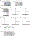
Extended Data Fig 1.. Analyses of mutant recombinant and semisynthetic NEDD4 and NEDD4L.
(A) In vitro ubiquitination assays comparing activity of recombinantly expressed NEDD4L protein with D955N or D955A mutations (P6-P8). The unmodified NEDD4L band was quantified by densitometry. The percentage of unmodified NEDD4L species versus time zero in each case is listed below the lanes (n=3). (B) In vitro ubiquitination assay comparing the enzyme activity of recombinantly expressed NEDD4 proteins including WT (P1), ΔD897/D900E (P9), D900E (P2), and D900A (P4). The unmodified NEDD4 band was quantified by densitometry. The percentage of unmodified NEDD4 species in each case is listed below the lanes (n=2). (C) In vitro ubiquitination assays comparing enzyme activity of recombinantly expressed NEDD4 WT (P1), T893C (P10), with semisynthetic NEDD4 with C-terminal carboxylate (P11). The unmodified NEDD4 band was quantified by densitometry and the percentage of unmodified NEDD4 species is listed below the lanes (n=2). (D) LC-MS spectrum of NEDD4 D900-phospho-serine (P12) made by expressed protein ligation. (E) LC-MS spectrum of NEDD4 D900-homo-cysteine (P13) made by expressed protein ligation. (F) LC-MS spectrum of NEDD4 D900-sulfonyl-alanine (P14) made by expressed protein ligation. (G) LC-MS spectrum of NEDD4 D900-d-Asp (P15) made by expressed protein ligation. (H) LC-MS spectrum of NEDD4 D900-d-Glu (P16) made by expressed protein ligation. (I) In vitro ubiquitination assay comparing the activity of NEDD4 D900 substitutions to d-Asp (dD, P15) or d-Glu (dE, P16). The unmodified NEDD4 bands were quantified by densitometry. The percentages of unmodified NEDD4 species versus time zero are listed below the graph (n=2).
Extended Data Fig 2.
Extended Data Fig 2.. Chemical rescue of NEDD4 mutant.
(A) Chemical rescue assays of NEDD4 D900A (P4) with acetate, difluoroacetate (DFA, pKa 1.3) and trifluoroacetate (TFA). The α-ubiquitin western blot signal from the highest concentration of each reagent was used for densitometry quantification and plotted relative to the acetate-rescued NEDD4 D900A activity in the bar graph (n=3). For the catalytically defective D900A NEDD4–1, the assays were conducted at 37°C with 5 μM E2 to enhance the baseline activities. The same reaction conditions were used for other chemical rescue assays below. (B) Chemical rescue assays of NEDD4 D900A (P4) with acetate, fluoroacetate (FA, pKa 2.6) and trifluoroacetate (TFA). Results were analyzed by densitometry and plotted relative to the acetate-rescued NEDD4 D900A activity in the bar graph (n=3). (C) Chemical rescue assays of NEDD4 D900A (P4) with formate (pKa 3.8), acetate and propionate (pKa 4.9). Results were analyzed by densitometry and plotted relative to the acetate-rescued NEDD4 D900A activity in the bar graph (n=3). (D) Chemical rescue assays of NEDD4 D900A (P4) with acetate and phosphate (pKa 6.8). Results were analyzed by densitometry and plotted to reflect the relative activity in the bar graph (n=3). (E) Chemical rescue assay of NEDD4 D900A (P4) with acetate in deuterated water (2H2O). Autoubiquitination was analyzed by densitometry using the highest concentration of acetate in 2H2O. The quantification was plotted to compare the relative activity of NEDD4 in the absence and presence of acetate under 2H2O (n=4).
Extended Data Fig 3.
Extended Data Fig 3.. Characterization of semisynthetic NEDD4 and sequence comparison among NEDD4, HUWE1 and E6AP proteins.
(A) LC-MS spectrum of NEDD4 D900-carboxamide (P18). (B) LC-MS spectrum of NEDD4 ΔD900 V899-carboxylate (P19). (C) LC-MS spectrum of NEDD4 ΔD900 V899-carboxamide (P20). (D) LC-MS spectrum of NEDD4 ΔD900 V899-βVal (P21). (E) Sequence alignment of human NEDD4, HUWE1 and E6AP. The C-terminal residues are highlighted in the red box. Homologous residues of NEDD4 (V588) and HUWE1 (L4061) are highlighted with a red arrow.
Extended Data Fig 4.
Extended Data Fig 4.. Characterization of the C-terminal residue in HUWE1, E6AP, WWP2 and Rsp5 protein catalysis.
(A) Semisynthetic strategy to make HUWE1 HECT proteins containing different C-termini. (B) Semisynthetic strategy to make E6AP proteins containing different C-termini. (C) LC-MS spectrum of the HUWE1 HECT domain protein containing an A4374-carboxylate (P23) made by expressed protein ligation. (D) LC-MS spectrum of the HUWE1 HECT domain protein containing an A4374-carboxamide (P24) made by expressed protein ligation. (E) Ubiquitination assays of E6AP protein with UbcH5b E2 protein. Semisynthetic E6AP proteins containing carboxylate or carboxamide C termini (P25 and P26). The assays were conducted at 37°C with 5 μM E2. Quantification of ubiquitination smear western blot signals was used to compare the relative activities (n=3). (F) In vitro ubiquitination assays comparing activity of recombinantly expressed WWP2 protein with E870Q or E870A mutations (P33-P35). The unmodified WWP2 band was quantified by densitometry and the percentage of unmodified species is listed below the lanes (n=3). (G) Representative AlphaFold2 multimer structure of the Rsp5-Ub-WBP2 complex (without Rosetta refinement). Three ubiquitins were used to capture the Rsp5 in the L-shape containing Ub in the active site. Two ubiquitin molecules engage the allosteric ubiquitin-binding site (Ub1) and E2 binding site (Ub2) on the N-lobe, whereas the third ubiquitin (Ub3) sits in the E3 active site to mimic the Ub-loaded state, reminiscent of the previously reported Ub-loaded Rsp5 crystal structure trapped in the L-shape (PDB: 4LCD). (H) The predicted aligned error (PAE) plot for the AlphaFold2 structure of the Rsp5-Ub-WBP2 complex shown in panel G. (I) Superimposed structures of the Rsp5-Ub-WBP2 complex from in silico modeling with the Rsp5-Ub-Sna3 crystal structure (PDB: 4LCD). Despite differences in the bound substrates (WBP2 versus Sna3) and rounds of structural refinements (for Rsp5-Ub-WBP2), the two structures align reasonably well (RMSD of 1.113 Å). The recognition of different substrates likely causes orientational differences in the flexbile WW3 domain between the two structures, as WW3 provides a principal substrate binding platform for proline-rich motif in substrates.
Extended Data Fig 5.
Extended Data Fig 5.. MST analysis of E3/E3-Ub binding to E2 or substrate.
(A) In vitro ubiquitination comparing the relative activity of NEDD4 WT versus the single Cys form of NEDD4. The single Cys form contains three Cys to Ser replacements. The percentage of unmodified NEDD4 species in each case is listed below the lanes (n=2). (B) In vitro ubiquitination comparing the relative activity of WWP2 WT versus single Cys form of WWP2. The single Cys form of WWP2 contains seven Cys to Ser/Ala replacements. The percentage of unmodified WWP2 species in each case is listed below the lanes. The single Cys WWP2 demonstrates robust activity, more active than the WT, which might result from loosened linker autoinhibition (n=2). (C) Chemical details of NEDD4 Cys ubiquitin linkages. The native thioester linkage, vinyl thioether linkage (made with a Ub propargyl probe), and hydrazide mimic linkage (made using the Ub hydrazide method) are compared. (D) MST analysis of UbcH5b E2 protein binding with NEDD4-Ub prepared with a Ub-Prg activity-based probe to form a vinyl-thioester linkage (P41). The MST bound fractions as a function of E2 protein concentrations are shown. The equilibrium dissociation constant Kd values were calculated from three repeats using a quadratic binding model (n=3). (E) MST analysis of UbcH5b E2 protein binding with NEDD4-Ub prepared with Ub-hydrazide with the last Ub Gly76 deleted (P42) to approximate the linker length of a native Ub-Cys thioester linkage (n=3). (F) MST analysis of UbcH7 E2 binding with WWP2 or WWP-Ub (P39 and P40) (n=2). (G) MST analysis of UbcH5b E2 binding with NEDD4 HECT or HECT-Ub (P44 and P45). The Kd values were calculated from repeats using a quadratic binding model (n=2). (H) MST analysis of UbcH5b E2 binding with NEDD4 HECT or HECT-Ub (P44 and P45) in the presence of WT Ub at 100 μM (n=2). (I) MST analysis of UbcH5b E2 binding with NEDD4 HECT or HECT-Ub (P44 and P45) in the presence of UbV (5 μM) (n=2). (J) Fluorescence anisotropy analysis of UbV binding to NEDD4 or NEDD4-Ub (n=2). (K) MST analysis of substrate NDP52 (P46) binding to NEDD4 (P36) or NEDD4-Ub (P37). The Kd values were calculated from two repeats using a quadratic binding model (n=2). (L) MST analysis of substrate WBP2 binding with NEDD4 or NEDD4-Ub (P36 and P37). The Kd values were calculated from repeats using a quadratic binding model (n=2). (M) Fluorescence anisotropy analysis of NEDD4 or NEDD4-Ub protein binding to E2 (UbcH5b). The E2 protein is labeled with fluorescein (P50) and the Kd was calculated using a quadratic binding model (n=2). (N) Fluorescence anisotropy analysis of NEDD4 protein binding to E2 (UbcH5b)-Ub. The E2-Ub protein is labeled with fluorescein (P51) and the Kd was calculated using a quadratic binding model (n=2). (O) MST analysis of E2-E3 interactions. NEDD4 is labeled with Cy5 at its N-terminus. E2(UbcH5b)-Ub (P49) is generated using the ubiquitin hydrazide mimic method (n=2).
Extended Data Fig 6.
Extended Data Fig 6.. MST analysis of HUWE1, E6AP and NEDD4 binding to E2 and NMR strategy to study HECT conformational switch.
(A) MST analysis of UbcH5b E2 binding with HUWE1 HECT or HUWE1 HECT-Ub (P52 and P53). HUWE1 HECT-Ub (P53) was prepared with a ubiquitin Prg probe (n=3). (B) MST analysis of UbcH5b binding with E6AP or E6AP-Ub (P54 and P55). E6AP-Ub (P55) was prepared with a ubiquitin Prg probe (n=3). (C) MST analysis of UbcH7 binding with E6AP or E6AP-Ub (P54 and P55) (n=3). (D) MST analysis of UbcH5b E2 binding to NEDD4 (P36) under high salt (1.6M NaCl) condition. The Kd values were calculated from two repeats using a quadratic binding model (n=2). (E) MST analysis of UbcH5b E2 binding to NEDD4-Ub (P37) under high salt (1.6M NaCl) condition. The K d values were calculated from two repeats using a quadratic binding model (n=2). (F) Ubiquitination assays comparing WT NEDD4 with ΔC-tail-5aa and V588A NEDD4 mutants (P56 and P59). The unmodified NEDD4 band was quantified by densitometry. The percentage of unmodified NEDD4 is listed below each lane (n=3). (G) Semisynthetic scheme for preparing NEDD4 or NEDD4-Ub containing fluoro-Phe896-NEDD4 proteins used in NMR study. (H) Ubiquitination assays of NEDD4 prepared by expressed protein ligation bearing different fluoro-Phe896 regioisomers containing C terminal tails. The NEDD4 proteins containing 2-fluoro-Phe896 (P63), 3-fluoro-Phe896 (P64), and 4-fluoro-Phe896 (P65) were prepared and compared with unligated NEDD4 (aa188–893, P66) for autoubiquitination activity (n=2). (I) Quantification of fluoro-Phe896 containing NEDD4 activity. The ubiquitin western blot signals of the autoubiquitination smear were used to compare the relative activities of these NEDD4 proteins (n=2). (J) A proposed structural model shows the HECT domain undergoes a transition from the T-conformation to the L conformation upon ubiquitin transfer from E2~Ub to the E3 catalytic Cys. During this T-to-L conformation change, E2 protein is ejected to allow the turnover of the ubiquitination process.
Extended Data Fig 7.
Extended Data Fig 7.. Maximum likelihood tree from HECT proteins.
Bootstrap values are displayed as black circles. The tree representing the NEDD4 family is labeled and highlighted in red. Human HECT E3 proteins are colored in red branches.
Extended Data Fig 8.
Extended Data Fig 8.. Unclasped view of a eukaryotic phylogenetic tree shown in Figure 6A.
The leaves of tree represent the species that contain HECT E3 ligases that were used in this study.
Extended Data Fig 9.
Extended Data Fig 9.. NEDD4 family phylogeny tree from maximum likelihood tree analysis.
The species that contain NEDD4 family ligases are listed in this relationship comparison. These species are highlighted in the evolutionary tree of eukaryotes in Figure 6A.
Fig 1:
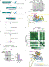
Fig 1:. Enzymatic analysis of NEDD4 C-terminal Asp mutants.
(A) Protein ubiquitination catalyzed by E1, E2 and HECT family E3 ligases. (B) Sequence alignment of human NEDD4 family HECT E3 and other representative HECT E3 ligases. The triangle marks the catalytic Cys. Human NEDD4 HECT E3 ligases share the acidic C-terminus residues Asp or Glu. (C) In vitro ubiquitination assays of NEDD4 D900 mutants (P1-P5). NEDD4 autoubiquitination is visualized by Coomassie staining. The unmodified NEDD4 band was quantified by densitometry. The percentage of unmodified NEDD4 species in each case is listed below the lanes (n=2). (D) In vitro ubiquitination assays with SDS-PAGE run with or without the presence of beta-mercaptoethanol. In the presence of beta-mercaptoethanol, the thioester forms of E2~Ub and E3~Ub are not preserved while they are stable in the absence of beta-mercaptoethanol (n=2). (E) Applying expressed protein ligation to make semisynthetic NEDD4 proteins containing different Asp900 substitutions by ligating recombinantly expression NEDD4 protein with various C-terminal peptides. (F) In vitro ubiquitination assays comparing the activity of NEDD4 D900 substitutions to phospho-serine (pS, P12), homo-cysteine (hC, P13) and sulfonyl-Ala (sA, P14). Quantification of the ubiquitin western blot signals was used to compare the relative activities and plotted the bar graph (n=3).
Fig 2:
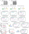
Fig 2:. Chemical rescue of NEDD4 mutants.
(A) Chemical rescue assays of D900A NEDD4 mutant (P4) or WT NEDD4 (P1) with acetate (pKa 4.8) or trifluoroacetic acid (TFA, pKa 0.2). NaCl was used for comparison to assess the effects of ionic strength. The concentrations of rescue reagents (low to high) were 0.3, 0.6, and 0.9 M (n=2). For the catalytically defective D900A NEDD4–1, the assays were conducted at 37°C with 5 μM E2 to enhance the baseline activities. (B) Densitometry quantification of NEDD4 activity in chemical rescue assays. The α-ubiquitin western blot signal from the highest concentration of each reagent was used for quantification to compare the relative enzyme activity (n=2). (C) Brønsted plot showing the log of relative NEDD4 activities vs. pKa values of the rescue reagents. Linear regression gave a slope of 0.39 ± 0.02. The propionate rescue point is shown in orange but not included in the slope calculation because of its greater steric bulk. (D) Chemical rescue assays of D900A NEDD4 with substrate WBP2. The fluorescent signals corresponding to WBP2 and its different ubiquitinated states are shown. The assays were conducted at 37°C with 5 μM E2. The unmodified WBP2 fluorescence intensity was quantified and plotted to compare the ubiquitination rate of WBP2 by E3 ligase (n=3). Data are presented as mean values ± SEM. (E) In silico model of a hypothetical ubiquitinated HUWE1-NEDD4 chimeric protein. In the HUWE1-Ub crystal structure (PDB: 6XZ1), the C-terminal tail of HUWE1 was replaced by the related NEDD4 residues (green) and the structure was modeled using Rosetta. The C-terminal Asp900 (in green) highlights both the sidechain and backbone carboxylates in this model. The catalytic Cys867 and the ubiquitin C-terminal Gly76 are depicted as a thioester intermediate and are highlighted. The distance between the side chain carboxylate of Asp900 and catalytic thiol in this model was measured to be 4.1 Å.
Fig 3:
Fig 3:. Analysis of the C-terminal backbone carboxylate in HECT enzymes.
(A) Ubiquitination assays of WT NEDD4, T893C NEDD4 (recombinant protein) and semisynthetic NEDD4s with carboxylate or carboxamide C-termini (P1, P10, P11, P18). The unmodified NEDD4 bands were quantified by densitometry and values were displayed below each lane (n=2). (B) Substrate WBP2 ubiquitination assay with recombinant WT, semisynthetic NEDD4 with carboxylate (CO2H) or carboxamide (CONH2) C-termini, and T893C NEDD4. The fluorescent signals corresponding to WBP2 and its different ubiquitinated states are shown. The unmodified WBP2 fluorescence intensity was quantified and plotted to compare the ubiquitination rate of WBP2 by E3 ligase (n=3). Data are presented as mean values ± SEM. (C) Ubiquitination assays of ΔD900 (V899) NEDD4 with carboxylate and carboxamide C-termini (P19 and P20), V899βVal NEDD4 (P21), as well as D900A NEDD4 (P4). For the catalytically defective ΔD900 variants, the assays were conducted at 37°C with 5 μM E2 to enhance the baseline activities. Quantification of ubiquitin western blot signals was used to compare the relative activities (n=3). Data are presented as mean values ± SEM. (D) Substrate WBP2 ubiquitination assay with NEDD4 proteins of V899 carboxylate or carboxamide, V899βVal, and D900A. The fluorescent signals corresponding to WBP2 and its different ubiquitinated states are shown. The unmodified WBP2 fluorescence intensity was quantified and plotted to compare the ubiquitination rate of WBP2 by E3 ligase (n=3). Data are presented as mean values ± SEM. (E) Ubiquitination of WT HUWE1 recombinant HECT protein (P22) and semisynthetic HUWE1 with carboxylate or carboxamide C-termini (P23 and P24). The assays were conducted at 37°C with 5 μM E2. Quantification of free di-ubiquitin western blot signals was used to compare the relative activities of HUWE1 forms (n=4). Data are presented as mean values ± SEM. (F) Ubiquitination assays of E6AP protein with UbcH7 E2 protein. Semisynthetic E6AP proteins containing carboxylate or carboxamide C termini (P25 and P26). The assays were conducted at 37°C with 5 μM E2. Quantification of ubiquitin smear western blot signals was used to compare the relative activities (n=4). Data are presented as mean values ± SEM.
Fig 4:
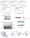
Fig 4:. The versatility of carboxylate usage in Rsp5 E3 ligase.
(A) Autoubiquitination assays of Rsp5 protein forms. Semisynthetic Rsp5 proteins of Glu809, Gln809 or Ala809 with carboxylate or carboxamide C-termini (P27-P32) were assayed and compared for their relative activities (n=4). (B) Densitometry quantification of Rsp5 autoubiquitination. The Coomassie-stained autoubiquitination smear at final time points (60 min) were quantified by densitometry to compare the relative activities of the different C-terminal forms of Rsp5 protein (n=4). Data are presented as mean values ± SEM. (C) In silico model of Rsp5-Ub intermediate in complex with its substrate WBP2. Alphafold2 and Rosetta were used. The Rsp5 C-lobe and N-lobe are colored in pink and blue, respectively. The C terminal tail of Rsp5 is colored in green. The incoming WBP2 substrate is colored in yellow. (D) A detailed view of the catalytic center of the Rsp5-Ub-WBP2 complex. The distance between Lys222 of WBP2 to the catalytic Cys777 (4.9 Å) and Glu809 backbone carboxylate (2.6 Å) are highlighted. (E) A proposed enzyme mechanism for Lys ubiquitination catalyzed by HECT E3 ligase. The HECT terminal carboxylate serving as catalytic base/acid is colored in blue. The catalytic Cys is colored in fuchsia. The ubiquitin G76 residue is shown in orange. The substrate Lys is highlighted in teal.
Fig 5:
Fig 5:. Analysis of protein-protein interaction using ubiquitination intermediates (E3-Ub or E2-Ub).
(A) Reaction scheme of chemical ubiquitination to afford ubiquitin hydrazide linked to a protein of interest. (B) MST analysis of NEDD4 or WWP2 binding to E2 protein. NEDD4 or WWP2 and their ubiquitinated forms (P36–37 or P39–40) are labeled with Cy5 at their N-termini, and titrated with a gradient of UbcH5b E2 protein. The MST bound fractions as a function of E2 protein concentrations are shown. The equilibrium dissociation constant Kd values were calculated using a quadratic binding model (n=3 for NEDD4 and n=2 for WWP2). For NEDD4 binding, data are presented as mean values ± SEM. (C) Molecular interactions of the HECT domain in its L conformation. The key HUWE1 residue in the original PDB 6XZ1 structure are labeled to demonstrate the detailed interactions. The ubiquitin C-terminus is colored in green. The HECT C tail is shown in blue. The Leu4061, the key residue that interacts with Phe4371 (green) of C-terminal tail is colored in gray. (D) MST analysis of E2 binding with NEDD4 or NEDD4-Ub mutants (n=3). The NEDD4 mutants include the C-tail mutant (ΔC-tail-5aa, P57 and P58), V588A (P60 and P61) and NEDD4 chemically ubiquitinated with mutant Ub (R72A/R74A) hydrazide (P62). Data are presented as mean values ± SEM. (E) 19Fluoro (19F) probe at the C-terminus of NEDD4 to assess the conformational state of the HECT domain. The 19F probe exists in two different chemical environments as the C-tail is flexible and solvent exposed in the T-conformation or constrained and packed in the L-conformation. (F) 19F NMR analysis of 3-fluoro-Phe896-NEDD4 (P64), 3-fluoro-Phe896-NEDD4-Ub (P67) and free C-tail 8 aa peptide with 3-fluoro-Phe896. The free/flexible peptide dissolved in buffer gives a sharp peak at −113.5 ppm. The broad 19F NMR peak downfield of −113 ppm, is indicative of a rigid/constrained chemical environment for the 19F. In comparison, the 19F NMR peak of 3-fluoro-Phe896-NEDD4 (P64) shows an intermediate behavior, the peak is broad (peptide covalently linked to protein tumbles like a large molecule) but is not as shifted downfield as the corresponding peak in 3-fluoro-Phe896-NEDD4-Ub (P67).
Fig 6.
Fig 6.. Evolutionary analysis of HECT E3 ligases.
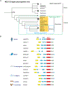
(A) A view of the eukaryotic phylogenetic tree with the last eukaryotic common ancestor (LECA) placed at the Unikonta/Bikonta split. The tree is collapsed into groups. The diameter of the circles indicates the number of species placed in each group. An uncollapsed view of this tree can be found in Extended Data Fig. 9. The evolutionary trend of the Rsp5-class HECT protein are highlighted in the green box. The later evolving examples of the NEDD4-class HECT proteins are highlighted with the blue box. (B) Representative domain architectures of NEDD4 homologs from yeast (S. cerevisiae) to human. The C2, WW, and HECT domains are colored in yellow, blue, and red, respectively. The lengths of the proteins and the position of domains are illustrated. The C-terminal residues are shown and the Asp-termini is colored in blue and Glu-termini is colored in green.

References

    1. Hershko A & Ciechanover A The ubiquitin system for protein degradation. Annu Rev Biochem 61, 761–807 (1992). 10.1146/annurev.bi.61.070192.003553 - DOI - PubMed
    1. Scheffner M, Nuber U & Huibregtse JM Protein ubiquitination involving an E1-E2-E3 enzyme ubiquitin thioester cascade. Nature 373, 81–83 (1995). 10.1038/373081a0 - DOI - PubMed
    1. Komander D & Rape M The ubiquitin code. Annual review of biochemistry 81, 203–229 (2012). - PubMed
    1. Swatek KN & Komander D Ubiquitin modifications. Cell research 26, 399–422 (2016). - PMC - PubMed
    1. Harper JW & Schulman BA Cullin-RING Ubiquitin Ligase Regulatory Circuits: a Quarter Century Beyond the F-box Hypothesis. Annual Review of Biochemistry 90 (2021). - PMC - PubMed

MeSH terms

Substances

LinkOut - more resources