

Monitoring correlates of SARS-CoV-2 infection in cell culture using a two-photon-active calcium-sensitive dye
- PMID: 39030477
- PMCID: PMC11264913
- DOI: 10.1186/s11658-024-00619-0
Monitoring correlates of SARS-CoV-2 infection in cell culture using a two-photon-active calcium-sensitive dye
Abstract
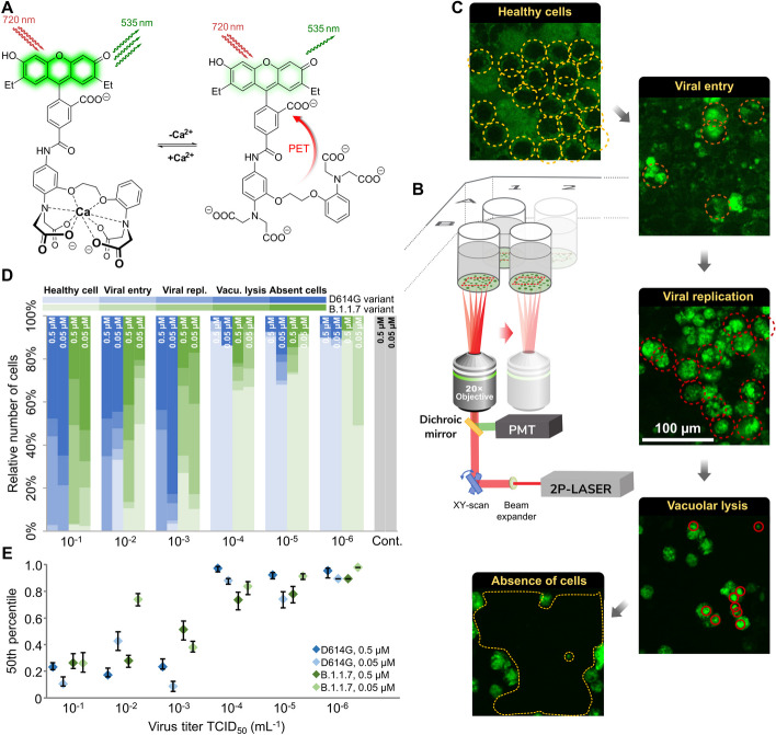
Background: The organism-wide effects of severe acute respiratory syndrome coronavirus 2 (SARS-CoV-2) viral infection are well studied, but little is known about the dynamics of how the infection spreads in time among or within cells due to the scarcity of suitable high-resolution experimental systems. It has been reported that SARS-CoV-2 infection pathways converge at calcium influx and subcellular calcium distribution changes. Imaging combined with a proper staining technique is an effective tool for studying subcellular calcium-related infection and replication mechanisms at such resolutions.
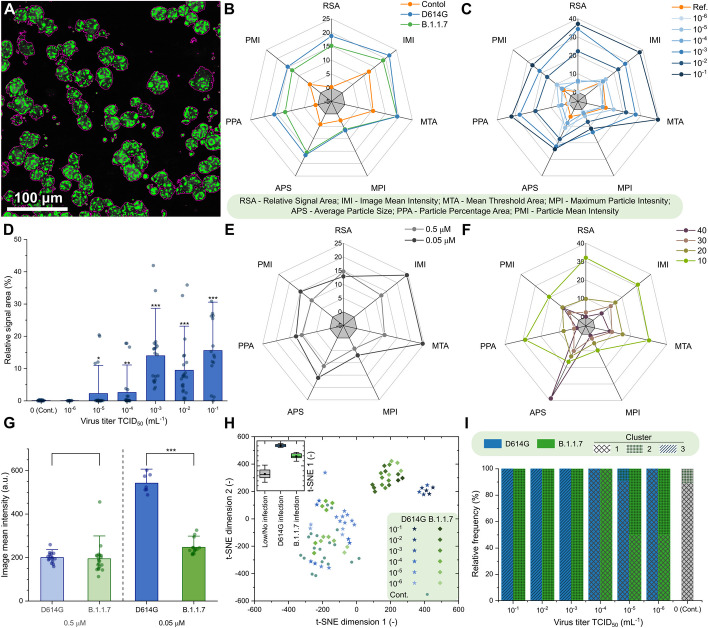
Methods: Using two-photon (2P) fluorescence imaging with our novel Ca-selective dye, automated image analysis and clustering analysis were applied to reveal titer and variant effects on SARS-CoV-2-infected Vero E6 cells.
Results: The application of a new calcium sensor molecule is shown, combined with a high-end 2P technique for imaging and identifying the patterns associated with cellular infection damage within cells. Vero E6 cells infected with SARS-CoV-2 variants, D614G or B.1.1.7, exhibit elevated cytosolic calcium levels, allowing infection monitoring by tracking the cellular changes in calcium level by the internalized calcium sensor. The imaging provides valuable information on how the level and intracellular distribution of calcium are perturbed during the infection. Moreover, two-photon calcium sensing allowed the distinction of infections by two studied viral variants via cluster analysis of the image parameters. This approach will facilitate the study of cellular correlates of infection and their quantification depending on viral variants and viral load.
Conclusions: We propose a new two-photon microscopy-based method combined with a cell-internalized sensor to quantify the level of SARS-CoV-2 infection. We optimized the applied dye concentrations to not interfere with viral fusion and viral replication events. The presented method ensured the proper monitoring of viral infection, replication, and cell fate. It also enabled distinguishing intracellular details of cell damage, such as vacuole and apoptotic body formation. Using clustering analysis, 2P microscopy calcium fluorescence images were suitable to distinguish two different viral variants in cell cultures. Cellular harm levels read out by calcium imaging were quantitatively related to the initial viral multiplicity of infection numbers. Thus, 2P quantitative calcium imaging might be used as a correlate of infection or a correlate of activity in cellular antiviral studies.
Keywords: Calcium sensors; Fluorescence imaging; SARS-CoV-2; Two-photon microscopy; Viral infections.
© 2024. The Author(s).
Conflict of interest statement
B.R. and G.K. are founders of Femtonics and members of its scientific advisory board. G.F. is now an AstraZeneca employee, but the work was conducted with no connection to A.Z. The other authors declare that no conflict of interest exists.
Figures
References
-
- Coronavirus Disease (COVID-2019) situation reports. WHO. 2022. https://covid19.who.int/. Accessed 20 Mar 2024.
MeSH terms
Substances
Supplementary concepts
Grants and funding
- KFI-18-2018-00097/Nemzeti Kutatási Fejlesztési és Innovációs Hivatal
- 2020-1.1.5-GYORSÍTÓSÁV-2021-00004/Nemzeti Kutatási Fejlesztési és Innovációs Hivatal
- 2018-1.3.1-VKE-2018-00032/Nemzeti Kutatási Fejlesztési és Innovációs Hivatal
- TKP2021-EGA-42/Nemzeti Kutatási Fejlesztési és Innovációs Hivatal
- 2020-2.1.1.-ED-2022-00208/Nemzeti Kutatási Fejlesztési és Innovációs Hivatal
LinkOut - more resources
Full Text Sources
Medical
Miscellaneous
