. 2024 Jul 19;25(1):703.
doi: 10.1186/s12864-024-10533-6.

Upstream CtrA-binding sites both induce and repress pilin gene expression in Caulobacter crescentus

Affiliations

Upstream CtrA-binding sites both induce and repress pilin gene expression in Caulobacter crescentus

Anurag Rijal et al. BMC Genomics. .

Abstract

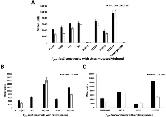
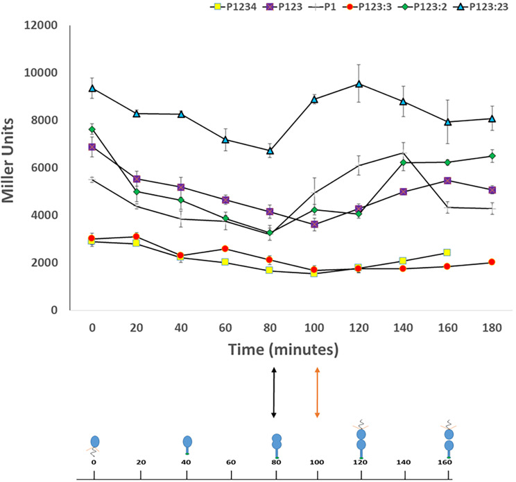
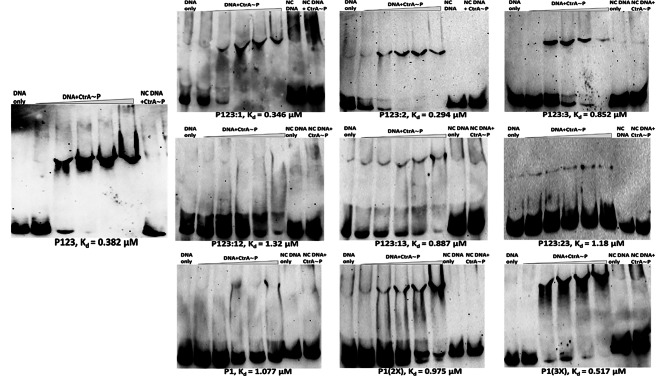
Pili are bacterial surface structures important for surface adhesion. In the alphaproteobacterium Caulobacter crescentus, the global regulator CtrA activates transcription of roughly 100 genes, including pilA which codes for the pilin monomer that makes up the pilus filament. While most CtrA-activated promoters have a single CtrA-binding site at the - 35 position and are induced at the early to mid-predivisional cell stage, the pilA promoter has 3 additional upstream CtrA-binding sites and it is induced at the late predivisional cell stage. Reporter constructs where these additional sites were disrupted by deletion or mutation led to increased activity compared to the WT promoter. In synchronized cultures, these mutations caused pilA transcription to occur approximately 20 min earlier than WT. The results suggested that the site overlapping the - 35 position drives pilA gene expression while the other upstream CtrA-binding sites serve to reduce and delay expression. EMSA experiments showed that the - 35 Site has lower affinity for CtrA∼P compared to the other sites, suggesting binding site affinity may be involved in the delay mechanism. Mutating the upstream inhibitory CtrA-binding sites in the pilA promoter caused significantly higher numbers of pre-divisional cells to express pili, and phage survival assays showed this strain to be significantly more sensitive to pilitropic phage. These results suggest that pilA regulation evolved in C. crescentus to provide an ecological advantage within the context of phage infection.

Keywords: Caulobacter crescentus; pilA; CtrA; Transcriptional regulation.


Conflict of interest statement

The authors declare no competing interests.

Figures

Fig. 1
Fig. 1
The pilA promoter architecture in C. crescentus with four CtrA-binding sites upstream of transcription start site. Site 1 (green) overlaps the − 35 position, Site 2 (red) is located 31 bp upstream of Site 1, Site 3 (maroon) is located 3 bp upstream of Site 2 and Site 4 (yellow) is positioned 78 bp upstream of Site 3. The nucleotide sequence for each binding site is provided between the extended lines with uppercase letters representing the half-sites. Site 1 is closest to the consensus CtrA-binding site TTAA-N7-TTAA with a single nucleotide deviation. The nucleotide sequences of the CtrA-binding sites in the pilA promoter was derived from [14].
Fig. 2
Fig. 2
Schematic representation of the PpilA architecture in the intact and mutated/artificial PpilA-lacZ constructs used for β-galactosidase assay. Each rectangle indicates a single half-site. Site 1 nucleotide sequence is indicated in green, Site 2 in red, Site 3 in maroon and Site 4 in yellow. The numbers below the arrows indicate the spacing between the CtrA-binding sites. Mutated sites are indicated by a stripes inside the respective rectangles representing the half-sites.
Fig. 3
Fig. 3
Sites 2, 3 and 4 repress PpilA activity, while Site 1 drives expression. β-galactosidase assays were done for various PpilA-lacZ constructs in C. crescentus NA1000 wild-type strain. A: Promoter activity for constructs with native, truncated, and mutated CtrA-binding sites. Other than the WT construct P1234, all constructs have Site 4 deleted. P123 has Site 4 deleted; P12 has Sites 3 and 4 deleted; P1 has Sites 2, 3 and 4 deleted; P123:1 has Site 1 mutated; P123:2 has Site 2 mutated; P123:3 has Site 3 mutated and P123:23 has Sites 2 and 3 mutated. Mutated sites had each TTAA half-site replaced by GGCC. The empty plac290 construct with a promoterless lacZ gene is the negative control. B: Comparison of promoter activity of wild-type construct with constructs where nucleotide sequence of Site 1 is used to replace the native sequence of Sites 2 and 3. P1(2X)N has the second copy of Site 1 at Site 2’s native position with other sites deleted. P1(3X)N has Sites 2 and 3 replaced by Site 1 sequence at native spacing. C: Comparison of promoter activity of wild-type with constructs having artificial spacing (6 bp) between duplicate and triplicate copies of Site 1. P1(2X) has second Site 1 copy 6 bp upstream of native Site 1 with other sites deleted. P1(3X) is similar to P1(2X) except that it has a third Site 1 copy 6 bp upstream of the second copy. P1(3X )31 has two additional Site 1 copies each 31 bp upstream of the previous copy.
Fig. 4
Fig. 4
Sites 2 and 3 increase PpilA sensitivity to CtrA levels. β-galactosidase assays were compared for the various PpilA-lacZ constructs between C. crescentus NA1000 wild-type strain and the low-CtrA abundance strain PC0227. Strain PC0227 contains a transposon insertion into the ctrA P2 promoter that causes a reduction in CtrA abundance, derived from strain YB3558. A: Promoter activity for constructs with native, truncated and mutated CtrA-binding sites. Other than the WT construct P1234, all constructs have Site 4 deleted. P123 has Site 4 deleted; P12 has Sites 3 and 4 deleted; P1 has Sites 2, 3 and 4 deleted; P123:1 has Site 1 mutated; P123:2 has Site 2 mutated; P123:3 has Site 3 mutated and P123:23 has Sites 2 and 3 mutated. Mutated sites had each TTAA half-site replaced by GGCC. The empty plac290 construct with a promoterless lacZ gene is the negative control. Similarly, the Pxyl-lacZ construct is a negative control for CtrA-dependent transcription, which shows no change in activity between WT and PC0227. B: Promoter activity for constructs where nucleotide sequence of Site 1 is used to replace the native sequence of Sites 2 and 3. P1(2X)N has the second copy of Site 1 at Site 2’s native position with other sites deleted. P1(3X)N has Sites 2 and 3 replaced by Site 1 sequence at native spacing. C: Promoter activity of constructs having artificial spacing (6 bp) between duplicate and triplicate copies of Site 1. P1(2X) has second Site 1 copy 6 bp upstream of native Site 1 with other sites deleted. P1(3X) is similar to P1(2X) except that it has a third Site 1 copy 6 bp upstream of the second copy. P1(3X  )31 has two additional Site 1 copies each 31 bp upstream of the previous copy.
Fig. 5
Fig. 5
Sites 2 and 3 delay PpilA expression. Gene expression of WT and mutated pilA promoter constructs were measured by β-galactosidase assay every 20 min during the course of the cell cycle. Generally, transcription was found to decrease initially, then increase in the predivisional cell stage. Transcription was found to initiate 20 min earlier for constructs P1, P123:2 and P123:23 at the early pre-divisional stage (indicated by black double-headed arrow) as compared to constructs P1234 (WT), P123 and P123:3 (indicated by orange double-headed arrow).
Fig. 6
Fig. 6
PilA accumulation occurs earlier and in greater quantity in P1234:23. PilA production was assessed over the course of the cell cycle for WT and mutated pilA promoters by anti-PilA western blots. Western blots were performed with uniform protein concentrations, antibody concentration, and exposure time. A: Representative western blots with synchronized cultures of P1234, P1234:2, P1234:3 and P1234:23 showing PilA accumulation over different timepoints during the cell cycle. Uncropped western blots have been included in Supplementary Figures S18-S21. B: Line graph depicting average PilA band pixel intensity values over different timepoints during the cell cycle for the above mentioned strains. The values are averages from 3 western blots done for 3 separate synchronized cultures. Similar to the β-galactosidase data, PilA was found to accumulate approximately 20 min earlier and in greater quantities for P1234:2 and P1234:23 (indicated by black double-headed arrow) compared to the other strains (indicated by orange double-headed arrow).
Fig. 7
Fig. 7
Sites 2 and 3 increase PpilA affinity to CtrA. EMSA experiments were performed with WT and mutated promoter fragments to assess the binding affinity of phosphorylated CtrA. All lanes contained 100 ng DNA and 1 ng Poly didC. NC indicates a negative control DNA fragment that has all half-sites in Sites 1, 2 and 3 mutated to GGCC and Site 4 is absent. The first lane had DNA only and the second to sixth lanes had DNA and 0.17 µM, 0.33 µM, 0.5 µM, 0.66 µM and 1.65 µM of phosphorylated CtrA respectively. The last two lanes were controls with NC DNA only and NC DNA with 1.65 µM of phosphorylated CtrA respectively. Constructs with Sites 2 and/or 3 generally had more robust shifts, indicating these sites had higher affinity for phosphorylated CtrA. The dissociation constant (Kd) for each construct has been listed at the bottom of the respective EMSA image. Gels have been cropped to remove background and uncropped gels have been included in Supplementary Figures S3-S12.
Fig. 8
Fig. 8
Pilus filament production occurs earlier in P1234:23, increasing sensitivity of C. crescentus to phage φCbK. Cells with WT or P1234:23 pilA promoters in the pilAT36C background were treated with biotin-PEG-maleimide to biotinylate the pili. Cells with labeled pili were bound to streptavidin magnetic beads. A: Cells were categorized as swarmer or predivisional based on the absense/presence respectively of a noticeable cell constriction. The P1234:23 strain had a much higher percentage of piliated predivisional cells bound to streptavidin-coated magnetic beads. B: Representative images showing predivisional and swarmer cells bound to magnetic beads. C: Cells were detached from beads and cell length was measured for roughly 7500 cells. Percentage abundance of cells belonging to respective length categories for WT and P1234:23 is shown in a histogram. The percentage abundance of cells of 5 μm or longer was consistently higher for P1234:23, indicating a greater proportion of predivisional cells attached to beads. D. Average percent survival of C. crescentus against φCbK infection. WT (P1234) strains showed significantly greater survival compared to P1234:23 with Sites 2 and 3 mutated.
Fig. 9
Fig. 9
Delayed pilus production prevents termination of C. crescentus cell lineages from φCbk phage infection. Schematic representation of the two different scenarios of C. crescentus lineage progression after φCbk phage infection based on timing of pilus filament expression. A: In cells with the wild-type pilA promoter (P1234), pilus filaments are predominantly expressed after cell division. In this case, φCbk can infect only the swarmer cells and the stalked cell can still continue the lineage by avoiding phage infection. B: In P1234:23 cells with Sites 2 and 3 mutated, a significantly higher proportion of predivisional cells produce pili, which results in φCbk infection before cell division preventing the entire cell lineage from entering another round of cell cycle.

References

    1. Fronzes R, Remaut H, Waksman G. Architectures and biogenesis of non-flagellar protein appendages in Gram-negative bacteria. EMBO J. 2008;27(17):2271–80. 10.1038/emboj.2008.155 - DOI - PMC - PubMed
    1. Scott JR, Zähner D. Pili with strong attachments: Gram-positive bacteria do it differently. Mol Microbiol. 2006;62(2):320–30. 10.1111/j.1365-2958.2006.05279.x - DOI - PubMed
    1. Jarrell KF, Stark M, Nair DB, Chong JPJ. Flagella and pili are both necessary for efficient attachment of Methanococcus maripaludis to surfaces. FEMS Microbiol Lett. 2011;319(1):44–50. 10.1111/j.1574-6968.2011.02264.x - DOI - PubMed
    1. Proft T, Baker EN. Pili in Gram-negative and Gram-positive bacteria - structure, assembly and their role in disease. Cell Mol Life Sci. 2009;66(4):613–35. 10.1007/s00018-008-8477-4 - DOI - PMC - PubMed
    1. Kaiser D. Bacterial motility: how do pili pull? Curr Biol. 2000;10(21):777–80. 10.1016/S0960-9822(00)00764-8 - DOI - PubMed

MeSH terms

LinkOut - more resources