

Myeloid-derived growth factor in diseases: structure, function and mechanisms
- PMID: 39030488
- PMCID: PMC11264862
- DOI: 10.1186/s10020-024-00874-z
Myeloid-derived growth factor in diseases: structure, function and mechanisms
Abstract
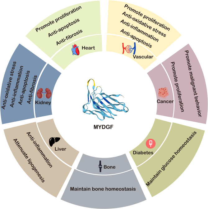
Myeloid-derived growth factor (MYDGF) is a novel secreted protein with potent antiapoptotic and tissue-repairing properties that is present in nearly 140 human tissues and cell lines, with the highest abundance in the oral epithelium and skin. Initially, MYDGF was found in bone marrow-derived monocytes and macrophages for cardioprotection and repair after myocardial infarction. Subsequent studies have shown that MYDGF plays an important role in other cardiovascular diseases (e.g., atherosclerosis and heart failure), metabolic disorders, renal disease, autoimmune/inflammatory disorders, and cancers. Although the underlying mechanisms have not been fully explored, the role of MYDGF in health and disease may involve cell apoptosis and proliferation, tissue repair and regeneration, anti-inflammation, and glycolipid metabolism regulation. In this review, we summarize the current progress in understanding the role of MYDGF in health and disease, focusing on its structure, function and mechanisms. The graphical abstract shows the current role of MYDGF in different organs and diseases (Fig. 1).
Keywords: Autoimmune/inflammatory disorders; Cancers; Cardiovascular diseases; Metabolic disorders; Myeloid-derived growth factor; Renal disease.
© 2024. The Author(s).
Conflict of interest statement
The authors have no relevant financial or non-financial interests to disclose.
Figures

Similar articles
-
Myeloid-Derived Growth Factor Protects Against Pressure Overload-Induced Heart Failure by Preserving Sarco/Endoplasmic Reticulum Ca2+-ATPase Expression in Cardiomyocytes.Circulation. 2021 Oct 12;144(15):1227-1240. doi: 10.1161/CIRCULATIONAHA.120.053365. Epub 2021 Aug 10. Circulation. 2021. PMID: 34372689
-
Myeloid-derived growth factor and its effects on cardiovascular and metabolic diseases.Cytokine Growth Factor Rev. 2024 Apr;76:77-85. doi: 10.1016/j.cytogfr.2023.12.005. Epub 2023 Dec 30. Cytokine Growth Factor Rev. 2024. PMID: 38185568 Review.
-
Mydgf promotes Cardiomyocyte proliferation and Neonatal Heart regeneration.Theranostics. 2020 Jul 11;10(20):9100-9112. doi: 10.7150/thno.44281. eCollection 2020. Theranostics. 2020. PMID: 32802181 Free PMC article.
-
Inflammatory Cell-Derived MYDGF Attenuates Endothelial LDL Transcytosis to Protect Against Atherogenesis.Arterioscler Thromb Vasc Biol. 2023 Nov;43(11):e443-e467. doi: 10.1161/ATVBAHA.123.319905. Epub 2023 Sep 28. Arterioscler Thromb Vasc Biol. 2023. PMID: 37767706
-
Cellular and molecular mechanisms of HGF/Met in the cardiovascular system.Clin Sci (Lond). 2015 Dec;129(12):1173-93. doi: 10.1042/CS20150502. Clin Sci (Lond). 2015. PMID: 26561593 Review.
Cited by
-
The Rapid Activation of MYDGF Is Critical for Cell Survival in the Acute Phase of Retinal Regeneration in Fish.Int J Mol Sci. 2025 Jul 27;26(15):7251. doi: 10.3390/ijms26157251. Int J Mol Sci. 2025. PMID: 40806387 Free PMC article.
-
Transcriptomic profiling of pancreatic neuroendocrine tumors: dysregulation of WNT, MAPK, PI3K, neddylation pathways and potential non-invasive biomarkers.PLoS One. 2025 Jun 16;20(6):e0325672. doi: 10.1371/journal.pone.0325672. eCollection 2025. PLoS One. 2025. PMID: 40522948 Free PMC article.
-
Understanding the Role of Adipokines in Cardiometabolic Dysfunction: A Review of Current Knowledge.Biomolecules. 2025 Apr 23;15(5):612. doi: 10.3390/biom15050612. Biomolecules. 2025. PMID: 40427505 Free PMC article. Review.
References
Publication types
MeSH terms
Grants and funding
- No.: 82270384/National Natural Science Foundation of China
- 20231321/Guangdong Provincial Bureau of Traditional Chinese Medicine research project
- SRSP2022016/Scientific Research Start Plan of Shunde Hospital, Southern Medical University
- SRSP2022012/Scientific Research Start Plan of Shunde Hospital, Southern Medical University
- SRSP2023017/Scientific Research Start Plan of Shunde Hospital, Southern Medical University
LinkOut - more resources
Full Text Sources
