Review
. 2024 Jul 19;21(1):60.
doi: 10.1186/s12987-024-00557-1.

Cellular and molecular mechanisms of the blood-brain barrier dysfunction in neurodegenerative diseases

Affiliations
Review

Cellular and molecular mechanisms of the blood-brain barrier dysfunction in neurodegenerative diseases

Tongli Chen et al. Fluids Barriers CNS. .

Abstract

Background: Maintaining the structural and functional integrity of the blood-brain barrier (BBB) is vital for neuronal equilibrium and optimal brain function. Disruptions to BBB performance are implicated in the pathology of neurodegenerative diseases.

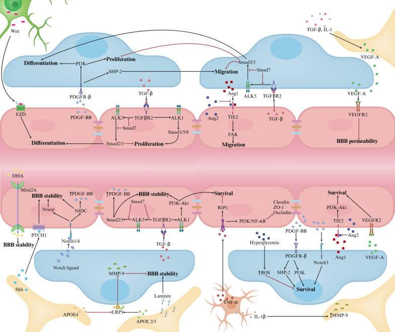
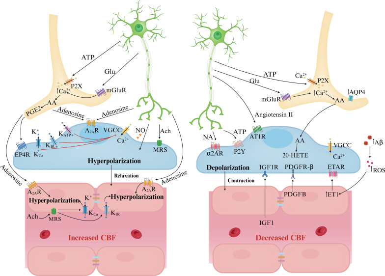
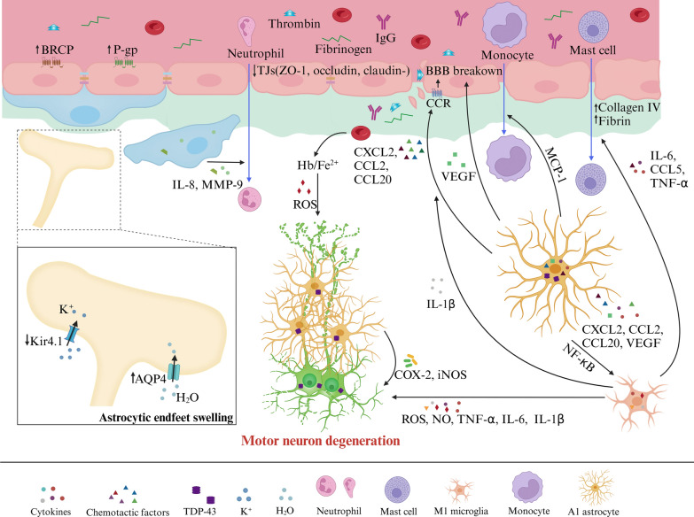
Main body: Early indicators of multiple neurodegenerative disorders in humans and animal models include impaired BBB stability, regional cerebral blood flow shortfalls, and vascular inflammation associated with BBB dysfunction. Understanding the cellular and molecular mechanisms of BBB dysfunction in brain disorders is crucial for elucidating the sustenance of neural computations under pathological conditions and for developing treatments for these diseases. This paper initially explores the cellular and molecular definition of the BBB, along with the signaling pathways regulating BBB stability, cerebral blood flow, and vascular inflammation. Subsequently, we review current insights into BBB dynamics in Alzheimer's disease, Parkinson's disease, amyotrophic lateral sclerosis, and multiple sclerosis. The paper concludes by proposing a unified mechanism whereby BBB dysfunction contributes to neurodegenerative disorders, highlights potential BBB-focused therapeutic strategies and targets, and outlines lessons learned and future research directions.

Conclusions: BBB breakdown significantly impacts the development and progression of neurodegenerative diseases, and unraveling the cellular and molecular mechanisms underlying BBB dysfunction is vital to elucidate how neural computations are sustained under pathological conditions and to devise therapeutic approaches.

Keywords: Blood–brain barrier; Cerebrovascular blood flow; Neurodegenerative diseases; Therapeutics; Vascular inflammation.


Conflict of interest statement

The authors declare no competing interests.

Figures

Fig. 1
Fig. 1
The NVU (capillary level). The NVU at the capillary level consists of vascular cells (ECs, pericytes, glial cells) and neurons. ECs, situated on the luminal side of blood vessels, are continuously sealed by intercellular junctional molecules like ZO proteins and claudins. The abluminal surface of ECs is enveloped by a basement membrane that incorporates pericytes and their projections. Astrocytic processes culminate in end-feet encircling the abluminal surface of the capillary vessel wall. The BBB functions to preserve neuronal health by preventing the unregulated influx of peripheral cells (such as red blood cells, leukocytes, and lymphocytes) and plasma proteins (e.g., fibrinogen, IgG). It also maintains low paracellular and transcellular permeability for molecules and ions while facilitating the delivery of essential oxygen and nutrients to the brain. Created with bioRender.com
Fig. 2
Fig. 2
Major signaling pathways regulating BBB stability. Pericyte-EC communications. PDGF-BB-PDGFRβ pathways regulate various pericyte functions (survival, migration, proliferation, differentiation) by activating phosphoinositide 3-Kinase (PI3K) and Src homology-2 domain-containing protein tyrosine phosphatase-2 (SHP-2). In ECs, TGF-β–TGFβR2 activates different cascades: Alk5-Smad2/3/4 complex boosts differentiation, curbs proliferation, Alk1-Smad1/5/8 complex enhances proliferation; and Alk1-PI3K–Akt pathway fortifies survival and BBB stability. In pericytes, Smad2/3 activation restrains proliferation and migration, promoting differentiation. Notch3 receptor engagement by Notch ligands strengthens BBB stability through Notch-regulated ankyrin repeat protein (Nrarp) or Notch intracellular domain (NICD) pathways when binding to Notch1 or Notch4. Excessive VEGF-A from pericytes and astrocytes weakens BBB by interacting with EC's VEGFR2. Ang1 binding to Tie2 receptors activates PI3K–Akt and inhibits β-catenin, supporting BBB stability, whereas Ang2 antagonizes this effect. Astrocyte-Pericyte/ EC communications. Astrocytes secrete APOE2 and APOE3 (not APOE4), which bind to pericyte LRP1 receptors, inhibiting the CypA–NFκB–MMP-9 pathway and bolstering BBB stability. Moreover, astrocytic Shh binds to PTCH1 receptors on ECs, playing a critical role in BBB stabilization. Astrocyte-derived laminin significantly supports BBB integrity. Neuron-EC communications. Neurons release Wnt, which upon binding to frizzled (FZD) receptors on ECs stimulates their differentiation. Microglia-EC Communications. Microglia release TNF-α and IL-1β, which cumulatively diminish the stability of BBB. Other factors influencing BBB integrity. Additional factors include MFSD2A's role in BBB formation and hyperglycemia-induced pericyte apoptosis via ROS
Fig. 3
Fig. 3
Major signaling pathways regulating CBF (capillary level). Increased CBF. Neuronal ATP and glutamate activate P2X receptors and metabotropic glutamate receptors (mGluRs) on astrocytes respectively, triggering Ca2+ influx. Elevated cytoplasmic Ca2+ stimulates COX-2 activity, leading to the conversion of AA into prostaglandin E2 (PGE2). PGE2 induces pericyte relaxation by binding to its receptor EP4 (EP4R), causing pericyte hyperpolarization. Similarly, acetylcholine released by neurons and adenosine from neurons and astrocytes target pericytes and ECs through muscarinic receptors and A2ARs respectively, resulting in hyperpolarization and relaxation, thereby increasing CBF. Furthermore, high extracellular K+ from neurons activates VGCCs, causing pericyte depolarization and contraction. Conversely, activation of channels like the inward rectifier potassium channel and KCa or KATP channels leads to K+ efflux and pericyte hyperpolarization, reducing Ca2+ entry through VGCCs. ACh or high extracellular K+ can stimulate KIR or KCa channels on ECs, causing endothelial hyperpolarization that propagates via gap junctions between ECs, enhancing CBF in a retrograde manner. Decreased CBF. Neurogenic ATP or NA binds to P2X or α2A receptors, respectively, on pericytes, resulting in cell depolarization and constriction, thereby reducing CBF. In astrocytes, AA is synthesized and then enters smooth muscle cells and pericytes where it transforms into 20-HETE, leading to pericyte depolarization, contraction, and a subsequent decrease in CBF. In AD models, excess Aβ triggers ROS generation in pericytes, upping ET-1 levels and inducing pericyte contraction and capillary constriction via ET-1 type A receptor (ETAR). Concurrently, EC-released PDGFB and blood-borne IGF1 interact with PDGFRβ and IGF1R, respectively, causing Ca2+ influx and pericyte depolarization. Moreover, brain pericyte AT1 receptors (AT1R) respond to neurogenic angiotensin II, amplifying contractile ability
Fig. 4
Fig. 4
Vascular inflammation. A Changes in TJs. Immune cells and molecules like LPS, TNF-α, IL-1β, ROS, and MMPs break TJs, while VEGF from astrocytes weakens them via PLCγ/PKCα/eNOS or PI3K/Akt/eNOS pathways when interacting with VEGFR on ECs. B Inflammation affects ECs. ROS disrupts the cytoskeleton, impairs Ca2+ influx, and damages membrane proteins, causing EC dysfunction. LPS harms ECs by inhibiting P-gp, promoting prostaglandin E2 synthesis, and triggering EC apoptosis through the MAPK pathway alongside TNF. Microglia recruited by CCL5-CCR5 signaling phagocytose ECs under chronic inflammation, worsening BBB breakdown. Activated T cells target ECs for apoptosis via the Fas pathway. C Modifications of transport pathways and receptors. Peripheral inflammation significantly impacts various transport pathways. Efflux transporters (e.g., P-gp) decrease, while influx transporters for insulin, monoamines, and lysosomal enzymes increase. Endothelium displays heightened IL-1, IL-6, RAGE, and TNF-α receptors, and reduced LRP, P-gp, and glutamate transporters. Pericytes downregulate LRP but upregulate PDGFβ and TLR. D Activation of Astrocyte and microglia. Astrocytes and microglia react to inflammation differently. Microglia polarize into pro-inflammatory M1 or anti-inflammatory M2 types, affecting synaptic function and neurogenesis. Reactive astrocytes triggered by M1 microglia cytokines lose their regulatory control over microglia, while M2 microglia-secreted cytokines dampen inflammation. Inflammatory cytokines released by microglia induce AQP4 upregulation, causing astrocytic end-feet swelling. E Infiltration of immune cells. Immune cell infiltration follows a multistep process involving capture, rolling, adhesion, crawling, and migration. Once infiltrated, lymphocytes adopt pro- or anti-inflammatory roles based on secreted cytokines
Fig. 5
Fig. 5
BBB dysfunction in AD. BBB breakdown in AD is characterized by TJ disruption, pericyte degeneration, and imbalanced Aβ clearance/production, impacting neuronal activity. ECs express receptors (LRP1, RAGE, FCRN) and transporters (P-gp, ABCA1) facilitating Aβ transcytosis across the BBB. Aβ complexes with APOE or IgG for enhanced recognition and clearance, with Aβ-APOE4-VLDLR interaction slowing endocytosis. Reduced Aβ clearance due to downregulated LRP1/P-gp and upregulated RAGE coincides with lower Glut1 expression, suggesting EC clearance impairment. Loss of AQP4 in perivascular astrocytes is associated with Aβ accumulation. Astrocytes, neutrophils, and BACE1 regulate Aβ secretion, while APOE4, ASC, fibrinogen, and Tregs contribute to Aβ plaque formation. Microglia cluster around plaques forming a protective barrier, influenced by fibrinogen, which activates EC IL-8. Infiltrated fibrinogen/IgG trigger microglia/astrocyte proliferation and release of various factors, including APOE, C1q, TNF-α, ROS, NO, and TGFβ, with IL-1β potentially phosphorylating tau. Neutrophils infiltrating the brain cause neurotoxicity via IL-17, NETs, and MPO. Aβ-specific CD4+ T cells exacerbate Aβ accumulation, microgliosis, inflammation, and cognition decline. Hemosiderin deposits from microhemorrhages boost ROS and vascular permeability, attracting inflammatory monocytes to amyloid deposits, and contributing to CAA. Pericyte-derived TNF-α/IL-1β reduces claudin-5, amplifies infiltration of vasoconstrictors Ang-2/EDN1, and constricts pericytes. Infiltration of Ang-2/EDN1, EC-derived Willebrand Factor (VWF), and pericyte loss reduce CBF. APOE4, mainly from astrocytes/pericytes, weakens APOE2/3 binding to LRP1 and activates CypA–MMP9 pathway, exacerbating BBB instability
Fig. 6
Fig. 6
BBB dysfunction in PD. BBB breakdown and dysfunction exist in the basal ganglia of PD patients. In ECs, excessive α-Syn PFFs induce the upregulation of LRP1-ICD, which, in turn, exacerbates αSyn PFF accumulation, and reduces the expression of endothelial TJs such as ZO-1 and occludin, and P-gp. Additionally, endothelial LRP1-ICD increases the expression of protease-activated receptor (PAR), leading to the release of proBDNF, which affects dopaminergic neurons and contributes to early neuronal apoptosis. Enhanced expression of the COL4A2 gene, encoding a subunit of type IV collagen, may alter the morphology and function of the basement membrane. Activated astrocytes secrete VEGFA and NO, causing downregulation of TJs. αSyn PFFs activate pericytes, microglia, and astrocytes, promoting the release of cytokines (IL-1-α, IL-1-β, IL-6, TNF-α, IFN-γ), MMP-9, and ROS, triggering inflammatory reactions and cellular degeneration in ECs, thereby increasing BBB permeability. Upregulated pro-inflammatory chemokines (CCL2, CCL10, CCL20, CXCL2) released by astrocytes attract immune cells from the peripheral circulation into the CNS, exacerbating the inflammatory response. Microglia activated by αSyn PFFs exhibit elevated expression of chemokine receptor CXCR4 and its ligand CXCL12, promoting dopaminergic neuron apoptosis. Furthermore, activated microglia release ROS, leading to pericyte necrosis. The disruption of the BBB allows the entry of plasma fibrinogen, RBCs, and other substances into the brain parenchyma, aggravating pathological processes
Fig. 7
Fig. 7
BBB dysfunction in ALS. Hallmark features of ALS include vascular dysfunction and motor neuron degeneration. The BBB breakdown in ALS is characterized by the loss of TJs including ZO-1, claudin-5, and occludin, along with degeneration of pericytes and ECs. This degeneration also involves swelling and detachment of astrocytic end-feet from vessels, elevated levels of BM components such as collagen IV and fibrin deposits, leading to the infiltration of blood-borne cells (red blood cells, neutrophils, monocytes, and mast cells), and plasma-derived proteins (fibrinogen, IgG, and thrombin) into the CNS. In ALS patients, augmented expression and activity of P-gp and BCRP on ECs have been observed. Decreased expression of the potassium channel Kir4.1 and an increased level of AQP4 in astrocytic end-feet are implicated in the swelling of these structures and detachment from the endothelium. Upregulation of TDP-43 in astrocytes promotes microgliosis through the NF-κB pathway. Activated microglia secrete IL-1β, which in turn stimulates astrocytes to release VEGF and pro-inflammatory chemokines including CXCL2, CCL2, and CCL20, resulting in BBB breakdown. Activated microglia contribute to oxidative stress in neurons through the release of ROS, NO, and pro-inflammatory cytokines (TNF-α, IL-6, and IL-1β), leading to motor neuron degeneration. COX-2 and iNOS released by astrocytes surrounding motor neurons, as well as free iron (Fe2+) and subsequent ROS production induced by extravasated RBCs, also contribute to this degeneration. Inflammatory reactive pericytes promote neutrophil transmigration via the release of IL-8 and MMP-9. Astrocytes secrete MCP-1, mediating monocyte migration into the CNS. Additionally, activated microglia release IL-6, CCL5, and TNF-α, which lead to the activation and infiltration of mast cells into the CNS

References

    1. Sweeney MD, Sagare AP, Zlokovic BV. Blood-brain barrier breakdown in Alzheimer disease and other neurodegenerative disorders. Nat Rev Neurol. 2018;14(3):133–50. 10.1038/nrneurol.2017.188 - DOI - PMC - PubMed
    1. Iadecola C. The neurovascular unit coming of age: a journey through neurovascular coupling in health and disease. Neuron. 2017;96(1):17–42. 10.1016/j.neuron.2017.07.030 - DOI - PMC - PubMed
    1. Vicario N, Parenti R. Connexins signatures of the neurovascular unit and their physio-pathological functions. Int J Mol Sci. 2022. 10.3390/ijms23179510. 10.3390/ijms23179510 - DOI - PMC - PubMed
    1. Hartmann DA, Berthiaume AA, Grant RI, Harrill SA, Koski T, Tieu T, et al. Brain capillary pericytes exert a substantial but slow influence on blood flow. Nat Neurosci. 2021;24(5):633–45. 10.1038/s41593-020-00793-2 - DOI - PMC - PubMed
    1. Berthiaume AA, Grant RI, McDowell KP, Underly RG, Hartmann DA, Levy M, et al. Dynamic remodeling of pericytes maintains capillary coverage in the adult mouse brain. Cell Rep. 2018;22(1):8–16. 10.1016/j.celrep.2017.12.016 - DOI - PMC - PubMed

MeSH terms

LinkOut - more resources