Case Reports
. 2024 Aug;11(8):2123-2137.
doi: 10.1002/acn3.52133. Epub 2024 Jun 21.

Muscle hypertrophy following acquired neurogenic injury: systematic review and analysis of existing literature

Affiliations
Case Reports

Muscle hypertrophy following acquired neurogenic injury: systematic review and analysis of existing literature

Camilla Mm Strano et al. Ann Clin Transl Neurol. 2024 Aug.

Abstract

Objectives: Neurogenic muscle hypertrophy (NMH) is a rare condition characterized by focal muscle hypertrophy caused by chronic partial nervous injury. Given its infrequency, underlying mechanisms remain poorly understood. Inspired by two clinical cases, we conducted a systematic review to gain insights into the different aspects of NMH.

Methods: We systematically searched online databases up until May 30, 2023, for reports of muscle hypertrophy attributed to acquired neurogenic factors. We conducted an exploratory analysis to identify commonly associated features. We also report two representative clinical cases.

Results: Our search identified 63 reports, describing 93 NMH cases, to which we added our two cases. NMH predominantly affects patients with compressive radiculopathy (68.4%), negligible muscular weakness (93.3%), and a chronic increase in muscle bulk. A striking finding in most neurophysiological studies (60.0%) is profuse spontaneous discharges, often hindering the analysis of voluntary traces. Some patients exhibited features consistent with more significant muscle damage, including higher creatine phosphokinase levels, muscle pain, and inflammatory muscle infiltration. These patients are sometimes referred to in literature as "focal myositis." Treatment encompassed corticosteroid, Botulinum Toxin A, decompressive surgery, antiepileptic medications, and nerve blocks, demonstrating varying degrees of efficacy. Botulinum Toxin A yielded the most favorable response in terms of reducing spontaneous discharges.

Interpretation: This systematic review aims to provide a clear description and categorization of this uncommon presentation of an often-overlooked neurological disorder. Though questions remain about the underlying mechanism, evidence suggests that aberrant fiber overstimulation along with increased workload that promotes focal damage may result in muscle hypertrophy. This may serve as a guide for therapeutic interventions.


Conflict of interest statement

The authors declare that they have no conflicts of interest to disclose.

Figures

Figure 1
Figure 1
Flowchart of systematic search and study selection. “Language” includes articles in French (n = 8), Spanish (n = 8), Italian (n = 2), Dutch (n = 2), Japanese (n = 1), and Portuguese (n = 1). “Congenital condition” includes Charcot Marie Tooth (n = 7), Spinal Muscular Atrophy (n = 4), occult spinal dysraphism (n = 2), neuromyotonia (n = 1), and unknown (n = 1). “Others” includes two categories: not full article (congress abstracts or teaching neuroimages, n = 12) and poorly characterized patients (n = 2).
Figure 2
Figure 2
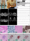
Clinical, neurophysiological, imaging, and pathological features of our patients. (A, B) Clinical appearance of left calf (Patient #1) and right arm (Patient #2) hypertrophy. (C–F) Spontaneous EMG activity recorded from right biceps brachii muscle of Patient #2. (G, H) MRI imaging of the calf muscles (Patient #1), T2‐weighted and T1 contrast enhancement, respectively. (I) MRI of the right forearm muscles from Patient #2 (right: T2‐weighted; left: T1 contrast enhanced). (J) MRI examination of the cervical roots from Patient #2 (T2‐weighted and T1 contrast enhanced, respectively). (K, L) Hematoxylin and eosin (H&E) and Nicotinamide Adenine Dinucleotide (NADH) staining of the left lateral gastrocnemius biopsy from Patient #1. (M, N) H&E and Adenosine triphosphatase (ATPase) staining at pH 9.4 of the right biceps brachii biopsy from Patient #2. (O) H&E staining of a endomysial inflammatory infiltrate in Patient #2. (P–R) Immunohistochemistry staining characterizing the infiltrate for CD3, CD20, and CD68, respectively.
Figure 3
Figure 3
Schematic pathogenesis of NMH. The diagram on the left depicts the established process of muscle atrophy resulting from chronic denervation. On the right, it illustrates the proposed mechanisms responsible for generating and sustaining NMH, supported in the literature by proof of principle.

References

    1. Ehmsen JT, Höke A. Cellular and molecular features of neurogenic skeletal muscle atrophy. Exp Neurol. 2020;331:113379. doi:10.1016/j.expneurol.2020.113379 - DOI - PubMed
    1. Gutmann L. AAEM minimonograph #46: neurogenic muscle hypertrophy. Muscle Nerve. 1996;19(7):811‐818. doi:10.1002/(SICI)1097-4598(199607)19:7<811::AID-MUS1>3.0.CO;2-C - DOI - PubMed
    1. Page MJ, McKenzie JE, Bossuyt PM, et al. The PRISMA 2020 statement: an updated guideline for reporting systematic reviews. BMJ. 2021;372:n71. doi:10.1136/bmj.n71 - DOI - PMC - PubMed
    1. Murad MH, Sultan S, Haffar S, Bazerbachi F. Methodological quality and synthesis of case series and case reports. BMJ Evid Based Med. 2018;23(2):60‐63. doi:10.1136/bmjebm-2017-110853 - DOI - PMC - PubMed
    1. Stålberg E, Van Dijk H, Falck B, et al. Standards for quantification of EMG and neurography. Clin Neurophysiol. 2019;130(9):1688‐1729. doi:10.1016/j.clinph.2019.05.008 - DOI - PubMed

LinkOut - more resources