Review
. 2024 Aug;11(32):e2403399.
doi: 10.1002/advs.202403399. Epub 2024 Jun 21.

Innovative Nanotechnology in Drug Delivery Systems for Advanced Treatment of Posterior Segment Ocular Diseases

Affiliations
Review

Innovative Nanotechnology in Drug Delivery Systems for Advanced Treatment of Posterior Segment Ocular Diseases

Yue Wu et al. Adv Sci (Weinh). 2024 Aug.

Abstract

Funduscopic diseases, including diabetic retinopathy (DR) and age-related macular degeneration (AMD), significantly impact global visual health, leading to impaired vision and irreversible blindness. Delivering drugs to the posterior segment of the eye remains a challenge due to the presence of multiple physiological and anatomical barriers. Conventional drug delivery methods often prove ineffective and may cause side effects. Nanomaterials, characterized by their small size, large surface area, tunable properties, and biocompatibility, enhance the permeability, stability, and targeting of drugs. Ocular nanomaterials encompass a wide range, including lipid nanomaterials, polymer nanomaterials, metal nanomaterials, carbon nanomaterials, quantum dot nanomaterials, and so on. These innovative materials, often combined with hydrogels and exosomes, are engineered to address multiple mechanisms, including macrophage polarization, reactive oxygen species (ROS) scavenging, and anti-vascular endothelial growth factor (VEGF). Compared to conventional modalities, nanomedicines achieve regulated and sustained delivery, reduced administration frequency, prolonged drug action, and minimized side effects. This study delves into the obstacles encountered in drug delivery to the posterior segment and highlights the progress facilitated by nanomedicine. Prospectively, these findings pave the way for next-generation ocular drug delivery systems and deeper clinical research, aiming to refine treatments, alleviate the burden on patients, and ultimately improve visual health globally.

Keywords: ROS scavenging; anti‐VEGF therapy; drug delivery; nanomedicine; posterior segment ocular diseases.


Conflict of interest statement

The authors declare no conflict of interest.

Figures

Figure 1
Figure 1
Multiple fabricated nanomaterials against different targets for the treatment of posterior segment ocular diseases. Created with BioRender.com.
Figure 2
Figure 2
Ocular drug delivery barriers. a) Graphic representation of the three‐layered structure of the tear film, including the lipid layer, aqueous layer, and mucus layer. Reproduced with permission.[ 56 ] Copyright 2022, Elsevier. b) The structure of the cornea reveals the presence of the corneal epithelium, stroma, and Descemet's membrane, and diagram depicting the tight junction of the corneal epithelium. Reproduced with permission.[ 59 ] Copyright 2021, Elsevier. c) Schematic of the hierarchical collagen structure of the sclera. Reproduced with permission. Five triple alpha‐chain tropocollagen molecules assemble into microfibrils, in which the axial stagger of individual molecules leads to gap/overlap regions that define the 67 nm axial D‐period. Varying numbers of near‐parallel microfibrils form collagen fibrils of diameters ranging from 25 to 230 nm.[ 63 ] Copyright 2020, Elsevier. d) Illustration of the lacrimal drainage system. Reproduced with permission.[ 65 ] Copyright 2020, Elsevier. e) Anatomy of the human eye. The structures responsible for maintaining the blood‐aqueous barrier, the outer blood‐retinal barrier formed by the RPE, and the inner blood‐retinal barrier are color‐coded as green, pink, and yellow, respectively. Reproduced with permission.[ 68 ] Copyright 2020, Elsevier. f) Anatomical localization of the inner and outer blood‐retinal barriers. Reproduced with permission.[ 69 ] Copyright 2021, Wiley. g) Schematic presentation of the inner and outer blood‐retinal barriers and their relative location. ECF = extracellular fluid. Reproduced with permission.[ 70 ] Copyright 2011, Wichtig Publishing Srl.
Figure 3
Figure 3
Lipid nanomaterials for ocular drug delivery. a) Diagram illustrates the structural composition of various types of lipid‐based nanoparticles, including liposomes, solid lipid nanoparticles (SLNs), and nanostructured lipid carriers. Reproduced under terms of the CC‐BY license.[ 71 ] Copyright 2020, published by MDPI. b) Schematic structure of LNPs encapsulating mRNA and c) Images depicting bioluminescence at 24 and 168 h and d) Bar graph displays the quantified expression, measured as average radiance, at different time points after subretinal injections of MC3 LNPs with a dosage of 400 ng mRNA per injection. n  =  5; mean ± SEM. ** p ≤ 0.01, *** p ≤ 0.001. Reproduced with permission.[ 82 ] Copyright 2019, Elsevier. e) The drug release of ATS‐SLNs and ATS‐SUS was evaluated in simulated tear fluid (pH 7.2) with the addition of 2% ethanol and f) The percentage of ATS‐SLNs and ATS‐SUS that passed through the porcine cornea at different time intervals (n  =  6). Reproduced with permission.[ 86 ] Copyright 2020, Springer. g) Etoposide concentrations in plasma and h) vitreous after intravitreal injection of etoposide solution (ETO) and etoposide loaded SLNs (ETO‐SLNs) in rats (mean ± SD, n  =  3).[ 90 ] Copyright 2019, Elsevier.
Figure 4
Figure 4
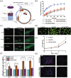
Polymeric nanomaterials. a–c) Chitosan oligosaccharide nanomicelles with functional properties are used to deliver dexamethasone to the ocular surface for topical medication administration. Reproduced with permission. a) Chitosan oligosaccharide‐valylvaline‐stearic acid nanomicelles were designed for topical ocular drug delivery, based on peptide transporter‐1 (PepT‐1) active targeting. b) In vitro drug release profiles of Dex solution, CSO‐SA nanomicelles, CSOVV‐SA (5:2) nanomicelles, CSO‐VV‐SA (5:4) nanomicelles, and HCO‐40/OC‐40/Dex mixed nanomicelles profiles in simulated tear fluid. c) Fluorescence microscopy of the cornea and conjunctiva of rabbit tissues, and sclera‐choroid‐retina of rat tissues after CSO‐VV‐SA (5:4)/Cou‐6 eyedrops administration. Reproduced with permission.[ 102 ] Copyright 2020, Elsevier. d–g) Evaluation of RPC growth rate in relation to Gel‐HA and Gel‐HA‐PDA hydrogels following a three‐day incubation period. d) Fluorescent images of GFP‐positive retinal progenitor cells (RPCs) illustrated that the Gel‐HA hydrogel group had the largest concentration of cells. e) The growth potency of RPCs was significantly greater in the Gel‐HA, as evidenced by the CCK‐8 assay. f) The mRNA expression levels of the cell proliferation marker Ki‐67 and the retinal progenitor‐related markers Nestin, Vimentin, and Pax‐6 were significantly higher in cells cultured on Gel‐HA hydrogel compared to the control group. g) Edu staining of RPCs in 3D hydrogels. Reproduced with permission.[ 106 ] Copyright 2019, Elsevier.
Figure 5
Figure 5
Metal and metal compound nanoparticles. a,b) AuNP's suppression on RPE cell dispersion a) BRPE cells, post‐trypsin treatment, were rinsed in Iscove's Modified Dulbecco's Medium (IMDM) with 10% FBS. Following a 30‐minute pre‐treatment with AuNPs, the cells were cultured on fibronectin‐coated wells (2 × 10⁵ cells per dish) within 35 × 10 mm culture dishes. At the 24‐hour mark, methanol‐fixed and Coomassie‐stained cells displayed inhibited adhesion and spread, both with and without VEGF and IL1‐β exposure. b) Statistical analysis of spread cell percentages across different views illustrated consistent results over three duplicated experiments, expressed as mean ± SEM (*p < 0.05 versus VEGF; **p < 0.05 versus IL‐1β; #p < 0.05 for both). Reproduced with permission.[ 111 ] Copyright 2010, Elsevier. c) Slit‐lamp biomicroscopic pictures were taken of rabbit eyes before the surgery (Pre group) and 3 days after experimentally inducing bacterial keratitis (BK group), followed by intrastromal administration of nanoparticles (Ag NPs or G‐Ag NPs). Reproduced with permission.[ 116 ] Copyright 2019, Elsevier. d) Schematic representation of the synthesis and e) The mechanism by which antioxidants are delivered and operate in RPE cells using injectable hydrogels loaded with Nanoceria. Reproduced with permission.[ 120 ] Copyright 2018, Wiley.
Figure 6
Figure 6
Carbon nanomaterials and quantum dot nanomaterials. a) Eyes injected with CNT‐FITC‐Bio or b) CTN‐FITC‐FA on days 1, 2, and 3 and control eye (×10, DAPID). Reproduced under terms of the CC‐BY license.[ 143 ] Copyright 2020, published by Knowledge E. c) Schematic illustration depicts the C18PGM as an innovative drug delivery device for the treatment of choroidal neovascularization. Reproduced with permission.[ 147 ] Copyright 2023, Wiley. d) The utilization of Super‐Cationic Carbon Quantum Dots as an eye drop formulation facilitates the opening of corneal epithelial tight junctions, enabling the effective topical therapy of bacterial keratitis. Reproduced with permission.[ 151 ] Copyright 2017, American Chemistry Society.
Figure 7
Figure 7
Hydrogel materials with superior biocompatibility for the treatment of ophthalmic diseases. a) The production of bioinspired hydrogels presents both challenges and potential, particularly when considering their physical functions over various length scales. Reproduced with permission.[ 155 ] Copyright 2020, American Chemistry Society. b–d) The multifunctional supramolecular filament hydrogel demonstrates strong therapeutic effectiveness in the rabbit model of experimental autoimmune uveitis (EIU). b) The self‐assembly of 2IPF‐DHB‐GFFYD to generate a supramolecular hydrogel. c) Slit‐lamp photos (e.g., iris hyperemia, miosis, exudation, hypopyon) of eyes and d) Quantification of inflammatory severity using clinical scores for each group. Reproduced with permission.[ 157 ] Copyright 2022, Wiley. e) Diagram illustrating the process of the hydrogel eye drops that are loaded with medication, as well as the use of these eye drops for the non‐invasive treatment of uveitis. Reproduced with permission.[ 158 ] Copyright 2021, Elsevier. f) Light‐cured hydrogel “T.E.S.T” for filling and repairing corneal defects combined with CXL. Reproduced with permission.[ 160 ] Copyright 2023, Elsevier.
Figure 8
Figure 8
Anti‐VEGF nanomaterials for posterior ocular diseases. a) Rats were subjected to in vivo imaging following intravitreal administration of CB and CB‐MVLs and b) The levels of Bev‐MVLs and Bev‐S were measured in the vitreous humor. Reproduced under terms of the CC‐BY license.[ 173 ] Copyright 2018, Taylor & Francis. c) The process of creating the Anti‐VEGF@BetP nanofiber hydrogel and its strategy for treating a mouse model of laser‐induced CNV, d) The mRNA levels of TNF‐α, NF‐κB p65, IL‐1β, and IL‐8 in choroidal‐retinal flat mounts reduced by the BetP‐gel, based on qPCR analysis, e) Representative IB4 staining images of individual lesions from the laser‐induced mouse CNV model at the 4th week after intravitreal injection with BetP‐Gel in comparison with saline, anti‐VEGF, and Anti‐VEGF@BetP‐Gel and f) Quantification of the CNV lesions. Reproduced with permission.[ 175 ] Copyright 2023, Wiley. g) The diagram depicts the process of the binding of VEGFR (BV) peptide with VEGFR and its effect on HUVECs for the inhibition of angiogenesis, h) CLSM images of HUVEC cells and L929 cells incubated with BV NPs (20 mmol L−1) for 4 h and i) SEM images of cell surfaces of HUVECs or L929 treated with BV NPs, and untreated cells as blank groups.[ 176 ] Copyright 2020, Elsevier.
Figure 9
Figure 9
ROS scavenging nanomaterials for posterior ocular diseases. a) ARPE19 cells were treated with CeO2NPs for 24 hours, resulting in a reduction of intracellular ROS levels. Oxidative stress was induced with tBH. Reproduced with permission.[ 180 ] Copyright 2023, American Chemical Society. b) Schematic illustration of the designed NP1 targeting the cell membranes, inhibiting necroptosis, and depleting ROS in acute glaucoma. Reproduced with permission.[ 182 ] Copyright 2022, American Chemical Society. c–e) Curcumin nanoparticles applied topically provide in vivo protection for retinal ganglion cell soma against cell loss induced by ocular hypertension. c) Diagram illustrating the in‐vivo experiment method. OHT rats were randomly assigned to one of three groups: no therapy, once‐daily administration of curcumin nanoparticles (CN) eye‐drops, or once‐daily administration of free curcumin (FC) eye drops. The treatment started two days before inducing IOP. Animals were euthanized three weeks later, and their retinas were prepared as flat mounts for labeling with Brn3a. d) The IOP of all animals subjected to OHT showed a substantial increase compared to the first measurement, up until 21 days post‐surgery. There was no disparity in IOP among the different treatment groups for OHT at any given time, indicating that any reported neuroprotective effects were not influenced by IOP. e) Elevated IOP in eyes with OHT only was linked to a decrease in RGC density by ≈23%. Treatment with CN but not FC significantly decreased the loss of RGCs. Reproduced under terms of the CC‐BY license.[ 184 ] Copyright 2018, published by Nature Publishing Group. f,g) ROS scavenging activities of PDA nanoparticles and the effects of Br@PDA on RGC survival and h) Axon regeneration 30 days after ONC. Reproduced under terms of the CC‐BY license.[ 185 ] Copyright 2021, published by Biomed Central.
Figure 10
Figure 10
Nanomaterials targeting macrophage polarization for posterior segment ocular diseases. a) Schematic presentation for analysis of M2 polarization by IL‐4‐loaded XL‐MSNs or soluble IL‐4 in vivo. Mice were injected intraperitoneally with soluble IL‐4 (Sol IL‐4) or 180 nm XL‐MSNs (200 µg per mouse) containing IL‐4 (XL‐MSNs IL‐4) at specified IL‐4 dosages (0, 10, 30, 100 ng per mouse). After a period of 3 days, cells located in the peritoneal cavity were collected. Peritoneal macrophages were then isolated by adhering them to culture plates. These macrophages were subsequently used to assess the expression of M2 genes. b) Schematic Presentations of Synthesis of Uniform Mesoporous Silica Nanoparticles with Extra‐Large Pores (XLMSNs). c) Their Application to IL‐4 Delivery for in Vivo M2 Macrophage Polarization. Reproduced with permission.[ 186 ] Copyright 2017, American Chemistry Society. d–f) FA transformed the phenotypes of microglia/macrophages, shifting them from a pro‐inflammatory state known as “M1” to an anti‐inflammatory one known as “M2”. d) Immunostaining of retinal whole‐mounts revealed that iba1+ microglia/macrophages expressing had elevated levels of iNOS (shown by white arrowheads) rather than Arg1 in the OIR retina. FA treatment resulted in an elevated quantity of Arg1+ iba1+ cells (shown by yellow arrowheads), whereas the amount of iNOS+ iba1+ cells dropped. e,f) The Western blot assay demonstrated that FA therapy resulted in a reduction of IL‐6 and TNF‐a levels, while simultaneously increasing the expression of IL‐10 in the OIR retinas. n = 6 mice. Reproduced under terms of the CC‐BY license.[ 189 ] Copyright 2022, published by Frontiers. g) Schematic structure of T‐155 prepared from tFNA and h) Micro RNA‐155 and and its ability to promote the polarisation of macrophages to the M1 type. Reproduced with permission.[ 192 ] Copyright 2022, Elsevier.

References

    1. Shen J., Gao H., Chen L., Jiang Y., Li S., Chao Y., Liu N., Wang Y., Wei T., Liu Y., Li J., Chen M., Zhu J., Liang J., Zhou X., Zhang X., Gu P., Chen Q., Liu Z., Sci. Adv. 2023, 9, eabq3104. - PMC - PubMed
    1. Campa C., Curr. Drug Targets 2020, 21, 1194. - PubMed
    1. Wang Z., Liu C.‐H., Huang S., Fu Z., Tomita Y., Britton W. R., Cho S. S., Chen C. T., Sun Y., Ma J., He X., Chen J., Sci. Adv. 2020, 6, eaba7457. - PMC - PubMed
    1. Heier J. S., Kherani S., Desai S., Dugel P., Kaushal S., Cheng S. H., Delacono C., Purvis A., Richards S., Le‐Halpere A., Connelly J., Wadsworth S. C., Varona R., Buggage R., Scaria A., Campochiaro P. A., Lancet 2017, 390, 50. - PubMed
    1. Baumal C. R., Spaide R. F., Vajzovic L., Freund K. B., Walter S. D., John V., Rich R., Chaudhry N., Lakhanpal R. R., Oellers P. R., Leveque T. K., Rutledge B. K., Chittum M., Bacci T., Enriquez A. B., Sund N. J., Subong E. N. P., Albini T. A., Ophthalmol. 2020, 127, 1345. - PubMed