. 2024 Aug:173:112228.
doi: 10.1016/j.jbiomech.2024.112228. Epub 2024 Jul 14.

Relationships between quantitative magnetic resonance imaging measures at the time of return to sport and clinical outcomes following acute hamstring strain injury

Affiliations

Relationships between quantitative magnetic resonance imaging measures at the time of return to sport and clinical outcomes following acute hamstring strain injury

Christa M Wille et al. J Biomech. 2024 Aug.

Abstract

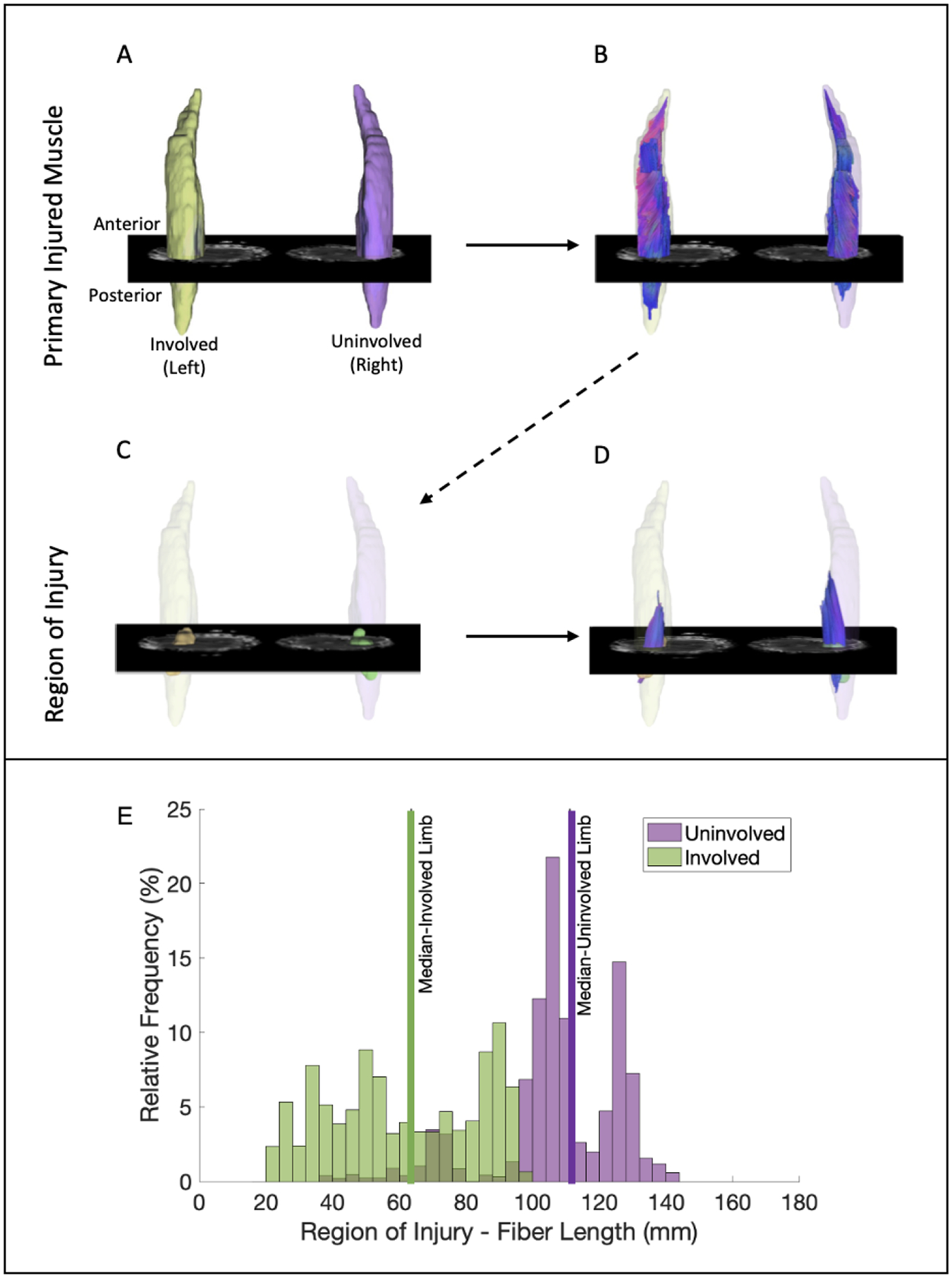
Hamstring strain injuries (HSI) are a common occurrence in athletics and complicated by high rates of reinjury. Evidence of remaining injury observed on magnetic resonance imaging (MRI) at the time of return to sport (RTS) may be associated with strength deficits and prognostic for reinjury, however, conventional imaging has failed to establish a relationship. Quantitative measure of muscle microstructure using diffusion tensor imaging (DTI) may hold potential for assessing a possible association between injury-related structural changes and clinical outcomes. The purpose of this study was to determine the association of RTS MRI-based quantitative measures, such as edema volume, muscle volume, and DTI metrics, with clinical outcomes (i.e., strength and reinjury) following HSI. Spearman's correlations and Firth logistic regressions were used to determine relationships in between-limb imaging measures and between-limb eccentric strength and reinjury status, respectively. Twenty injuries were observed, with four reinjuries. At the time of RTS, between-limb differences in eccentric hamstring strength were significantly associated with principal effective diffusivity eigenvalue λ1 (r = -0.64, p = 0.003) and marginally associated with mean diffusivity (r = -0.46, p = 0.056). Significant relationships between other MRI-based measures of morphology and eccentric strength were not detected, as well as between any MRI-based measure and reinjury status. In conclusion, this preliminary evidence indicates DTI may track differences in hamstring muscle microstructure, not captured by conventional imaging at the whole muscle level, that relate to eccentric strength.

Keywords: Diffusion Tensor Imaging; Hamstring Strain Injury; Reinjury; Return to Sport; Skeletal Muscle.


Conflict of interest statement

Declaration of competing interest The authors declare the following financial interests/personal relationships which may be considered as potential competing interests: Select authors of this manuscript declare relationships with the following companies: Dr. Bryan Heiderscheit declares a potential conflict of interest directly related to this work (research support to institution from NBA and GE HealthCare). The other authors (CMW, SAH, MRJ, KL, RK) of this manuscript declare no direct relationships with any companies, whose products or services may be related to the subject matter of the article].

Figures

Figure 1.
Figure 1.
Mean quantitative diffusion metrics were calculated within manually outlined regions of injury (ROI) on the injured limb defined by the hyperintense region of signal on T2-weighted imaging at the time of injury (TOI). Injury ROIs were registered to follow-up scans at return to sport (RTS) (peach arrow). For between-limb comparisons, ROIs were mirrored and manually registered to the uninvolved limb (white arrow). Data shown are from one slice of a representative participant. Biceps femoris short head (BFsh), biceps femoris long head (BFlh), semitendinosus (ST), semimembranosus (SM).
Figure 2.
Figure 2.
Representative imaging data from one participant demonstrating the process used to identify fascicles passing through the region of injury. A. Three-dimensional surface mesh generated from manual muscle boundary segmentation of the primary injured muscle (biceps femoris long head); B. Identification of all fascicles within the muscle boundary of interest; C. Three-dimensional surface mesh generated from manual segmentation of voxels containing increased signal intensity on the T2-weighted fluid sensitive sequence used to represent the region of injury and the mirrored, manually registered region on the uninvolved limb; D. Identification of fascicles within the primary injured muscle that pass through the region of injury on the involved limb relative to the uninvolved limb; E. Histogram of the relative frequency and median length of fascicles within the primary injured muscle on the involved limb (green) relative to the uninvolved limb (purple).
Figure 3.
Figure 3.
Participant inclusion criteria. All participants included in this analysis had unilateral evidence of injury on a T2-weighted magnetic resonance image (MRI) within 7 days of injury. Participants who sustained bilateral injuries linked to the same date/mechanism of injury were excluded from the analysis.
Figure 3.
Figure 3.
Participant inclusion criteria. All participants included in this analysis had unilateral evidence of injury on a T2-weighted magnetic resonance image (MRI) within 7 days of injury. Participants who sustained bilateral injuries linked to the same date/mechanism of injury were excluded from the analysis.
Figure 4.
Figure 4.
Representative imaging data from one participant with a prior left hamstring strain injury at return to sport, A.) T2-weighted, B. mean diffusivity, and C. primary eigenvalue maps along principal direction (λ1). In each subpanel, the left hamstring strain injury is represented by signal irregularities consistent with hyperintense signal intensity in the T2-weighted image (A) and mean diffusivity map (B) and irregularities in the color or direction of travel represented by the primary eigenvalue map along the principal direction (C).
Figure 5.
Figure 5.
Correlations in between-limb eccentric strength and between-limb diffusivity measures: A. Principal effective diffusivity eigenvalue λ1 and B. Mean diffusivity. Correlations with non-significant relationships between eccentric strength and other imaging measures are presented in Table 2.

References

    1. Alexander AL, Hurley SA, Samsonov AA, Adluru N, Hosseinbor AP, Mossahebi P, Tromp do PM, Zakszewski E, Field AS, 2011. Characterization of cerebral white matter properties using quantitative magnetic resonance imaging stains. Brain Connect 1, 423–446. - PMC - PubMed
    1. Andersson JL, Skare S, Ashburner J, 2003. How to correct susceptibility distortions in spin-echo echo-planar images: application to diffusion tensor imaging. Neuroimage 20, 870–888. - PubMed
    1. Andersson JLR, Sotiropoulos SN, 2016. An integrated approach to correction for off-resonance effects and subject movement in diffusion MR imaging. Neuroimage 125, 1063–1078. - PMC - PubMed
    1. Bayer ML, Hoegberget-Kalisz M, Jensen MH, Olesen JL, Svensson RB, Couppe C, Boesen M, Nybing JD, Kurt EY, Magnusson SP, Kjaer M, 2018. Role of tissue perfusion, muscle strength recovery, and pain in rehabilitation after acute muscle strain injury: A randomized controlled trial comparing early and delayed rehabilitation. Scand J Med Sci Sports 28, 2579–2591. - PubMed
    1. Berry DB, Regner B, Galinsky V, Ward SR, Frank LR, 2018. Relationships between tissue microstructure and the diffusion tensor in simulated skeletal muscle. Magn Reson Med 80, 317–329. - PMC - PubMed