. 2024 Sep:75:103268.
doi: 10.1016/j.redox.2024.103268. Epub 2024 Jul 17.

BSA-stabilized selenium nanoparticles ameliorate intracerebral hemorrhage's-like pathology by inhibiting ferroptosis-mediated neurotoxicology via Nrf2/GPX4 axis activation

Affiliations

BSA-stabilized selenium nanoparticles ameliorate intracerebral hemorrhage's-like pathology by inhibiting ferroptosis-mediated neurotoxicology via Nrf2/GPX4 axis activation

Xiao-Na Li et al. Redox Biol. 2024 Sep.

Abstract

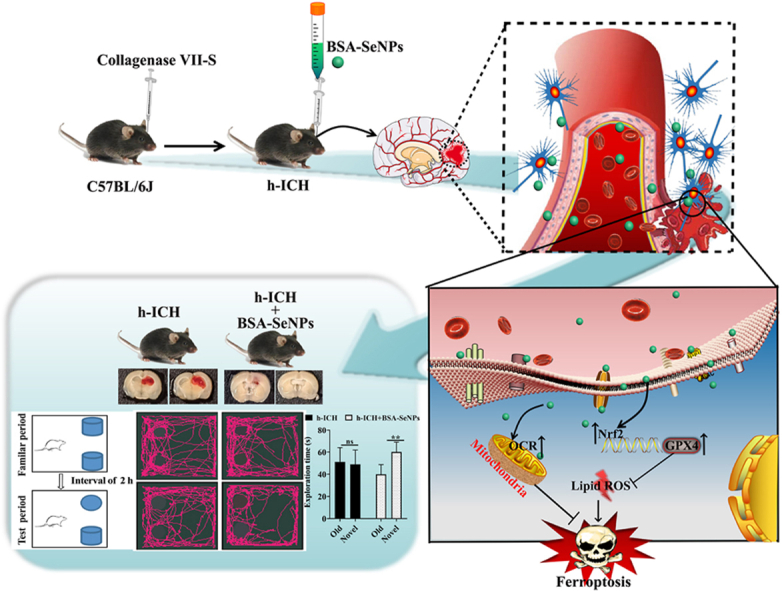
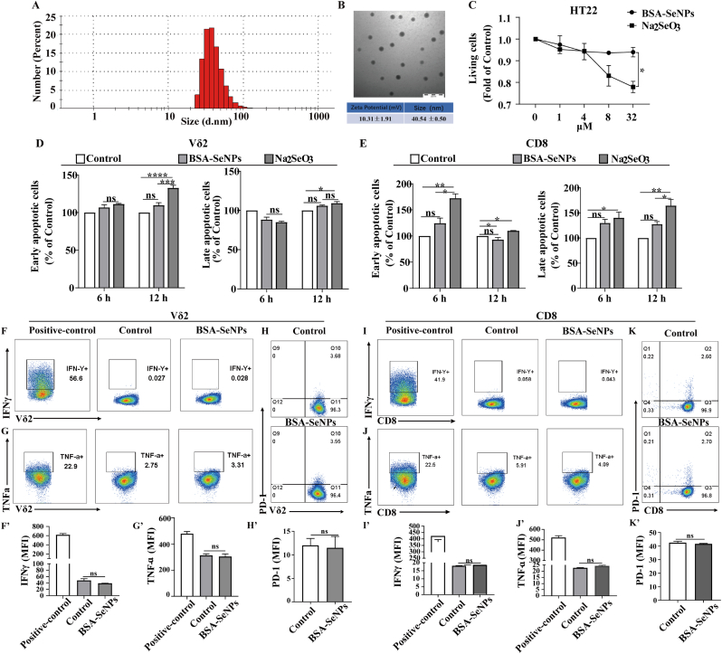
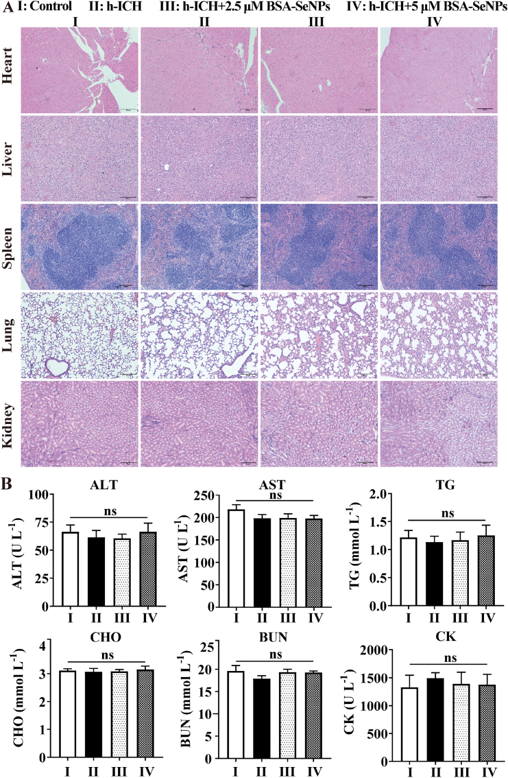
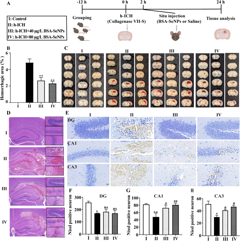
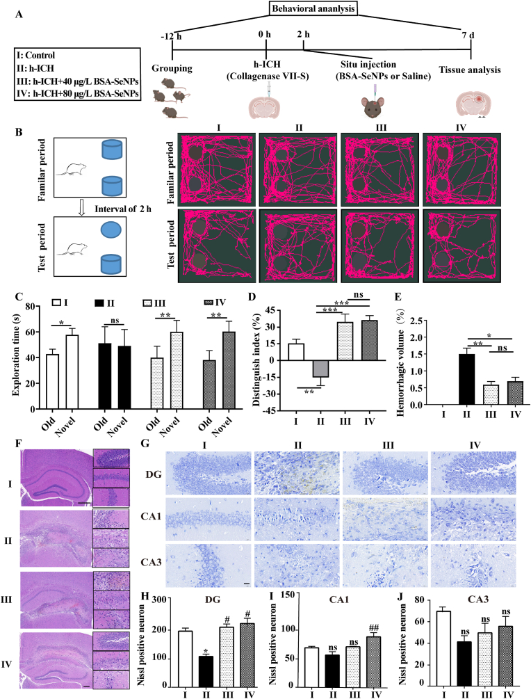
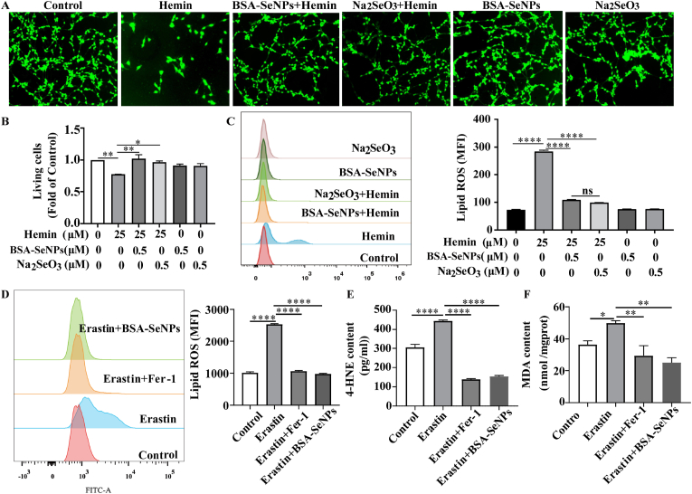
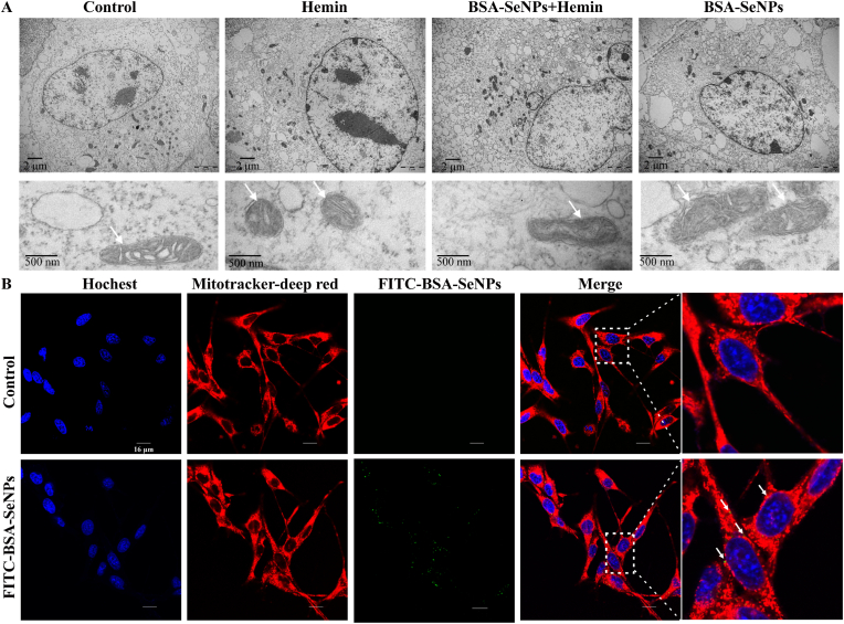
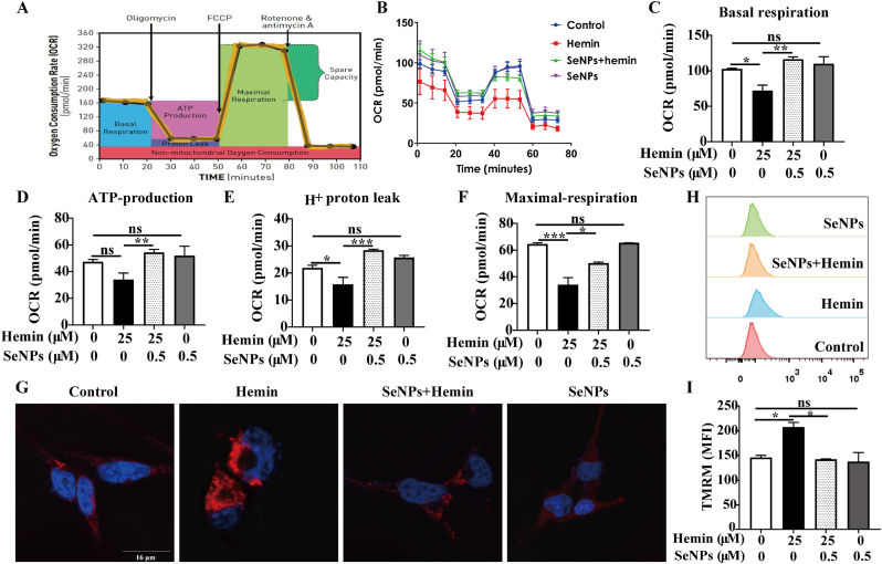
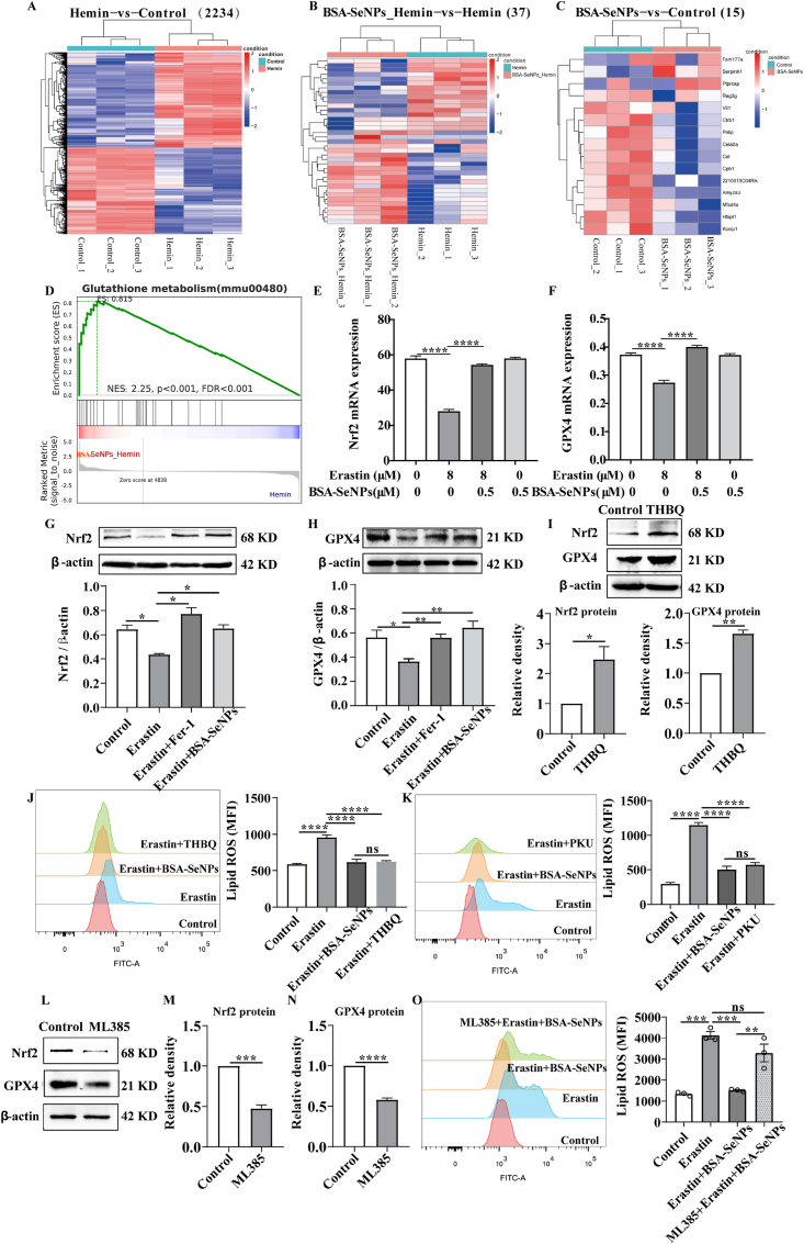
Intracerebral hemorrhage (ICH) is a prevalent hemorrhagic cerebrovascular emergency. Alleviating neurological damage in the early stages of ICH is critical for enhancing patient prognosis and survival rate. A novel form of cell death called ferroptosis is intimately linked to hemorrhage-induced brain tissue injury. Although studies have demonstrated the significant preventive impact of bovine serum albumin-stabilized selenium nanoparticles (BSA-SeNPs) against disorders connected to the neurological system, the neuroprotective effect on the hemorrhage stroke and the mechanism remain unknown. Therefore, based on the favorable biocompatibility of BSA-SeNPs, h-ICH (hippocampus-intracerebral hemorrhage) model was constructed to perform BSA-SeNPs therapy. As expected, these BSA-SeNPs could effectively improve the cognitive deficits and ameliorate the damage of hippocampal neuron. Furthermore, BSA-SeNPs reverse the morphology of mitochondria and enhanced the mitochondrial function, evidenced by mitochondrial respiration function (OCR) and mitochondrial membrane potential (MMP). Mechanistically, BSA-SeNPs could efficiently activate the Nrf2 to enhance the expression of antioxidant GPX4 at mRNA and protein levels, and further inhibit lipid peroxidation production in erastin-induced ferroptotic damages. Taken together, this study not only sheds light on the clinical application of BSA-SeNPs, but also provides its newly theoretical support for the strategy of the intervention and treatment of neurological impairment following ICH.

Keywords: BSA-selenium nanoparticles; Cognitive function; Ferroptosis; Intracerebral hemorrhage; Nrf2-GPX4 axis.


Conflict of interest statement

Declaration of competing interest All authors declare that there are no competing interests.

Figures

Image 1
Graphical abstract
Fig. 1
Fig. 1
Characterization of BSA-SeNPs and biocompatibility of BSA-SeNPs in vitro. (A–B) The distribution of zeta potential, particle size, and TEM images of BSA-SeNPs. (C) Hippocampal neuronal cells were treated with BSA-SeNPs and Na2SeO3 at concentrations of 1–32 μM, respectively, for 6 h. The living cells were detected by CCK-8. (D–E) Vδ2 and CD8 cells were double-stained with Annexin V and Propidium iodide (PI) after treatment with BSA-SeNPs (0.5 μM) or Na2SeO3 (0.5 μM) for 6 h or 12 h. (F–K) The expressions of TNF-a, IFN-γ, and PD-1 were detected by flow cytometry treatment with BSA-SeNPs (0.5 μM) for 6 h n = 3, ****P < 0.0001, ***P < 0.001, **P < 0.01, *P < 0.05, ns: no significance.
Fig. 2
Fig. 2
Biosafety of BSA-SeNPs was tested in vivo. (A) HE staining of heart, liver, spleen, lung and kidney (n = 4,x100). (B–G) Results of 6 blood biochemical tests (n = 4); ns, no significance.
Fig. 3
Fig. 3
Situ injection of selenium nanoparticles in hippocampus reduces the area of hematoma and the damage of hippocampus neuron in h-ICH mice. (A) Illustrations of the experimental grouping and the treatment procedures. (B–C) Statistical data and representative images of brain slices after treatment with BSA-SeNPs (40 μg/L and 80 μg/L) in h-ICH mouse mode (n = 4). (D) Representative images of HE staining in the hippocampus (n = 4, scale bar: 200 μm, 20 μm). (E–H) Images of Nissl staining (n = 3, scale bar: 20 μm) of treated hippocampus DG (F), CA1 (G), and CA3 (H) fields. *P < 0.05, **P < 0.01, vs Control; #P < 0.05, ##P < 0.01, vs h-ICH; ns, no significance.
Fig. 4
Fig. 4
BSA-SeNPs shield the hippocampus from neuronal injury and cognitive decline in the subacute phase of h-ICH. (A) Illustrations of the experimental grouping and the treatment procedures. (B–D) Schematic diagram of the testing procedure (B), the exploration time (C), and the discrimination index (D) in the object recognition task (n = 4). (E) Hemorrhagic volume (F) Representative images of HE staining in the hippocampus (n = 4, scale bar: 200 μm, 20 μm). (G–J) Images of Nissl staining (n = 4) of treated hippocampus (DG, CA1, and CA3 fields, scale bar: 20 μm), DG: dentate gyrus. *P < 0.05, vs Control; #P < 0.05, vs h-ICH; ns, no significance.
Fig. 5
Fig. 5
BSA-SeNPs inhibited hemin/erastin-induced ferroptotic damages in vitro. Hippocampal neuronal cells were co-treated with hemin or erastin and BSA-SeNPs for 6 h. (A–B) The relative living cell viability was imaged and detected by CCK-8 and after co-treatment with hemin and BSA-SeNPs (0.5 μM) or Na2SeO3 (0.5 μM) for 6 h. (C–D) The content of lipid ros was examined by flow cytometry. (E–F) The content of 4-HNE, MDA were respectively detected by cellular mouse 4-HNE (4-Hydroxynonenal) elisa kit and MDA elisa kit. ****P < 0.0001, **P < 0.01, *P < 0.05.
Fig. 6
Fig. 6
Effects of BSA-SeNPs on mitochondrial morphology. (A) Transmission electron microscopy (TEM) images of hippocampal neuronal cells treated with Hemin (6 h), BSA-SeNPs + Hemin (6 h), BSA-SeNPs (6 h). Shrunken mitochondria were marked as single white arrowheads (upper scale bar: 2 μm, bottom scale bar: 500 nm). (B) Internalization of BSA-SeNPs into mitochondria of hippocampal neurons was imaged by confocal microscopy (scale bar: 16 μm). The nucleus of hippocampal neuron was stained with Hoechst (blue); mitochondria were stained with Mitotracker Red (red).
Fig. 7
Fig. 7
Effects of BSA-SeNPs on mitochondrial function and respiration in hippocampal neurons. (A) The profile of the Agilent Seahorse XF Cell Mito Stress Test [28] illustrates the essential elements of mitochondrial function. (B–F) Key parameters of mitochondrial function: basal respiration (C), ATP-production (D), H+ proton leak (E), maximal respiration (F) of Mitochondria respiration in hippocampal neurons before and after BSA-SeNPs treatment were detected by Seahorse XF96 Mito Stress Kit. (G–I) Mitochondrial membrane potential (TMRM staining) of hippocampal neurons before and after BSA-SeNPs treatment was analyzed by confocal microscopy (G, scale bar: 16 μm) and flow cytometry (H–I). ***P < 0. 001, **P < 0.01, *P < 0.05, ns: no significance.
Fig. 8
Fig. 8
BSA-SeNPs reduced GPX4-mediated lipid peroxidation by activating Nrf2. (A–C) Differences in gene expression level cluster analysis. Red represented relatively high expression protein-coding genes, blue represented protein-coding genes expressed in relatively low. Hemin vs Control (2234);BSA-SeNPs_Hemin vs Hemin (37);BSA-SeNPs vs Control (15). (D) GSEA plots of the gene sets upon the BSA-SeNPs_Hemin and Hemin group. (E, F) The expressions of GPX4 and Nrf2 mRNA in hippocampal neuronal cells were assessed by q-PCR. (G–I) The protein expressions of GPX4 and Nrf2 were assessed by Western blot analysis after treatment with THBQ ((5 μM, 24h). (J–K) The level of lipid ROS was assessed by flow cytometry after co-treated with BSA-SeNPs (0.5 μM, 6h), THBQ, PKU ((10 μM, 24h) respectively in erastin (8 μM, 6h)-exposed hippocampal neurons. (L–M) The protein expressions of Nrf2 (M) and GPX4 (N) were assessed by Western blot analysis after treatment with mL385 (5 μM, 24 h). (O) Lipid ROS level was assessed by flow cytometry after co-treated with mL385 and BSA-SeNPs in erastin-exposed hippocampal neurons. *P < 0.05, **P < 0.01, ****P < 0.0001, ns: no significance.

References

    1. Lim T.C., Mandeville E., Weng D., Wang L.S., Kurisawa M., Leite-Morris K., et al. Hydrogel-based therapy for brain repair after intracerebral hemorrhage. Translational stroke research. 2020;11:412–417. - PubMed
    1. Qureshi AI, Mendelow AD, Hanley DF. Intracerebral haemorrhage. Lancet. 2009;373(9675):1632–1644. - PMC - PubMed
    1. Zille M, Farr TD, Keep RF, Römer C, Xi G, Boltze J. Novel targets, treatments, and advanced models for intracerebral haemorrhage. EBioMed. 2022 Feb;76 - PMC - PubMed
    1. Loan JJ, Kirby C, Emelianova K, Dando OR, Poon MT, Pimenova L, Hardingham GE, McColl BW, Klijn CJ, Al-Shahi Salman R, Schreuder FH, Samarasekera N. Secondary injury and inflammation after intracerebral haemorrhage: a systematic review and meta-analysis of molecular markers in patient brain tissue. J. Neurol. Neurosurg. Psychiatr. 2022 Feb;93(2):126–132. - PMC - PubMed
    1. Puy L, Parry-Jones AR, Sandset EC, Dowlatshahi D, Ziai W, Cordonnier C. Intracerebral haemorrhage. Nat. Rev. Dis. Primers. 2023 Mar 16;9(1):14. - PubMed

MeSH terms

LinkOut - more resources