. 2024 Sep:130:103954.
doi: 10.1016/j.mcn.2024.103954. Epub 2024 Jul 20.

iPSC-induced neurons with the V337M MAPT mutation are selectively vulnerable to caspase-mediated cleavage of tau and apoptotic cell death

Affiliations

iPSC-induced neurons with the V337M MAPT mutation are selectively vulnerable to caspase-mediated cleavage of tau and apoptotic cell death

Panos Theofilas et al. Mol Cell Neurosci. 2024 Sep.

Abstract

Background: Tau post-translational modifications (PTMs) result in the gradual build-up of abnormal tau and neuronal degeneration in tauopathies, encompassing variants of frontotemporal lobar degeneration (FTLD) and Alzheimer's disease (AD). Tau proteolytically cleaved by active caspases, including caspase-6, may be neurotoxic and prone to self-aggregation. Also, our recent findings show that caspase-6 truncated tau represents a frequent and understudied aspect of tau pathology in AD in addition to phospho-tau pathology. In AD and Pick's disease, a large percentage of caspase-6 associated cleaved-tau positive neurons lack phospho-tau, suggesting that many vulnerable neurons to tau pathology go undetected when using conventional phospho-tau antibodies and possibly will not respond to phospho-tau based therapies. Therefore, therapeutic strategies against caspase cleaved-tau pathology could be necessary to modulate the extent of tau abnormalities in AD and other tauopathies.

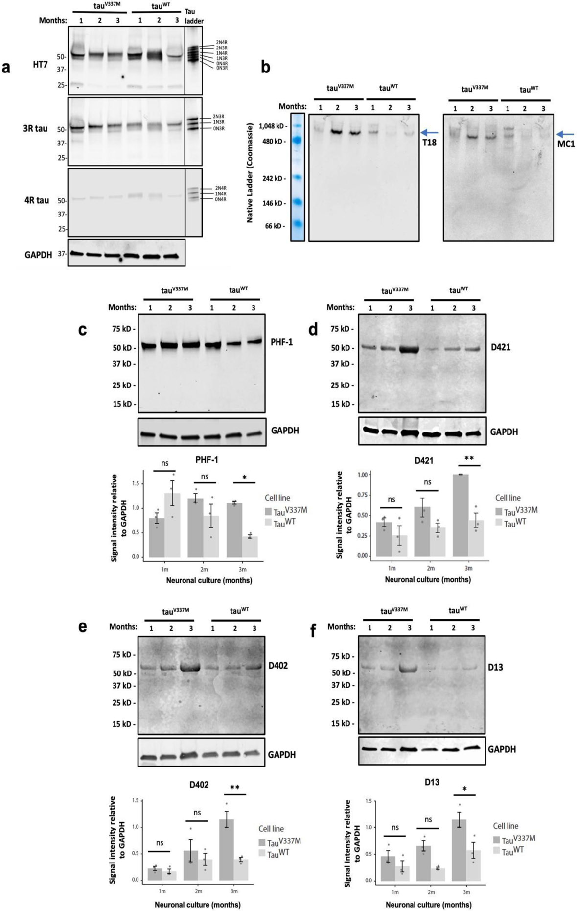
Methods: To understand the timing and progression of caspase activation, tau cleavage, and neuronal death, we created two mAbs targeting caspase-6 tau cleavage sites and probed postmortem brain tissue from an individual with FTLD due to the V337M MAPT mutation. We then assessed tau cleavage and apoptotic stress response in cortical neurons derived from induced pluripotent stem cells (iPSCs) carrying the FTD-related V337M MAPT mutation. Finally, we evaluated the neuroprotective effects of caspase inhibitors in these iPSC-derived neurons.

Results: FTLD V337M MAPT postmortem brain showed positivity for both cleaved tau mAbs and active caspase-6. Relative to isogenic wild-type MAPT controls, V337M MAPT neurons cultured for 3 months post-differentiation showed a time-dependent increase in pathogenic tau in the form of caspase-cleaved tau, phospho-tau, and higher levels of tau oligomers. Accumulation of toxic tau species in V337M MAPT neurons was correlated with increased vulnerability to pro-apoptotic stress. Notably, this mutation-associated cell death was pharmacologically rescued by the inhibition of effector caspases.

Conclusions: Our results suggest an upstream, time-dependent accumulation of caspase-6 cleaved tau in V337M MAPT neurons promoting neurotoxicity. These processes can be reversed by caspase inhibition. These results underscore the potential of developing caspase-6 inhibitors as therapeutic agents for FTLD and other tauopathies. Additionally, they highlight the promise of using caspase-cleaved tau as biomarkers for these conditions.

Keywords: Active caspase-6; FTLD; Neoepitope antibody; Postmortem; Tau cleavage; Tauopathies; V337M MAPT mutation; iPSCs.


Conflict of interest statement

Declaration of competing interest Michelle R. Arkin is cofounder of Elgia Therapeutics, which is developing caspase-6 inhibitors for inflammatory diseases. The other authors have declared no conflict of interest.

Figures

Figure 1.
Figure 1.
Caspase-6 cleaved tau neoepitope antibody positivity in human postmortem brains with tau accumulation. (a) Schematic depicting two caspase-6 cleaved tau sites targeted by monoclonal neoepitope antibodies mAb.D13 (D13; 14-441) and mAb.D402 (D402; 1-402), at the N- and C-terminus of tau, respectively. The C-terminus D421 site of tau is targeted by the antibody TauC3 and cleaved by multiple caspases; (b) Brain sections from the temporal cortex of an individual with tauV337M and FTD, showing antibody positivity for active caspase-6 (brown) and caspase-cleaved tau (red), including TauC3 (D421) (i), mAb.D402 (ii), and mAb.D13 (iii). Asterisks indicate cells positive for both active caspase-6 and caspase-cleaved tau markers. Scale bars: 50 μm.
Figure 2.
Figure 2.
Timeline of iPSC differentiation to neurons following doxycycline-inducible expression of Ngn2. (a) Phase-contrast images represent individual stages of the differentiation protocol, including iPSCs (i), neuronal precursors (ii), immature neurons (iii), and mature neurons (iv); (b) IF characterization of neurons at 1-month post differentiation. Mutant (tauV337M) and control (tauWT) neurons were positive for cortical (green; i-iv), glutamatergic (green; v-vi), and neuronal (red; i-vi) markers. Nuclei were stained with DAPI (blue; i-vi). Scale bars: (a) i: 200 μm, ii-iv: 50 μm (b) 60 μm.
Figure 3.
Figure 3.
Time-dependent tau pathological changes in the tauV337M neurons detected by western blot analyses. (a) Expression of total tau (HT7) and tau isoforms (3R and 4R tau) in tauV337M and tauWT neurons cultured from 1 to 3 months. Antibody specificity was confirmed by a recombinant tau ladder containing all six tau isoforms (right panels); (b) Oligomeric (T18) and conformational (MC1) tau levels were detected under non-denaturing conditions for preserving protein conformation. The molecular weight of the proteins was estimated using a protein standard for native electrophoresis stained with Coomassie blue (far-left panel) for band visualization (see also Fig. S4); (c-f) Semi-quantification of p-tau (PHF-1) and caspase- cleave tau (D421, D402, and D13) protein levels based on band intensities relative to GAPDH internal loading control (n ≥ 3 independent experiments; two-way ANOVA with post hoc Tukey test; ns, not significant: p > 0.05; *p < 0.05, **p < 0.01).
Figure 4.
Figure 4.
Caspase inhibition is neuroprotective against acute stress-induced cytotoxicity in the tauV337M neurons. Changes in cytotoxicity levels were measured by LDH release following a 48-hour treatment with STS and the pan-caspase inhibitor (z-VAD-fmk) in 3-month cultured neurons. The matrix heatmap illustrates p-values (color gradient) and significance levels (asterisks) between treatment groups. Dark gray shade represents tauV337M and light gray shade represents tauWT neurons (n=3 independent experiments; two-way ANOVA with post hoc Tukey test; *p < 0.05, **p<0.01, ***p<0.001, ****p<0.0001). RLU: Relative light units.
Figure 5.
Figure 5.
Stress-induced caspase activation in the induced neurons. Active caspase-6 (a) and caspase-3/7 levels (b) were examined in 3-month cultured neurons using caspase-specific cleavage substrates following treatment with vehicle, STS, and STS/z-VEID-fmk (a caspase-6 inhibitor) or STS/z-VAD-fmk (a pan-caspase inhibitor). The matrix heatmap illustrates p-values (color gradient) and significance levels (asterisks) between treatment groups. Dark gray shade represents tauV337M and light gray shade represents tauWT neurons; (c-d) Semi-quantification of caspase-cleaved tau (D421) protein levels in neurons treated with STS or STS/z-VAD- fmk for 48h based on band intensities relative to GAPDH internal loading control (n ≥ 3 independent experiments; two-way ANOVA with post hoc Tukey test; *p < 0.05, **p<0.01, ***p<0.001, ****p<0.0001). RLU: Relative light units.
Figure 6.
Figure 6.
Caspase inhibition rescues stress-induced reduction of neurite length in the induced neurons. (a) IF staining of neurites (MAP2, red) and cell nuclei (DAPI, blue) following a 48h treatment with STS and caspase-inhibitor (z-VAD-fmk) in 3-month cultured neurons; (b) Image quantification of mean length of processes (based on a). Matrix heatmap illustrates p-values (color gradient) and significance levels (asterisks) between treatment groups. Dark gray shade represents tauV337M and light gray shade represents tauWT neurons (n ≥ 3 independent experiments; two-way ANOVA with post hoc Tukey test; *p < 0.05, **p<0.01, ***p<0.001, ****p<0.0001). Scale bar: 50 μm.

References

    1. Albrecht S, Bourdeau M, Bennett D, Mufson EJ, Bhattacharjee M, LeBlanc AC, 2007. Activation of caspase-6 in aging and mild cognitive impairment. Am. J. Pathol 170, 1200–1209. 10.2353/ajpath.2007.060974 - DOI - PMC - PubMed
    1. Alonso AC, Grundke-Iqbal I, Iqbal K, 1996. Alzheimer’s disease hyperphosphorylated tau sequesters normal tau into tangles of filaments and disassembles microtubules. Nat. Med 2, 783–787. 10.1038/nm0796-783 - DOI - PubMed
    1. Angel A, Volkman R, Royal TG, Offen D, 2020. Caspase-6 Knockout in the 5xFAD Model of Alzheimer’s Disease Reveals Favorable Outcome on Memory and Neurological Hallmarks. Int. J. Mol. Sci 21. 10.3390/ijms21031144 - DOI - PMC - PubMed
    1. Cotman CW, Poon WW, Rissman RA, Blurton-Jones M, 2005. The role of caspase cleavage of tau in Alzheimer's disease neuropathology. J. Neuropathol. Exp. Neurol 64, 104–112. 10.1093/jnen/64.2.104 - DOI - PubMed
    1. Cusack CL, Swahari V, Hampton Henley W, Michael Ramsey J, Deshmukh M, 2013. Distinct pathways mediate axon degeneration during apoptosis and axon-specific pruning. Nat. Commun 4, 1876. 10.1038/ncomms2910 - DOI - PMC - PubMed

LinkOut - more resources