. 2024 Jul 20;22(1):429.
doi: 10.1186/s12951-024-02694-z.

Determining M2 macrophages content for the anti-tumor effects of metal-organic framework-encapsulated pazopanib nanoparticles in breast cancer

Affiliations

Determining M2 macrophages content for the anti-tumor effects of metal-organic framework-encapsulated pazopanib nanoparticles in breast cancer

Zhijie Xu et al. J Nanobiotechnology. .

Abstract

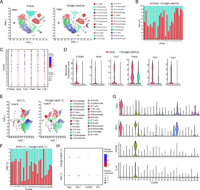
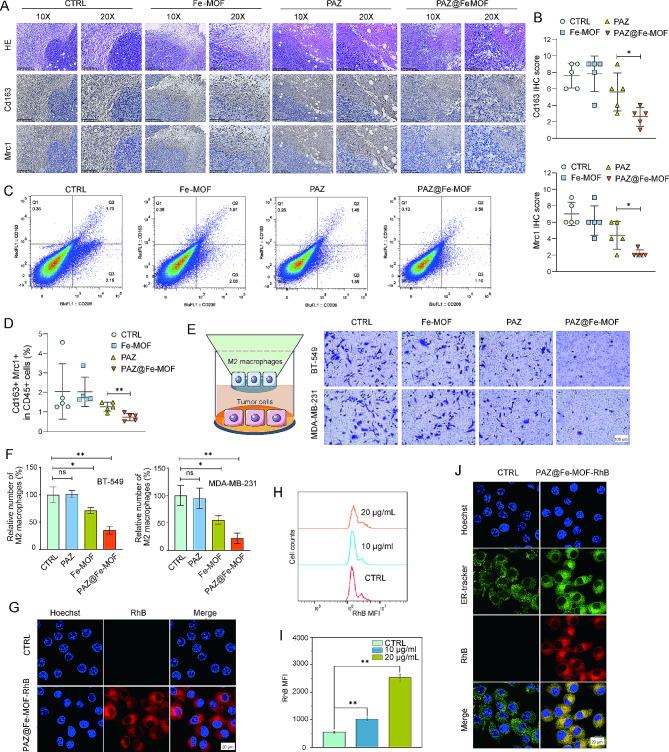
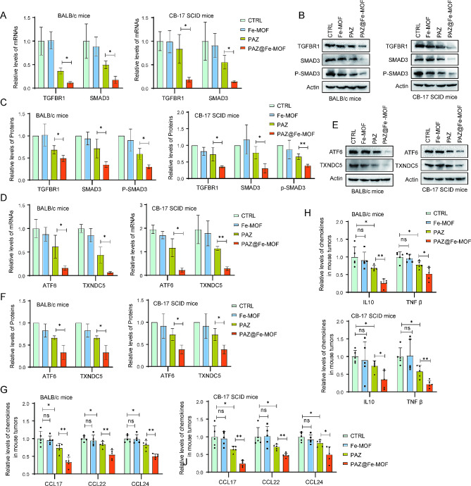
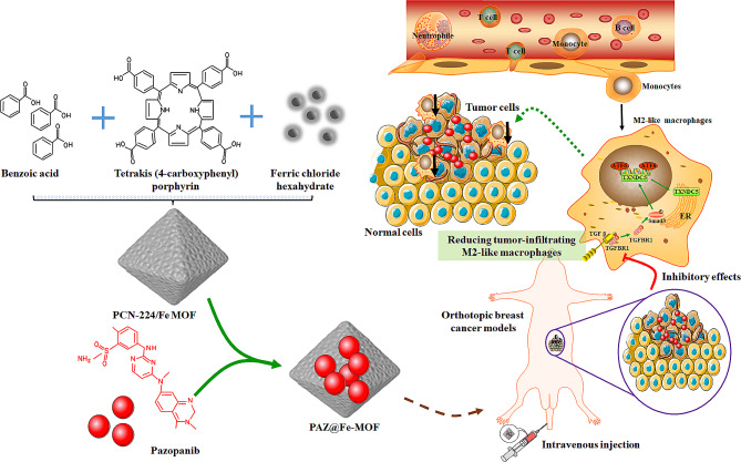
Pazopanib (PAZ), an oral multi-tyrosine kinase inhibitor, demonstrates promising cytostatic activities against various human cancers. However, its clinical utility is limited by substantial side effects and therapeutic resistance. We developed a nanoplatform capable of delivering PAZ for enhanced anti-breast cancer therapy. Nanometer-sized PAZ@Fe-MOF, compared to free PAZ, demonstrated increased anti-tumor therapeutic activities in both syngeneic murine 4T1 and xenograft human MDA-MB-231 breast cancer models. High-throughput single-cell RNA sequencing (scRNAseq) revealed that PAZ@Fe-MOF significantly reduced pro-tumorigenic M2-like macrophage populations at tumor sites and suppressed M2-type signaling pathways, such as ATF6-TGFBR1-SMAD3, as well as chemokines including CCL17, CCL22, and CCL24. PAZ@Fe-MOF reprogramed the inhibitory immune microenvironment and curbed tumorigenicity by blocking the polarization of M2 phenotype macrophages. This platform offers a promising and new strategy for improving the cytotoxicity of PAZ against breast cancers. It provides a method to evaluate the immunological response of tumor cells to PAZ-mediated treatment.

Keywords: Breast cancer; Immune microenvironment; M2-like macrophages; Metal-organic framework; Pazopanib.


Conflict of interest statement

The authors declare no competing interests.

Figures

Fig. 1
Fig. 1
Characterizations of PAZ@Fe-MOF. (A) The TEM observation indicated that the successful preparation of PAZ@Fe-MOF. The particle size was about 100 nm. Pronounced metal junctions of iron ions on its surface could be observed. (B) The elemental mapping of PAZ@Fe-MOF. (C) The XPS detection results of PAZ@Fe-MOF for the elemental analysis. (D) The hydordynamic size of PAZ@Fe-MOF was about 122.6 nm, as detected by malvern laser particle size analyzer. (E) The zeta-potential of Fe-MOF and PAZ@Fe-MOF was measured by malvern laser particle size analyzer. (F) The FTIR spectrum analysis of PAZ@Fe-MOF. (G) UV-vis assay identified the crystal structure of Fe-MOF and PAZ@Fe-MOF. (H) The TGA analysis of Fe-MOF and PAZ@Fe-MOF. (I) X-Ray diffraction (XRD) experiments confirmed the Fe-MOF and PAZ@Fe-MOF crystal pattern. (J) The hydrated particle size of prepared nanoparticles varied little within 7 days. (K) The Zeta-potential of Fe-MOF and PAZ@Fe-MOF kept unchanged within 7 days. (L) The collapse of PAZ@Fe-MOF nanoparticles under different pH conditions detected by TEM
Fig. 2
Fig. 2
In vivo distribution of PAZ@Fe-MOF in orthotopic breast tumor models. (A) The distribution of PAZ@Fe-MOF-RhB was analyzed by in vivo imaging. The circle indicates tumor site. (B) Quantitative results of PAZ@Fe-MOF-RhB staining at tumor tissues from A. (C) The fluorescence distribution of PAZ@Fe-MOF-RhB in tumor tissue, kidney lung, spleen, liver and heart. (D) Quantitative results of PAZ@Fe-MOF-RhB staining at tumor tissues from C. (E) Representative images of PAZ@Fe-MOF-RhB staining in tumor tissue section
Fig. 3
Fig. 3
PAZ@Fe-MOF displayed the suppression of tumor growth in orthotopic breast tumor models. (A) Experimental design for the procedures of 4T1 orthotopic mice models. (B) The representative small-animal PET-CT images of tumoral 18F-FDG distribution. The circle indicates tumor site. (C) ROIs indicated the the tumor size. (D) The tumor volume measured by PET-CT. (E-F) The representative images of 4T1 orthotopic breast tumor models at the end of experiments. (G) The tumor weight in 4T1 orthotopic breast tumor models treated with Fe-MOF, PAZ or PAZ@Fe-MOF. (H) The tumor growth curves in 4T1 orthotopic breast tumor models treated with Fe-MOF, PAZ or PAZ@Fe-MOF. (I) The body weight curves in 4T1 orthotopic breast tumor models treated with Fe-MOF, PAZ or PAZ@Fe-MOF. (J) Experimental design for the procedures of MDA-MB-231 orthotopic tumor models. (K) The representative images of MDA-MB-231 orthotopic tumor models at the end of experiments. (L) The tumor weight in MDA-MB-231 orthotopic tumor models treated with Fe-MOF, PAZ or PAZ@Fe-MOF. (M) The tumor growth curves in MDA-MB-231 orthotopic tumor models treated with Fe-MOF, PAZ or PAZ@Fe-MOF. (N) The body weight curves in MDA-MB-231 orthotopic tumor models treated with Fe-MOF, PAZ or PAZ@Fe-MOF. ns: not significant, *: p-value < 0.05, **: p-value < 0.01
Fig. 4
Fig. 4
scRNAseq identified the effect of PAZ@Fe-MOF on monocyte-derived macrophages. (A-B) The changes of twenty-six subpopulations in blood after PAZ@Fe-MOF treatment. (C) Bubble plots indicated the expression of monocytic makers in each single cell cluster. (D) Violin plots indicated the changes of monocytic makers after PAZ@Fe-MOF treatment. (E-F) The changes of different cell clusters in tumor tissues after PAZ@Fe-MOF treatment. (G) Violin plots of M2-like macrophage makers for each cluster. (H) Bubble plots indicated the changes of M2-like macrophage makers after PAZ@Fe-MOF treatment
Fig. 5
Fig. 5
Validation of decreased M2-like macrophages following PAZ@Fe-MOF treatment. (A) Representative immunostaining images of Mrc1 and Cd163 on mice specimens. Scale bars are indicated. (B) Protein levels of Mrc1 and Cd163 were quantified in mice specimens with different treatment. (C-D) Flow cytometry indicated the effects of PAZ@Fe-MOF on the tumor-infiltrating Cd163 + Mrc1 + cells. (E-F) Infiltration of M2-like macrophages in human breast cancer cells MDA-MB-231 treated with the indicated conditions. (G) PAZ@Fe-MOF-RhB nanoagents could be absorbed by IL-4-stimulated RAW264.7 cells. (H-I) The mean fluorescence intensity was quantified. (J) ER was labeled by the ER-tracker. In IL-4-stimulated RAW264.7 cells, the co-localization of PAZ@Fe-MOF with ER were observed with confocal microscopy. *: p-value < 0.05, **: p-value < 0.01
Fig. 6
Fig. 6
Inhibitory effect of PAZ@Fe-MOF on TGFBR1-SMAD3-ATF6-TXNDC5 signaling axis. (A) Syngeneic murine 4T1 and xenograft human MDA-MB-231 breast cancer models were treated with Fe-MOF, PAZ or PAZ@Fe-MOF. Tumor tissues were collected and processed for RNA isolation. The mRNA expression of TGFBR1 and SMAD3 was analyzed using qRT-PCR. (B-C) Tumor tissues were lysates and then blotted with indicated antibodies. (D) The mRNA expression of ATF6 and TXNDC5 in tumor tissues was analyzed using qRT-PCR. (E-F) Tumor tissues were lysates and then blotted with indicated antibodies. (G-H) The effect of PAZ@Fe-MOF on M2-type chemokines (CCL17, CCL22 and CCL24) and anti-inflammatory factors (IL10 and TGF β) in tumor tissue homogenates
Fig. 7
Fig. 7
Schematic diagram of how PAZ@Fe-MOF nanoparticles affect the immunological microenvironment in breast cancers. Upon treatment with PAZ@Fe-MOF, the pro-tumor M2-like macrophage populations were significantly down-regulated in tumor sites, subsequently reversing the inhibitory immune microenvironment and blocking tumorigenicity

References

    1. Sicking M, Falke I, Löblein MT, Eich HT, Götte M, Greve B, et al. The Musashi RNA-binding proteins in female cancers: insights on molecular mechanisms and therapeutic relevance. Biomark Res. 2023;11(1):76. 10.1186/s40364-023-00516-2 - DOI - PMC - PubMed
    1. Ye F, Dewanjee S, Li Y, Jha NK, Chen Z-S, Kumar A, et al. Advancements in clinical aspects of targeted therapy and immunotherapy in breast cancer. Mol Cancer. 2023;22(1):105. 10.1186/s12943-023-01805-y - DOI - PMC - PubMed
    1. Singh D, Assaraf YG, Gacche RN. Long non-coding RNA mediated drug resistance in breast cancer. Drug Resist Updates. 2022;63:100851.10.1016/j.drup.2022.100851 - DOI - PubMed
    1. Chellappan DK, Chellian J, Ng ZY, Sim YJ, Theng CW, Ling J, et al. The role of pazopanib on tumour angiogenesis and in the management of cancers: a review. Biomed Pharmacother. 2017;96:768–81. 10.1016/j.biopha.2017.10.058 - DOI - PubMed
    1. Harris PA, Stafford JA. Discovery of Pazopanib: a Pan Vascular endothelial growth factor kinase inhibitor. 2009:57–77.

MeSH terms

LinkOut - more resources