. 2024 Jul 20;15(1):6124.
doi: 10.1038/s41467-024-50477-9.

Enhanced disulphide bond stability contributes to the once-weekly profile of insulin icodec

Affiliations

Enhanced disulphide bond stability contributes to the once-weekly profile of insulin icodec

František Hubálek et al. Nat Commun. .

Abstract

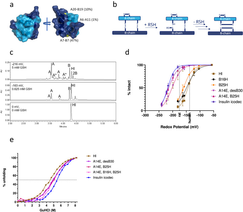
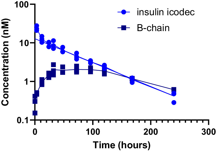
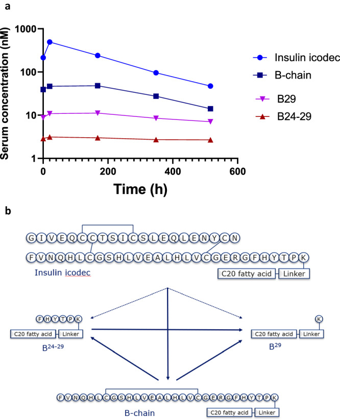
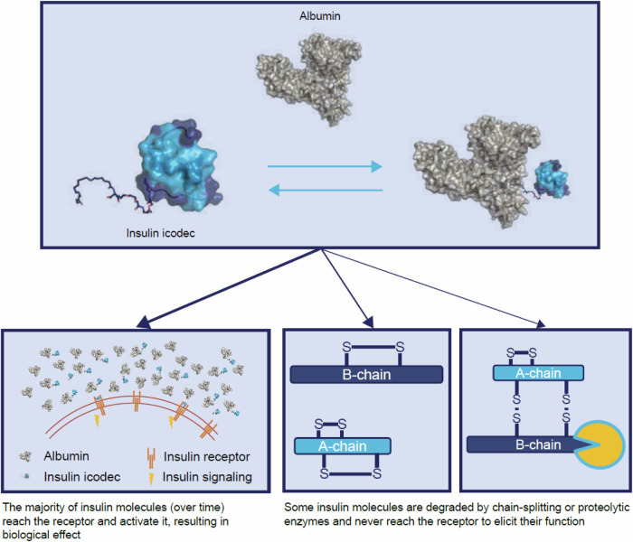
Insulin icodec is a once-weekly insulin analogue that has a long half-life of approximately 7 days, making it suitable for once weekly dosing. The Insulin icodec molecule was developed based on the hypothesis that lowering insulin receptor affinity and introducing a strong albumin-binding moiety would result in a long insulin half-life, provided that non-receptor-mediated clearance is diminished. Here, we report an insulin clearance mechanism, resulting in the splitting of insulin molecules into its A-chain and B-chain by a thiol-disulphide exchange reaction. Even though the substitutions in insulin icodec significantly stabilise insulin against such degradation, some free B-chain is observed in plasma samples from minipigs and people with type 2 diabetes. In summary, we identify thiol-disulphide exchange reactions to be an important insulin clearance mechanism and find that stabilising insulin icodec towards this reaction significantly contributes to its long pharmacokinetic/pharmacodynamic profile.


Conflict of interest statement

All authors are present employees of Novo Nordisk A/S and are shareholders of Novo Nordisk A/S.

Figures

Fig. 1
Fig. 1. Thiol–disulphide reaction and stability of insulin icodec.
a Solvent exposure of disulphide bonds in insulin icodec. Colour scheme: light blue, insulin A-chain; dark blue, insulin B-chain; yellow, sulphur atoms forming disulfide bonds. Relative solvent exposure for sulphur atoms in each disulphide bond is indicated. b Schematic representation of thiol–disulphide exchange reaction leading to insulin chain-splitting. RSH represents a thiol group reacting with a disulphide bond in insulin. c High-performance liquid chromatography chromatograms showing human insulin after 4-h incubations at 37 °C under different redox conditions as described in “Methods”. Top panel represents the incubation of human insulin without added glutathione, the middle panel shows human insulin incubated with 0.625 mM GSH and 1 mM GSSG representing a state where ~50% of HI is degraded and the bottom panel shows human insulin incubated with 6.3 mM GSH and 1 mM GSSG. A corresponds to A-chain isoforms containing two internal disulfide bonds; B corresponds to B-chain containing an internal disulphide bond; 2B corresponds to two insulin B-chains forming a dimer via two disulfide bonds; HI corresponds to human insulin. Additional peak assignment and MS spectra of the corresponding species are shown in Supplementary Fig. 2. d Stability of selected insulin analogues exposed to varying redox potential as described in the Methods section, showing mean ± SD, n = 3. See the text for a definition of insulin substitutions. The two dashed lines represent a glutathione redox potential in rat and human plasma, respectively. e Stability of selected insulin analogues exposed to varying concentrations of guanidinium hydrochloride as described in “Methods”, n = 1. Source data are provided as a Source Data file.
Fig. 2
Fig. 2. X-ray structure of insulin icodec.
a Cartoon representation of the trimeric arrangement of insulin icodec in the crystal. The dimer is coloured in orange and dark blue, for A-chain and B-chain, respectively. The additional icodec molecule is coloured in yellow (A-chain) and purple (B-chain). b Superposition of icodec molecule 1 with molecule 3 of the icodec trimer (left) and icodec molecule 1 with human insulin (right). Colours for icodec are as in (a); human insulin is in light yellow (A-chain) and cyan (B-chain). Residues A14, B16 and B25, which differ between human insulin and icodec are depicted in stick representation. c Interactions (dashed lines) around the A20-B19 disulphide bond for insulin icodec molecule 1 (1st from the left), insulin icodec molecule 3 (2nd from the left), human insulin (3rd from the left) and OI338 (4th from the left).
Fig. 3
Fig. 3. In vitro rat plasma stability of selected insulin analogues.
The disappearance of intact insulin analogues upon incubation in rat plasma (a), with the corresponding appearance of insulin B-chains (b), n = 2. All insulin B-chains contain an internal disulphide bond connecting Cys 7 with Cys 19, assessed by the exact monoisotopic masses. See text for definition of insulin substitutions. Source data are provided as a Source Data file.
Fig. 4
Fig. 4. In vitro plasma stability of selected insulin analogues.
Disappearance of intact insulin analogues upon incubation in human (a), dog (b) and minipig (c) plasma, n = 2. The corresponding appearance of B-chains are shown in Supplementary Fig. 5. See text for definition of insulin substitutions.
Fig. 5
Fig. 5. Pharmacokinetics of insulin icodec administered intravenously to minipigs.
Disappearance of insulin icodec (circles) and appearance of insulin icodec B-chain (containing a disulphide bond connecting Cys 7 and Cys 19; squares) in plasma are shown, n = 3. Source data are provided as a Source Data file.
Fig. 6
Fig. 6. Pharmacokinetics of insulin icodec and major metabolites in human serum samples.
a Pharmacokinetic profiles of insulin icodec (light blue circles), insulin icodec B-chain (containing a disulphide bond connecting Cys 7 and Cys 19; dark blue squares), B29 acylated metabolite (purple inverted triangles) and B24–B29 acylated metabolite (brown triangles) at steady state (after the fifth weekly subcutaneous dose) in people with diabetes. Each time point represents a single value obtained from a pooled plasma sample from 12 human subjects. b Possible metabolic pathways leading to formation of the observed metabolites (Supplementary Table 3). The solid lines represent the most likely paths while the dashed lines represent alternative paths to the observed degradation products of insulin icodec. Source data are provided as a Source Data file and in Supplementary Table 4.
Fig. 7
Fig. 7. Fate of insulin icodec in plasma.
Scheme showing the fate of insulin icodec in plasma.

References

    1. Muttenthaler, M., King, G. F., Adams, D. J. & Alewood, P. F. Trends in peptide drug discovery. Nat. Rev. Drug Discov.20, 309–325 (2021). - PubMed
    1. Peyrot, M., Barnett, A. H., Meneghini, L. F. & Schumm-Draeger, P. M. Insulin adherence behaviours and barriers in the multinational global attitudes of patients and physicians in insulin therapy study. Diabet. Med.29, 682–689 (2012). - PMC - PubMed
    1. Rosenstock, J. & Del Prato, S. Basal weekly insulins: the way of the future! Metabolism126, 154924 (2022). - PubMed
    1. Kjeldsen, T. B. et al. Molecular engineering of insulin icodec, the first acylated insulin analog for once-weekly administration in humans. J. Med Chem.64, 8942–8950 (2021). - PubMed
    1. Nishimura, E. et al. Molecular and pharmacological characterization of insulin icodec: a new basal insulin analog designed for once-weekly dosing. BMJ Open Diabet. Res. Care9, e002301 (2021). - PMC - PubMed

MeSH terms