. 2024 Sep;79(9):2502-2523.
doi: 10.1111/all.16231. Epub 2024 Jul 20.

Elucidating allergic reaction mechanisms in response to SARS-CoV-2 mRNA vaccination in adults

Affiliations

Elucidating allergic reaction mechanisms in response to SARS-CoV-2 mRNA vaccination in adults

Mihir M Shah et al. Allergy. 2024 Sep.

Erratum in

Abstract

Background: During the COVID-19 pandemic, novel nanoparticle-based mRNA vaccines were developed. A small number of individuals developed allergic reactions to these vaccines although the mechanisms remain undefined.

Methods: To understand COVID-19 vaccine-mediated allergic reactions, we enrolled 19 participants who developed allergic events within 2 h of vaccination and 13 controls, nonreactors. Using standard hemolysis assays, we demonstrated that sera from allergic participants induced stronger complement activation compared to nonallergic subjects following ex vivo vaccine exposure.

Results: Vaccine-mediated complement activation correlated with anti-polyethelyne glycol (PEG) IgG (but not IgM) levels while anti-PEG IgE was undetectable in all subjects. Depletion of total IgG suppressed complement activation in select individuals. To investigate the effects of vaccine excipients on basophil function, we employed a validated indirect basophil activation test that stratified the allergic populations into high and low responders. Complement C3a and C5a receptor blockade in this system suppressed basophil response, providing strong evidence for complement involvement in vaccine-mediated basophil activation. Single-cell multiome analysis revealed differential expression of genes encoding the cytokine response and Toll-like receptor (TLR) pathways within the monocyte compartment. Differential chromatin accessibility for IL-13 and IL-1B genes was found in allergic and nonallergic participants, suggesting that in vivo, epigenetic modulation of mononuclear phagocyte immunophenotypes determines their subsequent functional responsiveness, contributing to the overall physiologic manifestation of vaccine reactions.

Conclusion: These findings provide insights into the mechanisms underlying allergic reactions to COVID-19 mRNA vaccines, which may be used for future vaccine strategies in individuals with prior history of allergies or reactions and reduce vaccine hesitancy.

Keywords: COVID‐19; COVID‐19 vaccines; SARS‐CoV‐2; mRNA vaccines; polyethylene glycol; vaccine allergy.


Conflict of interest statement

Competing Interests:

All authors declare no competing financial interests.

Figures
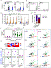
Figure 1.
Figure 1.. Allergic subjects can be further sub-divided into those with low or high serum-induced basophil responses (‘low basophil responders’ vs ‘high basophil responders’).
(A) Proportion of CD63+CRTh2+ basophils in response to anti-IgE (5 μg/mL) or grass pollen allergen (Phleum pratense; Phl p) in enriched basophils (left panel, n=3) or PBMCs (right panel; n=6). Basophil activation was evaluated in cells that were left untreated, IgE-stripped with 4% lactic acid or IgE-stripped and resensitized with indicator serum from a grass pollen highly allergic individual (GP IS, grass pollen-specific IgE >100kUA/L). (B) Concentration-ranging effect of COVID-19 vaccine (BNT162b2), PEG (1 μg/mL) and P80 (1 μg/mL), on CD63+CRTh2+ basophil activation in enriched basophil or PBMCs. In this validation experiment, we performed this side-by-side basophil vs. PBMC comparison in 5 allergic serum samples using 2 donor basophils. (C) Concentration-ranging effect of COVID-19 vaccine (BNT162b2 or mRNA-1273), PEG and P80 on non-allergics (n=13) and allergics (n=19). (D) Proportion of individuals in the allergic groups who had a reaction within ≤ 5 min, ≤ 20 min or ≤ 120 min. (E) Allergic individuals can be sub-divided into those with low (n=11) or high (n=8) basophil response upon resensitization with serum alone. (F) Unbiased clustering analysis FlowSOM showed differences in metaclusters (MC) 9, 10 and 11 in allergics with low or high basophil response following stimulation with serum alone. FlowSOM was performed on concatenated files. (G) Heatmap representing the expression of CD63, CD203c, C5aR and C3aR in all MC. Blue and red denotes low and high expression, respectively. (H) Population abundance for MC 9, 10 and 11 in allergics with low or high basophil response following stimulation with serum alone, vaccine (15 ug/mL), PEG2000 or P80. Data are presented as mean ± SEM. Violin plots are presented with its media represented in black line. Mann-Whitney U Test, * P<0.05, ** P<0.01.
Figure 2.
Figure 2.. Anti-PEG antibody levels and COVID-19 mRNA vaccine-mediated hemolytic activity.
(A) Concentration of anti-PEG-IgE in serum of non-allergic (n=13, blue) and allergic individuals (n=19, orange) from the earliest timepoint of blood collection shortly after receiving the first dose of vaccine (Median: 11.5 days, Range 0 – 86 days). (B) Dot plot depicting concentration of anti-PEG-IgG in serum of non-allergic (n=13, blue) and allergic individuals (n=19), split into high (n=8, green) and low (n=11, pink) basophil responses, as characterized in Fig. 1. (C) Dot plot depicting concentration of anti-PEG-IgM in serum of non-allergic (n=13, blue) and allergic individuals (n=19), split into high (n=8, green) and low (n=11, pink) basophil responses, as characterized in Fig. 1. Values were compared between the two groups at the same relative times using two-tailed Mann-Whitney test. (D) Serum was incubated with BNT162b2 vaccine for 30 min to trigger complement activation. Residual complement activity in the serum following incubation with BNT162b2 vaccine is expressed as average residual hemolytic activity (Avg RHA). Dot plots showing Avg RHA in non-allergic (n = 13, blue) and allergic individuals (n = 19), split into high (n=8, green) and low (n=11, pink) basophil responses, as characterized in Fig. 1. Values were compared between the two groups at the same relative times using two-tailed Mann-Whitney test. (E) A subgroup of sera from allergic individuals were depleted of IgG (Dpl) and subjected to the hemolysis assay. Depletion was confirmed by the absence of IgG heavy and light chains by Western blotting (data not shown). Antibody depletion diminished complement activation, resulting in higher average RHAs in the majority of sera tested. Wilcoxon signed rank test, * P<0.05.
Figure 3.
Figure 3.. C5a and C3a receptors play a role in serum+vaccine-induced basophil activation.
(A) Proportion of C5aR and C3aR on the surface of enriched basophil (black bar) and PBMC (white bar) presented as a percentage or geo mean fluorescence intensity (MFI) value. (B) Effect of C5aR (75 nM) and C3aR (1 μM) antagonist on vaccine induced CD63+CRTh2+ basophil activation in enriched basophil (black bar) and PBMC (white bar). (C) Effect of complement antagonist (C5aRA and C3aRA) on serum+vaccine-induced basophil activation in non-allergics (n=13; blue bar), allergics with low basophil response (n=11; magenta bar) or high basophil response (n=8; green bar). (D) Unbiased clustering analysis FlowSOM showed differences in metaclusters (MC) 9, 10, 11 and 12 in non-allergics and allergics (low or high basophil response) targeted by complement receptor antagonists C5aRA and C3aRA. FlowSOM was performed on concatenated files. (E) Population abundance for MC 9, 10, 11 and 12 in non-allergics or allergics with low or high basophil response following stimulation with serum+vaccine, or in the presence of C5aRA and C3aRA. Data are presented as mean ± SEM. Violin plots are presented with its media represented in black line. Kruskal-Wallis Test, * P<0.05, ** P<0.01, *** P<0.001.
Figure 4.
Figure 4.. Hemolysis assay and correlation with anti-PEG antibody.
(A) Higher anti-PEG IgG levels correlated with higher complement activation, and reflected in lower Avg RHA in allergic, high basophil responders (R2 = 0.5813, P = 0.028). (B) No correlation between anti-PEG IgM levels and Avg RHA (R2 = 0.056, P = 0.573) in allergic, high basophil responders. (C) Complement activation generated complement anaphylatoxin C3a following incubation of serum with COVID-19 mRNA vaccine leading to basophil activation, which was partially blocked by complement C3a receptor antagonist (C3aRA) (R2 = 0.5680, P = 0.031). (D) There is low correlation (R2= 0.2182, P = 0.243) between basophil response in the presence of C5aRA and Avg RHA in the allergic, high basophil responders.
Figure 5.
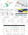
Figure 5.. Singe cell multiome analyses of individuals with differing basophil responses to COVID vaccines.
(A) A total of 1 non-reactor, 3 low-(basophil) BAT reactors and 3 high-(basophil) BAT reactors were chosen for single cell ATAC sequencing based on results from the basophil activation tests. The PBMCs were collected from blood and stimulated with autologous patient serum collected after first dose of the vaccine for 20 minutes at 37C before single cell multiome sequencine. (B) Weighted nearest neighbor (WNN) UMAPs of predicted cell types based on azimuth classifications (level 1). UMAP of cells classified from different patient responses. Proportions of predicted cell types for each patient group. (C) RNA transcriptome correlation analysis based on sctransformed normalized data recapitulated across different cell types of stimulation condition. (D) Inferred gene activity correlation analysis based on scATAC-seq data captures similar profiles across treatment conditions.
Figure 6.
Figure 6.. Single cell multiome analyses on PBMCs identified contribution of the myeloid compartment on high basophil responders and low basophil responders.
(A) Weighted nearest neighbor (WNN) UMAPs of predicted cell types based on azimuth classifications. Four subsets of cells within the myeloid compartment were identified (classical and non-classical monocytes, cDC2s and pDCs). (B) Top genes highly expressed within each clusters of cells identified. (C) Volcano plot to illustrate differentially expressed genes between high basophil responders versus low basophil responders versus non-allergics. (D) Heatmap and violin plots denoting a selection of genes differentially expressed within the myeloid compartment of differen study subjects. (E) Pathway enrichment analysis in high basophil responders vs. low basophil responders, high basophil responders vs non-allergics and low basophil responders vs non-allergics. (F) Peak callings to illustrate accessible regions within the IL-13, IL-4, and IL-1B loci of high basophil responders, low basophil responders and non-allergics.
Figure 7.
Figure 7.
Schematic representing vaccine-induced molecular pathway in high basophil responders and low basophil responders.

References

    1. Polack FP et al. Safety and Efficacy of the BNT162b2 mRNA Covid-19 Vaccine. New England Journal of Medicine 383, 2603–2615 (2020). doi: 10.1056/NEJMoa2034577 - DOI - PMC - PubMed
    1. Baden LR et al. Efficacy and Safety of the mRNA-1273 SARS-CoV-2 Vaccine. New England Journal of Medicine 384, 403–416 (2021). doi: 10.1056/NEJMoa2113017 - DOI - PMC - PubMed
    1. Allergic Reactions Including Anaphylaxis After Receipt of the First Dose of Pfizer-BioNTech COVID-19 Vaccine — United States, December 14–23, 2020. Centers for Disease Control and Prevention (2021). Available at: https://www.cdc.gov/mmwr/volumes/70/wr/mm7002e1.htm. (Accessed: 1st September 2021)
    1. Allergic Reactions Including Anaphylaxis After Receipt of the First Dose of Moderna COVID-19 Vaccine — United States, December 21, 2020–January 10, 2021. Centers for Disease Control and Prevention (2021). Available at: https://www.cdc.gov/mmwr/volumes/70/wr/mm7004e1.htm. (Accessed: 1st September 2021).
    1. Banerji A et al. mRNA Vaccines to Prevent COVID-19 Disease and Reported Allergic Reactions: Current Evidence and Suggested Approach. The Journal of Allergy and Clinical Immunology: In Practice 9, 1423–1437 (2021). doi: 10.1016/j.jaip.2020.12.047 - DOI - PMC - PubMed

MeSH terms