. 2024 Oct 2;32(10):3331-3345.
doi: 10.1016/j.ymthe.2024.07.012. Epub 2024 Jul 20.

Preclinical studies of gene replacement therapy for CDKL5 deficiency disorder

Affiliations

Preclinical studies of gene replacement therapy for CDKL5 deficiency disorder

Gregory Voronin et al. Mol Ther. .

Abstract

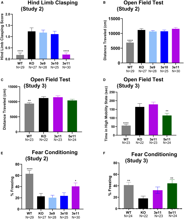
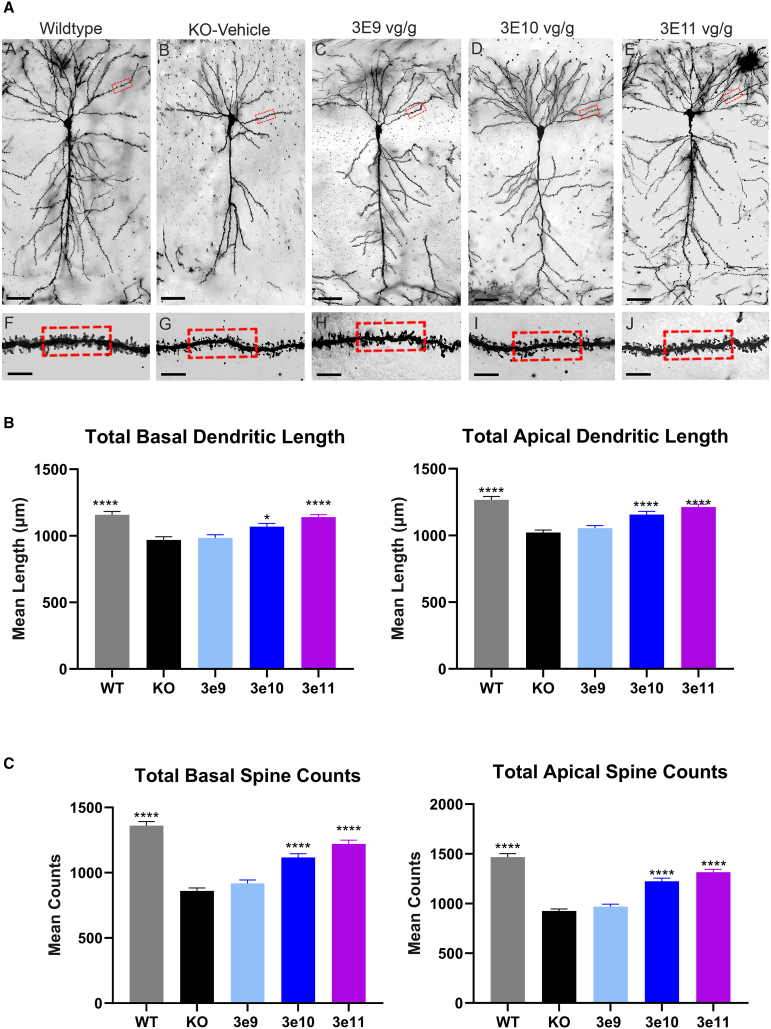
Cyclin-dependent kinase-like 5 (CDKL5) deficiency disorder (CDD) is a rare neurodevelopmental disorder caused by a mutation in the X-linked CDKL5 gene. CDKL5 is a serine/threonine kinase that is critical for axon outgrowth and dendritic morphogenesis as well as synapse formation, maturation, and maintenance. This disorder is characterized by early-onset epilepsy, hypotonia, and failure to reach cognitive and motor developmental milestones. Because the disease is monogenic, delivery of the CDKL5 gene to the brain of patients should provide clinical benefit. To this end, we designed a gene therapy vector, adeno-associated virus (AAV)9.Syn.hCDKL5, in which human CDKL5 gene expression is driven by the synapsin promoter. In biodistribution studies conducted in mice, intracerebroventricular (i.c.v.) injection resulted in broader, more optimal biodistribution than did intra-cisterna magna (i.c.m.) delivery. AAV9.Syn.hCDKL5 treatment increased phosphorylation of EB2, a bona fide CDKL5 substrate, demonstrating biological activity in vivo. Our data provide proof of concept that i.c.v. delivery of AAV9.Syn.hCDKL5 to neonatal male Cdkl5 knockout mice reduces pathology and reduces aberrant behavior. Functional improvements were seen at doses of 3e11 to 5e11 vector genomes/g brain, which resulted in transfection of ≥50% of the neurons. Functional improvements were not seen at lower doses, suggesting a requirement for broad distribution for efficacy.

Keywords: AAV9; CDD; CDKL5; biodistribution; drug discovery and development; gene therapy; molecular therapeutics; mouse models of disease; pharmacodynamics; route of administration.


Conflict of interest statement

Declaration of interests G.V., J.N., J.G., J.S., P.L., M.P., S.D., L.C., Y.V., M.J.K., L.P., E.F., S.S., B.R., P.Y., C.S., J.P., K.S., S.J., J.L., A.M., E.M.W., Z.W., and M.W. are or were at the time the work employees of PTC Therapeutics. M.C.W. is the President and CEO of NeuroDigiTech and was paid by PTC Therapeutics for this work.

Figures

None
Graphical abstract
Figure 1
Figure 1
Increased biodistribution in Cdkl5 knockout mice after i.c.v. injection compared to i.c.m. injection at postnatal day 0 Symbols represent the mean ± SEM of 4 mice per group. (A–G) Comparison of biodistribution of CDKL5 protein levels in brain tissues 35–37 days after i.c.v. or i.c.m. administration of AAV9.Syn.hCDKL5 vector. (H) Correlation between vector DNA and gene expression, hCDKL5 mRNA. Symbols represent DNA (qPCR) and RNA (RT-qPCR) values for individual animals. (I) Increases in phospho-EB2 with increasing CDKL5 protein levels in the hippocampus. Symbols in (H) and (I) represent individual animals. Dose is reported as vg/g brain weight. ∗∗∗p < 0.001, ∗∗p < 0.01, ∗p < 0.05 (Student’s t test comparing i.c.v. to comparable i.c.m. dose).
Figure 2
Figure 2
Expression of hCDKL5 mRNA in neurons but not glial cells after administration of AAV9.Syn.hCDKL5 at postnatal day 0 Three mice per group were evaluated. Images of the cortex are shown at 40× magnification. (A) Expression of hCDKL5 (pink) mRNA in NeuN protein positive neurons (green) after i.c.v. administration at 3.6e11 vg/g. (B) Lack of expression of hCDKL5 mRNA (pink) in glial fibrillary acidic protein (GFAP)-positive glial cells (green). (C) Comparison of distribution after i.c.v. or i.c.m. administration. Shown are the percentage of cells in the hemisphere that are double-positive for hCDKL5 mRNA and for NeuN. The probes are selective for human CDKL5 mRNA and do not cross-react with murine Cdkl5 mRNA. Regions across the entire brain were evaluated.
Figure 3
Figure 3
AAV9.Syn.hCDKL5 improved behavior of Cdkl5 KO mice (A) Hindlimb clasp measured at 11 weeks of age. Bars represent the mean score ± SEM (study 2). (B) Open-field distance traveled (cm) (study 2) measured at 15 weeks. (C) Open-field distance traveled (cm) (study 3) measured at 14 weeks. (D) Open-field highly mobile state (study 3) measured at 14 weeks. (E) Fear conditioning post training percent freezing (study 2) measured at 17 weeks. (F) Fear conditioning (higher doses) post training percent freezing (study 3) measured at 16 weeks. Dose values on x axis are in vg/g brain delivered i.c.v. Number per group is shown below the x axis. Statistics show comparison to Cdkl5 KO vehicle-dosed mice evaluated by a one-way ANOVA followed by Dunnett’s multiple comparison test (p ≤ 0.05) where ∗∗∗∗p < 0.0001, ∗∗∗p ≤ 0.001, ∗∗p ≤ 0.01, and ∗p ≤ 0.05.
Figure 4
Figure 4
Dendritic morphology is improved after treatment with AAV9.Syn.hCDKL5 (A) Normalization of dendritic morphology. Representative images of CA1 hippocampal pyramidal cells across groups. Compared to WT mice, Cdkl5 KO mice exhibited significant reductions in dendritic morphology (upper panel; scale bar, 50 μm) as well as the spines of CA1 pyramidal basal dendrites (box in the lower panel; scale bar, 5 μm). (B and C) Bars show the mean ± SEM (8 mice/group) for basal and apical pyramidal cell dendritic length (B) and spine count (C). Dose values on the x axis are in vg/g brain. Statistics show comparison to Cdkl5 KO vehicle-dosed mice evaluated by a one-way ANOVA followed by Dunnett’s multiple comparison test (p ≤ 0.05) where ∗∗∗∗p < 0.0001, ∗∗∗p ≤ 0.001, ∗∗p ≤ 0.01, and ∗p ≤ 0.05.
Figure 5
Figure 5
Biodistribution of AAV9.Syn.hCDKL5 in nonhuman primates AAV9.hSyn.CDKL5 vector after i.c.v. administration is detectable throughout the brain (A) and periphery (B) in nonhuman primates. Values represent the mean ± SEM. The number above the bar represents the number of animals for which data were available for that tissue. Biodistribution of vector measured in the CNS using qPCR.

References

    1. Fehr S., Wilson M., Downs J., Williams S., Murgia A., Sartori S., Vecchi M., Ho G., Polli R., Psoni S., et al. The CDKL5 disorder is an independent clinical entity associated with early-onset encephalopathy. Eur. J. Hum. Genet. 2013;21:266–273. - PMC - PubMed
    1. Fehr S., Wong K., Chin R., Williams S., de Klerk N., Forbes D., Krishnaraj R., Christodoulou J., Downs J., Leonard H. Seizure variables and their relationship to genotype and functional abilities in the CDKL5 disorder. Neurology. 2016;87:2206–2213. - PubMed
    1. Olson H.E., Demarest S.T., Pestana-Knight E.M., Swanson L.C., Iqbal S., Lal D., Leonard H., Cross J.H., Devinsky O., Benke T.A. Cyclin-Dependent Kinase-Like 5 Deficiency Disorder: Clinical Review. Pediatr. Neurol. 2019;97:18–25. - PMC - PubMed
    1. Rodak M., Jonderko M., Rozwadowska P., Machnikowska-Sokołowska M., Paprocka J. CDKL5 Deficiency Disorder (CDD)-Rare Presentation in Male. Children (Basel, Switzerland) 2022;9:1806. - PMC - PubMed
    1. Rusconi L., Salvatoni L., Giudici L., Bertani I., Kilstrup-Nielsen C., Broccoli V., Landsberger N. CDKL5 expression is modulated during neuronal development and its subcellular distribution is tightly regulated by the C-terminal tail. J. Biol. Chem. 2008;283:30101–30111. - PMC - PubMed

MeSH terms

Supplementary concepts

LinkOut - more resources