. 2024 Jul 21;15(1):6152.
doi: 10.1038/s41467-024-50454-2.

Glutathione synthesis in the mouse liver supports lipid abundance through NRF2 repression

Affiliations

Glutathione synthesis in the mouse liver supports lipid abundance through NRF2 repression

Gloria Asantewaa et al. Nat Commun. .

Abstract

Cells rely on antioxidants to survive. The most abundant antioxidant is glutathione (GSH). The synthesis of GSH is non-redundantly controlled by the glutamate-cysteine ligase catalytic subunit (GCLC). GSH imbalance is implicated in many diseases, but the requirement for GSH in adult tissues is unclear. To interrogate this, we have developed a series of in vivo models to induce Gclc deletion in adult animals. We find that GSH is essential to lipid abundance in vivo. GSH levels are highest in liver tissue, which is also a hub for lipid production. While the loss of GSH does not cause liver failure, it decreases lipogenic enzyme expression, circulating triglyceride levels, and fat stores. Mechanistically, we find that GSH promotes lipid abundance by repressing NRF2, a transcription factor induced by oxidative stress. These studies identify GSH as a fulcrum in the liver's balance of redox buffering and triglyceride production.


Conflict of interest statement

I.S.H. reports financial support from Kojin Therapeutics. All other authors declare no competing interests.

Figures

Fig. 1
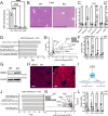
Fig. 1. GSH synthesis is required for the survival of adult animals.
A Schematic of the whole-body inducible Gclc knockout mouse model. Knockout of the whole-body Gclc was induced by a 5-day daily 160 mg/kg tamoxifen administration. Mice were sacrificed 12–15 days following tamoxifen administration. B Relative expression of Gclc mRNA in tissues from the KO (n = 4 (spleen); n = 5 (all other annotated tissues)) compared to WT (n = 5) mice 12–15 days following tamoxifen administration. Expression levels were normalized to the expression of the reference gene Rps9. C Representative immunoblot analysis of GCLC protein in the liver, kidney, and lung of WT and KO mice 12–15 days following tamoxifen administration. Data shown are representative of at least 3 replicates. D Relative GSH abundance in the liver, kidney, and lungs of WT (n = 4) and KO (n = 4) mice 12–15 days following tamoxifen administration. A two-way ANOVA with subsequent Šidák’s multiple comparisons test was used to determine statistical significance (WT vs. KO: Liver P value = 0.0031, Kidney P value = 0.0099, Lung P value = 0.2054). E Percent survival of WT (n = 9) and KO (n = 11) mice following tamoxifen treatment. Loss of greater than 20% body weight resulted in a humane endpoint for mice. F Percent change in body weight in WT (n = 18) and KO (n = 17) mice at death. Initial weight measurements were collected on Day 0 of tamoxifen administration, and final weight measurements were collected at endpoints. An unpaired two-tailed t test was used to determine statistical significance (WT vs. KO P value < 0.0001). Indicated n values represent biologically independent samples from mice. ns = not significant, *P value < 0.05, **P value < 0.01, ****P value < 0.0001. (A) created with BioRender.com, released under a Creative Commons Attribution-NonCommercial-NoDerivs 4.0 International license.
Fig. 2
Fig. 2. Liver tissue from Gclc KO mice has induced NRF2 target genes and repressed lipogenic gene expression.
A GSH quantity (normalized to mg tissue weight) in the liver, kidney, lung, and brain from Gclc WT mice (n = 4). A one-way ANOVA with subsequent Dunnett’s multiple comparisons test was used to determine statistical significance (Liver vs. Kidney P value < 0.0001, Liver vs. Lung P value < 0.0001, Liver vs. Brain P value < 0.0001). B Representative H&E-stained liver slides in WT and KO mice. Scale bars = 500 µm. Data shown is representative of at least three replicates. C Relative abundance of GSH precursors and their related metabolites in the serum of WT (n = 4) and KO (n = 4) mice. A two-way ANOVA with subsequent Šidák’s multiple comparisons test was used to determine statistical significance (WT vs. KO: Cysteine P value = 0.9987, Glutamate P value = 0.0278, Glycine P value = 0.9993, Hypotaurine P value = 0.0004, Glutamine P value > 0.9999, Serine P value = 0.0105). D Gene Set Enrichment Analysis (GSEA) of oxidative stress-related pathways in the liver of KO (n = 4) compared to WT (n = 4) mice. Enrichment p-values were calculated using an adaptive multi-level split Monte Carlo scheme and were corrected for multiple testing using Benjamini and Hochberg false discovery rate. E Proteomic analysis of upregulated liver proteins in KO (n = 6) compared to WT (n = 6) mice. Black data points = proteins with P value < 0.05 and log2 fold change >1. Red data points = annotated NRF2 target proteins. F Relative mRNA expression of annotated NRF2 target genes in the liver of WT (n = 4) and KO (n = 4) mice. Expression levels were normalized to the expression of the reference gene Rps9. A two-way ANOVA with subsequent Šidák’s multiple comparisons test was used to determine statistical significance (WT vs. KO: Nqo1 P value < 0.0001, Hmox1 P value < 0.0001, Txnrd1 P value = 0.2255, Gclm P value = 0.7934). G Representative immunoblot analysis of NQO1 in the liver of WT and KO mice. Data shown is representative of at least three replicates. H Representative immunofluorescence images of the liver from WT and KO mice stained with an antibody against NQO1. Scale bars = 200 µm. Data shown is representative of at least three replicates. I Schematic of the proposed mechanism of NRF2-dependent repression of lipogenic gene expression. J GSEA of lipogenic-related pathways in the liver of KO (n = 4) compared to WT (n = 4) mice. Enrichment p values were calculated using an adaptive multi-level split Monte Carlo scheme and were corrected for multiple testing using Benjamini and Hochberg false discovery rate. K Proteomic analysis of downregulated liver proteins in KO (n = 6) compared to WT (n = 6) mice. Black data points = proteins with P value < 0.05 and log2 fold change > 1. Red data points = annotated lipogenic proteins. L Relative mRNA expression of annotated lipogenic genes in the liver of WT (n = 4) and KO (n = 4) mice. Expression levels were normalized to the expression of the reference gene Rps9. A two-way ANOVA with subsequent Šidák’s multiple comparisons test was used to determine statistical significance (WT vs. KO: Scd1 P value = 0.0160, Fasn P value = 0.0336, Acaa1b P value = 0.0162, Fabp1 P value = 0.0051). Indicated n values represent biologically independent samples from mice. Data are shown as mean ± SEM. ns = not significant, *P value < 0.05, **P value < 0.01, ****P value < 0.0001. (I) created with BioRender.com, released under a Creative Commons Attribution-NonCommercial-NoDerivs 4.0 International license.
Fig. 3
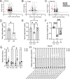
Fig. 3. The abundance of triglycerides is lower following GSH depletion in mice.
Lipidomic analysis of (A) adipose tissue, (B) liver tissue, and (C) serum in KO (n = 4) compared to WT (n = 4) mice. Black data points = lipid species with a p < 0.05 and log2 fold change >1. Red data points = triglycerides. D Triglyceride levels in the serum of WT (n = 4) and KO (n = 4) mice. An unpaired two-tailed t-test was used to determine statistical significance (WT vs. KO P value = 0.0119). E Triglyceride levels in the serum for WT (n = 4) and KO (n = 4) mice fed with either normal chow or a high-fat diet (HFD) for 14 days post-treatment with tamoxifen. A one-way ANOVA with subsequent Tukey’s multiple comparisons test was used to determine statistical significance (WT Normal Chow vs. KO Normal Chow P value = 0.0307, WT Normal Chow vs. KO HFD P value = 0.9683, KO Normal Chow vs. KO HFD P value = 0.0447). F Percent change in body weight of WT (n = 7) and KO mice fed with either normal chow (n = 7) or a high-fat diet (HFD; n = 9) 14 days post-treatment with tamoxifen. A one-way ANOVA with subsequent Tukey’s multiple comparisons test was used to determine statistical significance (WT Normal Chow vs. KO Normal Chow P value < 0.0001, WT Normal Chow vs. KO HFD P value =<0.0001, KO Normal Chow vs. KO HFD P value = 0.0006). G Relative abundance of select SCD1 fatty acids products in the liver from WT (n = 4) and KO (n = 4) mice. 16:0 = palmitic acid; 16:1;2 O= oxidized palmitoleic acid; 18:0 = stearic acid; 18:1 = oleic acid. A two-way ANOVA with subsequent Šidák’s multiple comparisons test was used to determine statistical significance (WT vs. KO: 16:0 P value = 0.0603, 16:1;2 O P value = 0.9991, 18:0 P value = 0.0377, 18:1 P value = 0.9268). H Relative abundance of select triglyceride species in the serum from WT (n = 4) and KO (n = 4) mice. A two-way ANOVA with subsequent Šidák’s multiple comparisons test was used to determine statistical significance (WT vs. KO for all comparisons indicated: **** P value < 0.0001). Indicated n values represent biologically independent samples from mice. Data are shown as mean ± SEM. ns not significant, * P value < 0.05, ** P value < 0.01, **** P value < 0.0001.
Fig. 4
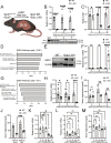
Fig. 4. Liver-specific GCLC expression sustains lipid synthesis and represses NRF2 activation.
A Schematic of inducible liver-specific Gclc deletion (L-KO). Liver-specific Gclc knockout was induced using the AAV-TBG-Cre. All mice were sacrificed 21 days post AAV-TBG-Cre injection unless otherwise indicated. B (Top) Relative abundance of Gclc mRNA in the liver of WT (n = 4) and L-KO (n = 4) mice, 1–3 weeks post-treatment with AAV-TBG-Cre. Expression levels of mRNA were normalized to the expression of the reference gene Rps9. A two-way ANOVA with subsequent Šidák’s multiple comparisons test was used to determine statistical significance (WT vs. L-KO: 1 week P value = 0.0002, 2 weeks P value < 0.0001, 3 weeks P value < 0.0001). (Bottom) Immunoblot analysis of GCLC protein in the liver of WT and L-KO mice 1–3 weeks post-treatment with AAV-TBG-Cre. C Relative abundance of GSH in liver tissue of WT (n = 4) and L-KO (n = 4) mice in 1–3 weeks following AAV-TBG-Cre treatment. A two-way ANOVA with subsequent Šidák’s multiple comparisons test was used to determine statistical significance (WT vs. L-KO: 1 week P value = 0.0424, 2 weeks P value < 0.0001, 3 weeks P value < 0.0001). D GSEA of oxidative stress-related pathways in the liver of L-KO (n = 4) compared to WT (n = 4) mice following treatment with AAV-TBG-Cre. Enrichment p values were calculated using an adaptive multi-level split Monte Carlo scheme and were corrected for multiple testing using Benjamini and Hochberg false discovery rate. E Representative immunoblot analysis of NRF2 in the liver of WT and L-KO mice following treatment with AAV-TBG-Cre. Data shown are representative of at least three replicates. F Relative mRNA levels of Nqo1 in WT (n = 8 for 1 and 2-week timepoints and n = 4 for 3-week timepoint) and L-KO (n = 6 for 1-week timepoint, n = 7 for 2-week timepoint and n = 4 for 3-week timepoint) mice,1–3 weeks following treatment with AAV-TBG-Cre. Expression levels were normalized to the expression of the reference gene Rps9. A two-way ANOVA with subsequent Šidák’s multiple comparisons test was used to determine statistical significance (WT vs. L-KO: 1 week P value = 0.9998, 2 weeks P value = 0.6378, 3 weeks P value < 0.0001). G GSEA of lipogenic-related pathways in the liver of KO (n = 4) compared to WT (n = 4) mice following treatment with AAV-TBG-Cre. Enrichment p values were calculated using an adaptive multi-level split Monte Carlo scheme and were corrected for multiple testing using Benjamini and Hochberg false discovery rate. H Relative mRNA levels of Scd1 in WT (n = 8 for 1 and 2-week timepoints and n = 4 for 3-week timepoint) and L-KO (n = 6 for 1-week timepoint, n = 7 for 2-week timepoint and n = 4 for 3-week timepoint) mice, 1–3 weeks following treatment with AAV-TBG-Cre. Expression levels were normalized to the expression of the reference gene Rps9. A two-way ANOVA with subsequent Šidák’s multiple comparisons test was used to determine statistical significance (WT vs. L-KO: 1 week P value = 0.0192, 2 weeks P value = 0.0222, 3 weeks P value = 0.0005). I Triglyceride levels in the serum of WT (n = 4) and L-KO (n = 4) mice in 1–3 weeks following treatment with AAV-TBG-Cre. A two-way ANOVA with subsequent Šidák’s multiple comparisons test was used to determine statistical significance (WT vs. L-KO: 1 week P value = 0.9995, 2 weeks P value = 0.0038, 3 weeks P value = 0.0069). J Triglyceride levels in the serum of L-KO mice fed normal chow (n = 4) and HFD (n = 7) 3 weeks following treatment with AAV-TBG-Cre. An unpaired two-tailed t-test was used to determine statistical significance (L-KO Normal Chow vs. L-KO High-fat Diet P value = 0.0004). Relative mRNA levels of (K) Nqo1 and (L) Scd1 in Gclc L-KO mice (n = 4) following treatment with GSH-ee (0, 50, 100, 500, and 1000 mg/kg) from days 17–20 post-AAV-TBG-Cre injection. mRNA levels were normalized to the expression of the reference gene Rps9. A one-way ANOVA with subsequent Dunnett’s multiple comparisons test was used to determine statistical significance (K): 0 mg/kg GSH-ee vs. 50 mg/kg GSH-ee P value = 0.9933, 0 mg/kg GSH-ee vs. 100 mg/kg GSH-ee P value = 0.9996, 0 mg/kg GSH-ee vs. 500 mg/kg GSH-ee P value = 0.2648, 0 mg/kg GSH-ee vs. 1000 mg/kg GSH-ee P value = 0.0454. L: 0 mg/kg GSH-ee vs. 50 mg/kg GSH-ee P value = 0.9816, 0 mg/kg GSH-ee vs. 100 mg/kg GSH-ee P value = 0.9664, 0 mg/kg GSH-ee vs. 500 mg/kg GSH-ee P value > 0.999, 0 mg/kg GSH-ee vs. 1000 mg/kg GSH-ee P value = 0.4731. M Triglyceride levels in the serum of Gclc L-KO mice (n = 4) following treatment with GSH-ee (0, 50, 100, 500, and 1000 mg/kg) from days 17–20 post-AAV-TBG-Cre injection. A one-way ANOVA with subsequent Dunnett’s multiple comparisons test was used to determine statistical significance (0 mg/kg GSH-ee vs. 50 mg/kg GSH-ee P value = 0.9663, 0 mg/kg GSH-ee vs. 100 mg/kg GSH-ee P value = 0.9879, 0 mg/kg GSH-ee vs. 500 mg/kg GSH-ee P value = 0.7552, 0 mg/kg GSH-ee vs. 1000 mg/kg GSH-ee P value = 0.9991). Indicated n values represent biologically independent samples from mice. Data are shown as mean ± SEM. ns = not significant, *P value < 0.05, **P value < 0.01, ***P value < 0.001, ****P value < 0.0001. (A) created with BioRender.com, released under a Creative Commons Attribution-NonCommercial-NoDerivs 4.0 International license.
Fig. 5
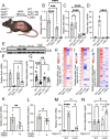
Fig. 5. GSH in the liver supports triglyceride levels in an NRF2-dependent manner.
A Schematic of inducible liver-specific Gclc deletion (Gclc L-KO), Nrf2 deletion (Nrf2 L-KO), and Gclc-Nrf2 deletion (L-DKO). Liver-specific knockout of genes was achieved using AAV-TBG-Cre. All mice were sacrificed 21 days post AAV-TBG-Cre treatment unless otherwise indicated. (B-D) Relative expression of (B) Gclc, (C) Nrf2 and (D) Nqo1 mRNA in the liver of WT (n = 4), Gclc L-KO (n = 4), Nrf2 L-KO (n = 4), and L-DKO (n = 6) mice following treatment with AAV-TBG-Cre. Expression levels were normalized to the expression of the reference gene Rps9. A one-way ANOVA with subsequent Dunnett’s multiple comparisons test (used in (B) and (C)) or Šidák’s multiple comparisons test (used in (D)) was used to determine statistical significance ((B): WT vs. Gclc L-KO P value < 0.0001, WT vs. Nrf2 L-KO P value = 0.0004, WT vs. L-DKO P value < 0.0001. C WT vs. Gclc L-KO P value = 0.7972, WT vs. Nrf2 L-KO P value < 0.0001, WT vs. L-DKO P value < 0.0001. D Gclc L-KO vs. L-DKO P value < 0.0001). E Representative immunoblot analysis of NRF2 in the liver of WT, Gclc L-KO, Nrf2 L-KO, and L-DKO mice following treatment with AAV-TBG-Cre. Data shown are representative of at least three replicates. F Relative expression of Scd1 mRNA in the liver of WT (n = 4), Gclc L-KO (n = 4), Nrf2 L-KO (n = 4), and L-DKO (n = 6) mice following treatment with AAV-TBG-Cre. Expression levels were normalized to the expression of the reference gene Rps9. A one-way ANOVA with subsequent Šidák’s multiple comparisons test was used to determine statistical significance (Gclc L-KO vs. L-DKO P value = 0.0866). G Serum triglyceride levels for WT (n = 12), Gclc L-KO (n = 14), Nrf2 L-KO (n = 6), and L-DKO (n = 8) mice 3 weeks post-treatment with AAV-TBG-Cre. A one-way ANOVA with subsequent Dunnett’s multiple comparisons test was used to determine statistical significance (Gclc L-KO vs. WT P value < 0.0001, Gclc L KO vs. Nrf2 L-KO P value = 0.0040, Gclc L-KO vs. L-DKO P value = 0.01421). H Relative expression of lipogenic genes mRNA in the liver of WT (n = 4), Gclc L-KO (n = 4), Nrf2 L-KO (n = 4), and L-DKO (n = 4) mice expressed as Log2 fold change. I Relative expression of proteins related to the lipogenic pathway in the liver of WT (n = 4), Gclc L-KO (n = 4), Nrf2 L-KO (n = 4) and L-DKO (n = 4) mice expressed as Log2 fold change. J Relative abundance of serum triglycerides in the liver of WT (n = 4), Gclc L-KO (n = 4), Nrf2 L-KO (n = 4) and L-DKO (n = 4) mice expressed as Log2 fold change. K Relative mRNA expression of Scd1 in the liver of WT mice following a 4-day treatment with either vehicle (n = 4) or CDDO-methyl (n = 8). Expression levels were normalized to the expression of the reference gene Rps9. An unpaired two-tailed t-test was used to determine statistical significance (Vehicle vs. CDDO-methyl P value = 0.0065). L Triglyceride levels in the serum of WT mice without AAV-TBG-Cre injection following a 4-day treatment with either vehicle (n = 4) or CDDO-methyl (n = 8). An unpaired two-tailed t test was used to determine statistical significance (Vehicle vs. CDDO-methyl P value = 0.0171). M Relative expression of Scd1 mRNA in the liver of WT (n = 4) and L-KO (n = 4) mice treated with either vehicle or T0901317 on days 17–20 post AAV-TBG-Cre injection. Expression levels were normalized to the expression of the reference gene Rps9. A one-way ANOVA with subsequent Šidák’s multiple comparisons test was used to determine statistical significance (vehicle vs. T0901317: WT P value < 0.0001, L-KO P value = 0.0462; WT/Vehicle vs. L-KO/T0901317 P value = 0.1254). N Triglyceride levels in the serum of WT (n = 4) and L-KO (n = 4) mice treated with either vehicle or T0901317 on days 17–20 post AAV-TBG-Cre injection. A one-way ANOVA with subsequent Šidák’s multiple comparisons test was used to determine statistical significance (vehicle vs. T0901317: WT P value = 0.9994, L-KO P value = 0.0560; WT/Vehicle vs. L-KO/T0901317 P value = 0.0155). Indicated n values represent biologically independent samples from mice. Data are shown as mean ± SEM. ns = not significant, *P value < 0.05, **P value < 0.01, ***P value < 0.001, ****P value < 0.0001. (A) created with BioRender.com, released under a Creative Commons Attribution-NonCommercial-NoDerivs 4.0 International license.
Fig. 6
Fig. 6. Prolonged Gclc deletion in the liver induces a sex-dependent requirement of NRF2.
A Schematic for extended monitoring of Gclc deletion (Gclc L-KO), Nrf2 deletion (Nrf2 L-KO), and Gclc-Nrf2 deletion (L-DKO) in mice. Liver-specific gene knockout was induced with AAV-TBG-Cre. Mice were sacrificed 10 weeks post AAV-TBG-Cre injection unless humane endpoints were reached earlier. B Percent survival for WT (n = 8), Gclc L-KO (n = 6), Nrf2 L-KO (n = 6) and L-DKO (n = 12) mice following treatment with AAV-TBG-Cre. C Percent survival for female (n = 6) and male (n = 7) L-DKO mice following treatment with AAV-TBG-Cre. D Hepatocellular Injury Score from H&E-stained slides of the liver from female WT (n = 5), Gclc L-KO (n = 8), Nrf2 L-KO (n = 3), L-DKO (n = 4) and male WT (n = 5), Gclc L-KO (n = 8), Nrf2 L-KO (n = 3), L-DKO (n = 7) mice. A two-way ANOVA with subsequent Tukey’s multiple comparisons test was used to determine statistical significance (Female: WT vs. Gclc L-KO P value = 0.2504, WT vs. Nrf2 L-KO P value = 0.9999, WT vs. L-DKO P value = 0.5546, Gclc L-KO vs. L-DKO P value > 0.9999; Male: WT vs. Gclc L-KO P value = 0.1238, WT vs. Nrf2 L-KO P value > 0.9999, WT vs. L-DKO P value < 0.0001, Gclc L-KO vs. L-DKO P value = 0.0006; Female L-DKO vs. Male L-DKO P value = 0.0020). E Representative H&E-stained slides of the liver from female and male WT, Gclc L-KO, Nrf2 L-KO, and L-DKO mice 10 weeks following treatment with AAV-TBG-Cre. Scale bars = 200 µm. Data shown are representative of at least 3 replicates. F Serum triglyceride concentration of WT (n = 14), Gclc L-KO (n = 19), Nrf2 L-KO (n = 5) and L-DKO (n = 17) mice 10 weeks following treatment with AAV-TBG-Cre. A one-way ANOVA with subsequent Tukey’s multiple comparisons test was used to determine statistical significance (WT vs. Gclc L-KO P value < 0.0001, WT vs. Nrf2 L-KO P value = 0.7838, WT vs. L-DKO P value < 0.0001, Gclc L-KO vs. L-DKO P value = 0.1525). G Epididymal fat adipose tissue (eWAT) mass normalized to body mass from WT (n = 10), Gclc L-KO (n = 16), Nrf2 L-KO (n = 6) and L-DKO (n = 17) mice 10 weeks post-treatment with AAV-TBG-Cre. A one-way ANOVA with subsequent Tukey’s multiple comparisons test was used to determine statistical significance (WT vs. Gclc L-KO P value = 0.0112, WT vs. Nrf2 L-KO P value = 0.9675, WT vs. L-DKO P value = 0.0515, Gclc L-KO vs. L-DKO P value = 0.8936). Indicated n values represent biologically independent samples from mice. Data are shown as mean ± SEM. ns = not significant, * P value < 0.05, ** P value < 0.01, *** P value < 0.001, **** P value < 0.0001. (A) created with BioRender.com, released under a Creative Commons Attribution-NonCommercial-NoDerivs 4.0 International license.

Update of

  • Glutathione supports lipid abundance in vivo.
    Asantewaa G, Tuttle ET, Ward NP, Kang YP, Kim Y, Kavanagh ME, Girnius N, Chen Y, Duncan R, Rodriguez K, Hecht F, Zocchi M, Smorodintsev-Schiller L, Scales TQ, Taylor K, Alimohammadi F, Sechrist ZR, Agostini-Vulaj D, Schafer XL, Chang H, Smith Z, O'Connor TN, Whelan S, Selfors LM, Crowdis J, Gray GK, Bronson RT, Brenner D, Rufini A, Dirksen RT, Hezel AF, Huber AR, Munger J, Cravatt BF, Vasiliou V, Cole CL, DeNicola GM, Harris IS. Asantewaa G, et al. bioRxiv [Preprint]. 2023 Feb 11:2023.02.10.524960. doi: 10.1101/2023.02.10.524960. bioRxiv. 2023. Update in: Nat Commun. 2024 Jul 21;15(1):6152. doi: 10.1038/s41467-024-50454-2. PMID: 36798186 Free PMC article. Updated. Preprint.

References

    1. Sies, H. et al. Defining roles of specific reactive oxygen species (ROS) in cell biology and physiology. Nat. Rev. Mol. Cell Biol.23, 499–515 (2022). 10.1038/s41580-022-00456-z - DOI - PubMed
    1. Sun, Y. et al. ROS systems are a new integrated network for sensing homeostasis and alarming stresses in organelle metabolic processes. Redox Biol.37, 101696 (2020). 10.1016/j.redox.2020.101696 - DOI - PMC - PubMed
    1. Sies, H. & Jones, D. P. Reactive oxygen species (ROS) as pleiotropic physiological signalling agents. Nat. Rev. Mol. Cell Biol.21, 363–383 (2020). 10.1038/s41580-020-0230-3 - DOI - PubMed
    1. Lennicke, C. & Cocheme, H. M. Redox metabolism: ROS as specific molecular regulators of cell signaling and function. Mol. Cell81, 3691–3707 (2021). 10.1016/j.molcel.2021.08.018 - DOI - PubMed
    1. Forman, H. J. & Zhang, H. Targeting oxidative stress in disease: promise and limitations of antioxidant therapy. Nat. Rev. Drug Discov.20, 689–709 (2021). 10.1038/s41573-021-00233-1 - DOI - PMC - PubMed